 mouse B7-H4 or (B) irrelevant protein and eGFP was stained with Goat Anti-Human/Mouse B7-H4 Biotinylated Polyclonal Antibody (Catalog # BAF2154) followed by APC-conjugated Streptavidin (F0050). Quadrant markers were set based on Goat IgG control antibody staining (Catalog # BAF108). View our protocol for Staining Membrane-associated Proteins.")
| Reactivity | MuSpecies Glossary |
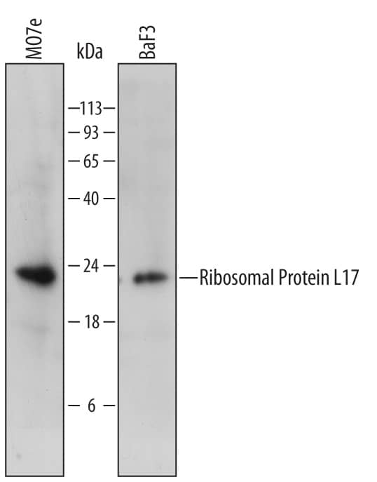
| Applications | WB, Flow |
| Clonality | Polyclonal |
| Host | Goat |
| Conjugate | Biotin |
| Concentration | LYOPH |
| Immunogen | Mouse myeloma cell line NS0-derived recombinant mouse B7-H4 Phe29-Pro258 Accession # Q7TSP5 |
| Specificity | Detects mouse B7-H4 in Western blots. In Western blots, less than 1% cross-reactivity with recombinant mouse (rm) B7‑1, rmB7‑2, rmB7‑H1, rmB7‑H2, rmB7‑H3, and rmPD‑L2 is observed. |
| Source | N/A |
| Isotype | IgG |
| Clonality | Polyclonal |
| Host | Goat |
| Gene | VTCN1 |
| Purity Statement | Antigen Affinity-purified |
| Innovator's Reward | Test in a species/application not listed above to receive a full credit towards a future purchase. |
| Dilutions |
|
|
| Readout System | ||
| Publications |
|
| Storage | Use a manual defrost freezer and avoid repeated freeze-thaw cycles.
|
| Buffer | Lyophilized from a 0.2 μm filtered solution in PBS with BSA as a carrier protein. |
| Preservative | No Preservative |
| Concentration | LYOPH |
| Reconstitution Instructions | Reconstitute at 0.2 mg/mL in sterile PBS. |
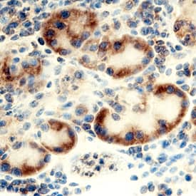
B7-H4, also known as B7x and B7S1, is a member of the B7 family of immune co-stimulatory proteins (1). Mature B7-H4 is a 50 kDa - 80 kDa glycosylated molecule with a 28 kDa protein core (2). Partial sensitivity to cleavage by PI-PLC suggests a possible GPI linkage of mouse B7-H4 to the cell membrane (3). The 230 amino acid (aa) extracellular region of B7-H4 contains one Ig-like V-set domain and one Ig-like C2-set domain which is followed by a hydrophobic C-terminal region (3 - 5). Within the ECD, mouse B7-H4 shares 90% and 99% aa sequence identity with human and rat B7-H4, respectively. It shares 21% - 29% aa sequence identity with B7-1, B7-2, B7-H1, B7-H2, B7-H3, and PD-L2. B7-H4 expression is induced on mitogen- or LPS-activated B cells, T cells, dendritic cells, monocytes, and macrophages (3, 4) and blocked by GM-CSF or IL-4 (6). It is also expressed on various normal epithelia and upregulated in several carcinomas and renal tubule epithelial cell lesions (2, 6 - 10). B7-H4 is expressed on the surface of macrophages but intracellularly in ovarian and breast cancer cells (2, 6 - 7). It is found in the serum and ascites fluid of cancer patients (11). B7-H4 binds an unidentified ligand on activated T cells which is distinct from BTLA, CD28, CTLA4, ICOS, and PD-1 (3 - 5, 12). Exposure to B7-H4 inhibits antigen-dependent induction of T cell proliferation and activation (3 - 5). Alternatively, B7-H4 expressing renal tubular epithelial cells promote T cell activation (8). Regulatory T cells, IL-6, and IL-10 induce B7-H4 expression on antigen presenting cells which fosters tumor growth by dampening the anti-tumor immune response (6, 13). B7-H4 also promotes the malignant transformation of epithelial cells by protecting them from apoptosis (2). Despite evidence for the involvement of B7-H4 in immune regulation, mice deficient in its expression do not show significant immune deficiencies, suggesting compensation by other molecules in vivo (14).
![Neutralization B7-1/CD80 Antibody (37711) [Unconjugated]](https://images.novusbio.com/images/antibody/B7-1_MAB140_Block_Neutralize_9024.jpg)
![Flow Cytometry B7-1/CD80 Antibody (37711) [Unconjugated]](https://images.novusbio.com/images/antibody/B71_MAB140_Flow_Cytometry_16025.jpg)
![Immunohistochemistry B7-1/CD80 Antibody (37711) [Unconjugated]](https://images.novusbio.com/images/mab140_human-b7-1-cd80-mab-clone-37711-4120241048542.jpg)


![Western Blot PD-L1 Antibody [Unconjugated]](https://images.novusbio.com/images/af1019_mouse-pd-l1-b7-h1-affinity-purified-polyclonal-ab-4120241048244.jpg)
![Flow Cytometry PD-L1 Antibody [Unconjugated]](https://images.novusbio.com/images/antibody/B7H1_AF1019_Flow_Cytometry_20531.jpg)
![In-situ Hybridization PD-L1 Antibody [Unconjugated]](https://images.novusbio.com/images/antibody/af1019_mouse-pd-l1-b7-h1-affinity-purified-polyclonal-ab-in-situ-hybridization-2352024211922..jpg)


![In-situ Hybridization PD-1 Antibody [Unconjugated]](https://images.novusbio.com/images/antibody/af1086_human-pd-1-affinity-purified-polyclonal-ab-in-situ-hybridization-235202421027..jpg)
![Western Blot PD-1 Antibody [Unconjugated]](https://images.novusbio.com/images/antibody/PD1_AF1086_Western_Blot_19120.jpg)


![Simple Western B7-H3/CD276 Antibody [Unconjugated]](https://images.novusbio.com/images/antibody/B7H3_AF1027_Simple_Western_20148.jpg)
![Flow Cytometry B7-H3/CD276 Antibody [Unconjugated]](https://images.novusbio.com/images/antibody/B7-H3_AF1027_Flow_Cytometry_8148.jpg)
![Western Blot B7-H3/CD276 Antibody [Unconjugated]](https://images.novusbio.com/images/antibody/B7H3_AF1027_Western_Blot_19947.jpg)
![B7-H4 Antibody [Biotin]](/sites/all/modules/enterprise-tech/et_datasheets/images/novus_guarantee.png "B7-H4 Antibody [Biotin]")
Secondary Antibodies |
Isotype Controls |
The concentration calculator allows you to quickly calculate the volume, mass or concentration of your vial. Simply enter your mass, volume, or concentration values for your reagent and the calculator will determine the rest.


![Bioactivity CTLA-4 [Unconjugated]](https://images.novusbio.com/images/protein/CTLA4_7268CT_2293.jpg)
![N/A IL-10 [Biotin]](https://images.novusbio.com/images/elisa/DATA_IL10_DY417_ELISA_2014.jpg)
![Bioactivity IL-2 [Unconjugated]](https://images.novusbio.com/images/202-il_recombinant-human-il-2-protein-bioactivity-174202314946.jpg)


![Agonist Activity CD28 Antibody (37407) [Unconjugated]](https://images.novusbio.com/images/antibody/CD28_MAB342_Functional_Assay_2393.jpg)
![Immunocytochemistry CD28 Antibody (37407) [Unconjugated]](https://images.novusbio.com/images/mab342_human-cd28-mab-clone-37407-immunocytochemistry-31102022134634.jpg)
![Bioactivity IL-4 [Unconjugated]](https://images.novusbio.com/images/protein/6507-ilcf_recombinant-human-il-4-cho-expressed-protein-cf-bioactivity-272020133214.jpg)


![Neutralization B7-H2/ICOSLG Antibody (136726) [Unconjugated]](https://images.novusbio.com/images/antibody/B7-H2_MAB165_Block_Neutralize_9035.jpg)
![Flow Cytometry B7-H2/ICOSLG Antibody (136726) [Unconjugated]](https://images.novusbio.com/images/antibody/B7-H2_MAB165_Flow_Cytometry_8343.jpg)
 or Normal Goat IgG Isotype Control Antibody (Catalog # AB-108-C, open histogram), followed by Phycoerythrin-conjugated Anti-Goat IgG Secondary Antibody (Catalog # F0107).")