 HAF005). A specific band was detected for ASGR1/ASGPR1 at approximately 42 kDa (as indicated). This experiment was conducted under reducing conditions and using Immunoblot Buffer Group 1." title="Western blot shows lysates of mouse liver tissue, human liver tissue (negative control), and HepG2 human hepatocellular carcinoma cell line (negative control). PVDF membrane was probed with 0.5 µg/mL of Rat Anti-Mouse ASGR1/ASGPR1 Monoclonal Antibody (Catalog # MAB27552) followed by HRP-conjugated Anti-Rat IgG Secondary Antibody (Catalog # HAF005). A specific band was detected for ASGR1/ASGPR1 at approximately 42 kDa (as indicated). This experiment was conducted under reducing conditions and using Immunoblot Buffer Group 1." />
| Reactivity | MuSpecies Glossary |
| Applications | WB, ELISA, IHC |
| Clone | 352803 |
| Clonality | Monoclonal |
| Host | Rat |
| Conjugate | Unconjugated |
| Immunogen | Mouse myeloma cell line NS0-derived recombinant mouse ASGR1/ASGPR1 Ser60-Asn284 Accession # NP_033844 |
| Specificity | Detects mouse ASGR1/ASGPR1 in direct ELISAs. |
| Source | N/A |
| Isotype | IgG2a |
| Clonality | Monoclonal |
| Host | Rat |
| Purity Statement | Protein A or G purified from hybridoma culture supernatant |
| Innovator's Reward | Test in a species/application not listed above to receive a full credit towards a future purchase. |
| Storage | Use a manual defrost freezer and avoid repeated freeze-thaw cycles.
|
| Buffer | Lyophilized from a 0.2 μm filtered solution in PBS with Trehalose. *Small pack size (SP) is supplied either lyophilized or as a 0.2 µm filtered solution in PBS. |
| Reconstitution Instructions | Reconstitute at 0.5 mg/mL in sterile PBS. |
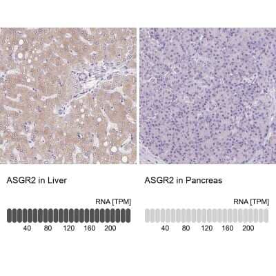
The mouse asialoglycoprotein receptor (ASGP-R) is an endocytic recycling receptor that belongs to the long-form subfamily of the C-type/Ca++-dependent lectin family (1‑3). It is a complex of two non‑covalently linked subunits, a major 42 kDa glycoprotein (ASGPR1), and a minor 51 kDa glycoprotein (ASGR2). The major mouse ASGP‑R subunit, ASGPR1, is synthesized as a 284 amino acid (aa) type II transmembrane (TM) protein that contains a 39 aa cytoplasmic region, a 21 aa TM segment, and a 224 aa extracellular domain (ECD) (4-6). The ECD contains two important structural regions. The first is a stalk region of 56 aa (aa’s 59-117) that contributes to non-covalent oligomerization. The second is a 118 aa, carbohydrate-binding, Ca++-dependent C-type lectin domain (aa’s 160-277) that is unusually stabilized by three Ca++ ions (3, 5). There are two potential alternate splice forms for ASGPR1. Both are TM and show a deletion of the C-type lectin domain. One is 113 aa in length and shows a deletion of aa’s 114-284 (7). The second is 132 aa in length and shows a deletion of aa’s 118-146 and aa’s 162-284 (8). Mouse ASGPR1 ECD is 89% and 79% aa identical to the ASGPR1 ECD in rat and human, respectively. The minor mouse ASGP-R subunit, ASGR2, is also a C-type lectin that shares the same structural organization as ASGR-1. It is 301 aa in length and has two 45 kDa and 51 kDa differentially-glycosylated isoforms (4, 6, 9). The ECD of ASGR2 is 50% aa identical to the ECD of ASGPR1. Although ASGPR1 and 2 can be expressed individually, a fully functional and stable ASGP-R requires simultaneous expression of both subunits (10-12). The stoichiometry of a functional ASGP-R is suggested to be either a 2:2, 3:1 or 3:2 ratio of ASGPR1:ASGR2 (13, 14). ASGPR1 is reported to bind Gal (nonreducing), GalNAc, and sialic acid alpha 2,6GalNAc (3, 15, 16). This is generally in the context of triantennary or tetraantennary configurations (2).
![Immunohistochemistry Albumin Antibody (188835) [Unconjugated] - Serum](https://images.novusbio.com/images/antibody/Albumin_MAB1455_Immunohistochemistry_23317.jpg)
![Immunocytochemistry Albumin Antibody (188835) [Unconjugated] - Serum](https://images.novusbio.com/images/antibody/Albumin_MAB1455_Immunocytochemistry__Immunofluorescence_17777.jpg)
![Intracellular Staining by Flow Cytometry Albumin Antibody (188835) [Unconjugated] - Serum](https://images.novusbio.com/images/antibody/Albumin_MAB1455_Flow_Cytometry_15110.jpg)
![Intracellular Staining by Flow Cytometry AKT [p Ser473] Antibody [Unconjugated] - Pan Specific](https://images.novusbio.com/images/antibody/Akt3_AF887_Flow_Cytometry_8283.jpg)
![Western Blot AKT [p Ser473] Antibody [Unconjugated] - Pan Specific](https://images.novusbio.com/images/af887_phospho-akt-s473-pan-specific-affinity-purified-pab-41202410485440.jpg)
![Western Blot AKT [p Ser473] Antibody [Unconjugated] - Pan Specific](https://images.novusbio.com/images/af887_phospho-akt-s473-pan-specific-affinity-purified-pab-8120255552843.jpg)


Secondary Antibodies |
Isotype Controls |
The concentration calculator allows you to quickly calculate the volume, mass or concentration of your vial. Simply enter your mass, volume, or concentration values for your reagent and the calculator will determine the rest.


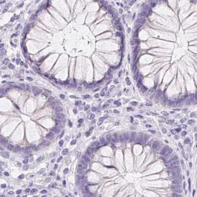
 CTS017) and counterstained with hematoxylin (blue). Specific staining was localized to cytoplasm in hepatocytes. View our protocol for
  MAB27551) coated on a Clear Polystyrene Microplate (Catalog #
 ![SDS-Page TNF-alpha [Unconjugated]](https://images.novusbio.com/images/protein/TNF-alpha_210-TA_256.jpg)
![Bioactivity TNF-alpha [Unconjugated]](https://images.novusbio.com/images/protein/TNFalpha_210TA_1658.jpg)
![SEC-MALS TNF-alpha [Unconjugated]](https://images.novusbio.com/images/210-ta_recombinant-human-tnf-alpha-protein-sec-mals-35202312244..jpg)


![Immunohistochemistry Caspase-3 Antibody [Unconjugated] - Active](https://images.novusbio.com/images/af835_human-mouse-active-caspase-3-affinity-purified-pab-41202410331943.jpg)
![Western Blot Caspase-3 Antibody [Unconjugated] - Active](https://images.novusbio.com/images/af835_human-mouse-active-caspase-3-affinity-purified-pab-812025554170.jpg)
![Western Blot Caspase-3 Antibody [Unconjugated] - Active](https://images.novusbio.com/images/af835_human-mouse-active-caspase-3-affinity-purified-pab-8120255534731.jpg)
![N/A Angiogenin [HRP]](https://images.novusbio.com/images/elisa/Angiogenin_DAN00_ELISA_28.jpg)
![N/A Angiogenin [HRP]](https://images.novusbio.com/images/elisa/DATA_Angiogenin_DAN00_ELISA_571.jpg)



![Western Blot ERK2 Antibody [Unconjugated]](https://images.novusbio.com/images/antibody/ERK2_AF1230_Western_Blot_5097.jpg)
![Knockout Validated ERK2 Antibody [Unconjugated]](https://images.novusbio.com/images/antibody/ERK2_AF1230_Knockout_Validated_22864.jpg)
![Immunohistochemistry ERK2 Antibody [Unconjugated]](https://images.novusbio.com/images/antibody/ERK2_AF1230_Immunohistochemistry_20696.jpg)
![N/A IL-6 [HRP]](https://images.novusbio.com/images/elisa/DATA_IL6_M6000_ELISA_936.jpg)
![N/A IL-6 [HRP]](https://images.novusbio.com/images/elisa/IL-6_M6000_ELISA_415.jpg)
![N/A IL-6 [HRP]](https://images.novusbio.com/images/m6000b_mouse-il-6-quantikine-elisa-kit-1752025024034.jpg)
![Immunohistochemistry Atrial Natriuretic Peptide/ANP Antibody [Unconjugated]](https://images.novusbio.com/images/antibody/Atrial_Natriuretic_Peptide_AF3366_Immunohistochemistry_14706.jpg)
![Simple Western Atrial Natriuretic Peptide/ANP Antibody [Unconjugated]](https://images.novusbio.com/images/antibody/Atrial_Natriuretic_Peptide_AF3366_Simple_Western_20228.jpg)
![Immunocytochemistry/ Immunofluorescence Atrial Natriuretic Peptide/ANP Antibody [Unconjugated]](https://images.novusbio.com/images/af3366_human-anp-affinity-purified-polyclonal-ab-41202412402519.jpg)


![Immunohistochemistry EphA3 Antibody [Unconjugated]](https://images.novusbio.com/images/antibody/EphA3_AF640_Immunohistochemistry_19184.jpg)
![Holo-Transferrin [Unconjugated]](/sites/all/modules/enterprise-tech/et_datasheets/images/novus_guarantee.png "Holo-Transferrin [Unconjugated]")


