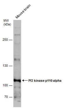
 or treated (+) with 100 nm Calyculin for 30 minutes, 1 µg/mL insulin for 5 minutes, or 100 ng/mL Recombinant Human PDGF-AB (Catalog # 222-AB) for 20 minutes. PVDF membrane was probed with 0.2 µg/mL of Mouse Anti-Human Phospho-Akt (S473) Pan Specific Monoclonal Antibody (Catalog # MAB887) followed by HRP-conjugated Anti-Mouse IgG Secondary Antibody (Catalog # HAF007). A specific band was detected for Phospho-Akt (S473) at approximately 65 kDa (as indicated). This experiment was conducted under reducing conditions and using Immunoblot Buffer Group 1.")
| Reactivity | Hu, MuSpecies Glossary |
| Applications | WB, Simple Western |
| Clone | 545007 |
| Clonality | Monoclonal |
| Host | Mouse |
| Conjugate | Unconjugated |
| Concentration | LYOPH |
| Immunogen | Phosphopeptide containing the human Akt (S473) site |
| Modification | p Ser473 |
| Specificity | Detects human and mouse Akt1, Akt2 and Akt3, when phosphorylated at S473, S474 and S472, respectively. |
| Source | N/A |
| Isotype | IgG1 |
| Clonality | Monoclonal |
| Host | Mouse |
| Gene | AKT1 |
| Purity Statement | Protein A or G purified from hybridoma culture supernatant |
| Innovator's Reward | Test in a species/application not listed above to receive a full credit towards a future purchase. |
| Dilutions |
|
|
| Application Notes | In Simple Western only 10-15 uL of the recommended dilution is used per data point. |
|
| Publications |
|
| Storage | Use a manual defrost freezer and avoid repeated freeze-thaw cycles.
|
| Buffer | Lyophilized from a 0.2 μm filtered solution in PBS with Trehalose. *Small pack size (SP) is supplied either lyophilized or as a 0.2 µm filtered solution in PBS. |
| Preservative | No Preservative |
| Concentration | LYOPH |
| Reconstitution Instructions | Sterile PBS to a final concentration of 0.5 mg/mL. |
![Simple Western EphB2 Antibody [Unconjugated]](https://images.novusbio.com/images2/EphB2_AF467_Simple_Western_21454.jpg)
![Western Blot EphB2 Antibody [Unconjugated]](https://images.novusbio.com/images2/EphB2_AF467_Western_Blot_21511.jpg)
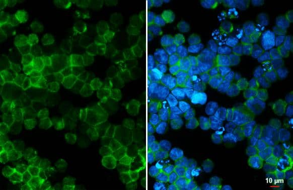
![Immunocytochemistry EphB2 Antibody [Unconjugated]](https://images.novusbio.com/images2/EphB2_AF467_Immunocytochemistry__Immunofluorescence_17288.jpg)
![Immunohistochemistry EGFR Antibody [Unconjugated]](https://images.novusbio.com/images2/EGF_R_AF231_Immunohistochemistry_20892.jpg)
![Immunocytochemistry EGFR Antibody [Unconjugated]](https://images.novusbio.com/images2/EGF_R_AF231_Immunocytochemistry__Immunofluorescence_21143.jpg)
![Western Blot EGFR Antibody [Unconjugated]](https://images.novusbio.com/images2/EGF_R_AF231_Western_Blot_19925.jpg)
![Western Blot PTEN Antibody [Unconjugated]](https://images.novusbio.com/images2/PTEN_AF847_Western_Blot_5922.jpg)
![Simple Western PTEN Antibody [Unconjugated]](https://images.novusbio.com/images2/16342.jpg)
![Knockout Validated PTEN Antibody [Unconjugated]](https://images.novusbio.com/images2/PTEN_AF847_Knockout_Validated_22996.jpg)


Secondary Antibodies |
Isotype Controls |
|
Insulin signaling in adipocytes: Carbohydrate-signaling transcription factor ChREBP is the link between lipolytic enzyme Hormone-Sensitive Lipase and lipogenic enzyme ELOVL6 By Jamshed Arslan, Pharm. D., PhD. Insulin resistance in adipocytes is a major feature of metabolic syndrome . Disrupted adipose tissue metabolism can lead to accumulation of lipid intermediates in insul... Read full blog post. |
|
mTOR Signaling and the Tumor Microenvironment By Yoskaly Lazo-Fernandez, PhD The mammalian target of rapamycin (mTOR) is a conserved serine/threonine kinase that, as a member of two distinct intracellular protein complexes, mTORC1 and mTORC2, regulates protein ... Read full blog post. |
|
Novel Insights into Hypoxia Induced AKT Signaling Hypoxia is a common feature of most tumors and is a product of rapid cell growth and poor vascularization1. When oxygen availability is low in the tumor environment, the hypoxia inducing transcription factors (HIFs) regulate a variety of signaling ... Read full blog post. |
|
Pathway Highlight: Three key factors that contribute to cellular heterogeneity in apoptosis Have you ever wondered why cells in the same population respond differently to an apoptotic stimulus? Apoptosis, a form of programmed cell death, is vital for the removal of unwanted or damaged cells. As with most cellular processes, too much or to... Read full blog post. |
|
The role of c-Fos in the regulation of the JC virus gene transcription c-Fos is a member of the AP-1 transcription factor family under the Fos protein family umbrella, alongside Fra-1, Fra-2 and Fos-B. Also in the AP-1 transcription family are the Jun proteins, c-Jun, Jun-B and Jun-D. Each member of the AP-1 transcri... Read full blog post. |
|
Pathway Highlight: Which caspase substrates contribute to the morphological features associated with apoptosis? Apoptosis, or programmed cell death, is controlled by a caspase signal cascade that activates downstream signals to induce the morphological changes used to differentiate apoptosis from other forms of cell death. Novus Biologicals offers a variet... Read full blog post. |
|
The use of apoptosis antibodies and controls in cell death research Apoptosis is a method of programmed cell death that is notably characterized by a morphological change in cellular nuclei and membrane appearance. Not to be confused with necrosis, apoptosis is a pathway that is induced by a variety of factors tha... Read full blog post. |
|
The role of Parkin and autophagy in retinal pigment epithelial cell (RPE) degradation The root of Parkinson’s disease (PD) points to a poorly regulated electron transport chain leading to mitochondrial damage, where many proteins need to work cohesively to ensure proper function. The two key players of this pathway are PINK1, ... Read full blog post. |
|
The effects of curcumin on IKB Alpha and the NFkB signaling pathway The IKK complex, or inhibitor of NFkB kinase, is composed of IKK alpha and IKK beta. These kinases are at the core of the NFkB signaling cascade. The NFkB family is made up of transcription factors that are kept inactive in the cytoplasm through... Read full blog post. |
|
AMPK Alpha 1 and lipid metabolism of adipocytes AMP-activated protein kinase (AMPK) is best known as a sensor of oxidative stress. AMPK is activated by increased intracellular AMP levels, which are a result of alterations in cellular metabolism from causes such as hypoxia, changes in ATP, sene... Read full blog post. |
The concentration calculator allows you to quickly calculate the volume, mass or concentration of your vial. Simply enter your mass, volume, or concentration values for your reagent and the calculator will determine the rest.
| Gene Symbol | AKT1 |


 or treated (+) with 100 ng/mL Recombinant Human IGF‑I (Catalog # 291-G1) for 20 minutes, loaded at 0.2 mg/mL. A specific band was detected for Phospho-Akt (S473) at approximately 65 kDa (as indicated) using 2 µg/mL of Mouse Anti-Human/Mouse Phospho-Akt (S473) Pan Specific Monoclonal Antibody (Catalog # MAB887). This experiment was conducted under reducing conditions and using the 12-230 kDa separation system.")
 score plot: Metabolomics profiling of control and ACPA-treated A549, H1299, H358, and H838 cells. b Changes in variable importance in projection (VIP) values for 19 metabolites in A549 cells. c, d, e Changes in VIP values for 20 metabolites in H1299, H358, and H838 cells. Significantly changed metabolites (*p < 0.05, indicated by arrows) were matched to apoptotic pathways. f, g, h, i Increase and decrease in several metabolites of ACPA-treated A549, H1299, H358, and H838 cells (*p < 0.05). j Simple Western showing total Akt, p-Akt (S473), total JNK46 and JNK54 and p-JNK46 and p-JNK54 (T183/Y185) in A549 cells at 24 hours after treatment with IC50 dose of ACPA. k Relative expression levels of Akt and p-Akt for control and ACPA-treated A549 cells after normalization by total vinculin protein. l Relative expression levels of JNK (46 and 54 kDa) and p-JNK for control and ACPA-treated A549 cells after normalization by total vinculin protein. *p < 0.05, Student’s t-test. All tests were done in quadruplicates. Image collected and cropped by CiteAb from the following publication (//pubmed.ncbi.nlm.nih.gov/33431819), licensed under a CC-BY license. Not internally tested by R&D Systems.")
 PTEN and pAKT levels in kidney cancer cell lines, IOD calculated relative to Vinculin (B) Relative to GusB expression of Pten in RNA isolates from kidney cancer and healthy kidney tissues (C) graphical workflow. Image collected and cropped by CiteAb from the following open publication (//pubmed.ncbi.nlm.nih.gov/35625614), licensed under a CC-BY license. Not internally tested by R&D Systems.")
 PTEN and pAKT levels in kidney cancer cell lines, IOD calculated relative to Vinculin (B) Relative to GusB expression of Pten in RNA isolates from kidney cancer and healthy kidney tissues (C) graphical workflow. Image collected and cropped by CiteAb from the following open publication (//pubmed.ncbi.nlm.nih.gov/35625614), licensed under a CC-BY license. Not internally tested by R&D Systems.")


![Western Blot ERK2 Antibody [Unconjugated]](https://images.novusbio.com/images2/ERK2_AF1230_Western_Blot_5097.jpg)
![Immunohistochemistry ERK2 Antibody [Unconjugated]](https://images.novusbio.com/images2/ERK2_AF1230_Immunohistochemistry_20696.jpg)
![Knockout Validated ERK2 Antibody [Unconjugated]](https://images.novusbio.com/images2/ERK2_AF1230_Knockout_Validated_22864.jpg)


![Simple Western PYK2/FAK2 [p Tyr402] Antibody (592918) [Unconjugated]](https://images.novusbio.com/images2/PYK2_MAB6210_Simple_Western_16639.jpg)
![Western Blot PYK2/FAK2 [p Tyr402] Antibody (592918) [Unconjugated]](https://images.novusbio.com/images2/PYK2_MAB6210_Western_Blot_9430.jpg)
![Western Blot PYK2/FAK2 [p Tyr402] Antibody (592918) [Unconjugated]](https://images.novusbio.com/images/mab6210_human-phospho-pyk2-y402-mab-clone-592918-western-blot-1212202585785.jpg)
![Immunohistochemistry-Paraffin TOR/mTOR [p Ser2448] Antibody - BSA Free](https://images.novusbio.com/images/TOR-mTOR-[p-Ser2448]-Antibody-Immunohistochemistry-Paraffin-NB600-607-img0005.jpg)
![Western Blot TOR/mTOR [p Ser2448] Antibody - BSA Free](https://images.novusbio.com/images/TOR-mTOR-[p-Ser2448]-Antibody-Western-Blot-NB600-607-img0006.jpg)
![Data TOR/mTOR [p Ser2448] Antibody - BSA Free](https://images.novusbio.com/images/TOR-mTOR-[p-Ser2448]-Antibody-N-A-NB600-607-img0008.jpg)
![Immunocytochemistry Insulin Antibody (182410) [Unconjugated]](https://images.novusbio.com/images2/Insulin_MAB1417_Immunocytochemistry_9376.jpg)
![Immunohistochemistry Insulin Antibody (182410) [Unconjugated]](https://images.novusbio.com/images2/mab1417_human-bovine-mouse-insulin-mab-clone-182410-immunohistochemistry-308202115145.jpg)
![SDS-PAGE TNF-alpha [Unconjugated]](https://images.novusbio.com/images2/TNF-alpha_210-TA_256.jpg)
![Bioactivity TNF-alpha [Unconjugated]](https://images.novusbio.com/images2/TNFalpha_210TA_1658.jpg)
![SEC-MALS TNF-alpha [Unconjugated]](https://images.novusbio.com/images/210-ta_recombinant-human-tnf-alpha-protein-sec-mals-35202312244..jpg)
![N/A VEGF [HRP]](https://images.novusbio.com/images2/DATA_VEGF_DVE00_ELISA_871.jpg)
![N/A VEGF [HRP]](https://images.novusbio.com/images2/DATA_VEGF_DVE00_ELISA_872.jpg)
![N/A VEGF [HRP]](https://images.novusbio.com/images2/VEGF_DVE00_ELISA_208.jpg)
![Immunocytochemistry Caspase-3 Antibody [Unconjugated] - Active](https://images.novusbio.com/images2/Caspase-3_AF835_Immunocytochemistry_6532.jpg)
![Immunohistochemistry Caspase-3 Antibody [Unconjugated] - Active](https://images.novusbio.com/images2/Caspase3_AF835_Immunohistochemistry_22976.jpg)
![Immunocytochemistry Caspase-3 Antibody [Unconjugated] - Active](https://images.novusbio.com/images2/Caspase-3_AF835_Immunocytochemistry_9340.jpg)
![SDS-PAGE IGF-I/IGF-1 [Unconjugated]](https://images.novusbio.com/images2/IGF-I_291-G1_40.jpg)
![Bioactivity IGF-I/IGF-1 [Unconjugated]](https://images.novusbio.com/images2/IGF-I_291-G1_41.jpg)
![Mass Spectrometry IGF-I/IGF-1 [Unconjugated]](https://images.novusbio.com/images2/IGF-I_291-G1_42.jpg)
![Western Blot JNK1 Antibody (228601) [Unconjugated]](https://images.novusbio.com/images2/JNK1_MAB17761_Western_Blot_5991.jpg)
![Western Blot JNK1 Antibody (228601) [Unconjugated]](https://images.novusbio.com/images2/JNK1_MAB17761_Western_Blot_6327.jpg)
![Immunocytochemistry JNK1 Antibody (228601) [Unconjugated]](https://images.novusbio.com/images2/JNK1_MAB17761_Immunocytochemistry__Immunofluorescence_20184.jpg)