HAF007). This experiment was conducted under reducing conditions and using
Immunoblot Buffer Group 4." class="big_lightbox">
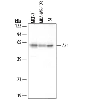
HAF007). This experiment was conducted under reducing conditions and using
Immunoblot Buffer Group 4." alt="Western blot shows lysates of MCF-7 human breast cancer cell line, MBA-MB-123 human breast cancer cell line, and TS1 mouse helper T cell line. PVDF membrane was probed with 0.2 µg/mL Mouse Anti-Human/Mouse/Rat Akt Pan Specific Monoclonal Antibody (Catalog # MAB2055) followed by HRP-conjugated Anti-Mouse IgG Secondary Antibody (Catalog #
HAF007). This experiment was conducted under reducing conditions and using
Immunoblot Buffer Group 4." class="big_thumb" />
1775-KS), recombinant human Akt2, and recombinant human Akt3 (5 ng/lane). PVDF membrane was probed with 0.2 µg/mL Mouse Anti-Human/Mouse/Rat Akt Pan Specific Monoclonal Antibody (Catalog # MAB2055) followed by HRP-conjugated Anti-Mouse IgG Secondary Antibody (Catalog #
HAF007). This experiment was conducted under reducing conditions and using
Immunoblot Buffer Group 4." class="big_lightbox">
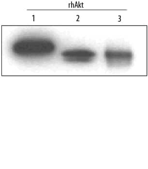
1775-KS), recombinant human Akt2, and recombinant human Akt3 (5 ng/lane). PVDF membrane was probed with 0.2 µg/mL Mouse Anti-Human/Mouse/Rat Akt Pan Specific Monoclonal Antibody (Catalog # MAB2055) followed by HRP-conjugated Anti-Mouse IgG Secondary Antibody (Catalog #
HAF007). This experiment was conducted under reducing conditions and using
Immunoblot Buffer Group 4." class="big_thumb" />
MAB6815). Both antibodies were used at 10 µg/mL for 3 hours at room temperature. Cells were stained using the NorthernLights™ 557-conjugated Anti-Mouse IgG Secondary Antibody (red; Catalog #
NL007) and counterstained with DAPI (blue). Specific staining was localized to plasma membranes and cytoplasm. View our protocol for
Fluorescent ICC Staining of Cells on Coverslips." class="big_lightbox">
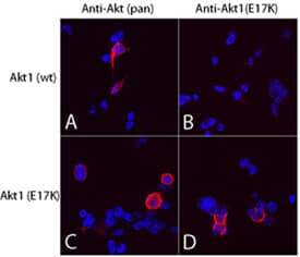
MAB6815). Both antibodies were used at 10 µg/mL for 3 hours at room temperature. Cells were stained using the NorthernLights™ 557-conjugated Anti-Mouse IgG Secondary Antibody (red; Catalog #
NL007) and counterstained with DAPI (blue). Specific staining was localized to plasma membranes and cytoplasm. View our protocol for
Fluorescent ICC Staining of Cells on Coverslips." class="big_thumb" />
VC001). Before incubation with the primary antibody, tissue was subjected to heat-induced epitope retrieval using Antigen Retrieval Reagent-Basic (
CTS013). Tissue was stained using DAB (brown) and counterstained with hematoxylin (blue). Specific staining was localized to cytoplasm in epithelial cells. Staining was performed using our IHC Staining with VisUCyte HRP Polymer Detection Reagents." class="big_lightbox">
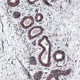
VC001). Before incubation with the primary antibody, tissue was subjected to heat-induced epitope retrieval using Antigen Retrieval Reagent-Basic (
CTS013). Tissue was stained using DAB (brown) and counterstained with hematoxylin (blue). Specific staining was localized to cytoplasm in epithelial cells. Staining was performed using our IHC Staining with VisUCyte HRP Polymer Detection Reagents." class="big_thumb" />
VC001). Before incubation with the primary antibody, tissue was subjected to heat-induced epitope retrieval using Antigen Retrieval Reagent-Basic (
CTS013). Tissue was stained using DAB (brown) and counterstained with hematoxylin (blue). Specific staining was localized to cytoplasm in lymphocytes. Staining was performed using our IHC Staining with VisUCyte HRP Polymer Detection Reagents." class="big_lightbox">
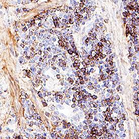
VC001). Before incubation with the primary antibody, tissue was subjected to heat-induced epitope retrieval using Antigen Retrieval Reagent-Basic (
CTS013). Tissue was stained using DAB (brown) and counterstained with hematoxylin (blue). Specific staining was localized to cytoplasm in lymphocytes. Staining was performed using our IHC Staining with VisUCyte HRP Polymer Detection Reagents." class="big_thumb" />
MAB0041, open histogram), followed by Phycoerythrin-conjugated Anti-Mouse IgG F(ab')
2Secondary Antibody (Catalog #
F0102B). To facilitate intracellular staining, cells were fixed with paraformaldehyde and permeabilized with saponin." class="big_lightbox">

MAB0041, open histogram), followed by Phycoerythrin-conjugated Anti-Mouse IgG F(ab')
2Secondary Antibody (Catalog #
F0102B). To facilitate intracellular staining, cells were fixed with paraformaldehyde and permeabilized with saponin." class="big_thumb" />
 120-HD) for 15 minutes and MCF-7 human breast cancer cell line untreated or treated with 100 ng/mL Recombinant Human IGF-1 (291-G1) for 15 minutes. PVDF membrane was probed with 0.5 µg/mL of Rabbit Anti-Human/Mouse/Rat Phospho-Akt (S473) Pan Specific Antigen Affinity-purified Polyclonal Antibody (Catalog # AF887), followed by HRP-conjugated Anti-Rabbit IgG Secondary Antibody (HAF008). A specific band was detected for Phospho-Akt (S473) Pan Specific at approximately 60 kDa (as indicated). This experiment was conducted under reducing conditions and using Immunoblot Buffer Group 1." alt="Western blot shows lysates of NIH-3T3 mouse embryonic fibroblast cell line untreated (-) or treated (+) with 100 ng/mL Human PDGF (120-HD) for 15 minutes and MCF-7 human breast cancer cell line untreated or treated with 100 ng/mL Recombinant Human IGF-1 (291-G1) for 15 minutes. PVDF membrane was probed with 0.5 µg/mL of Rabbit Anti-Human/Mouse/Rat Phospho-Akt (S473) Pan Specific Antigen Affinity-purified Polyclonal Antibody (Catalog # AF887), followed by HRP-conjugated Anti-Rabbit IgG Secondary Antibody (HAF008). A specific band was detected for Phospho-Akt (S473) Pan Specific at approximately 60 kDa (as indicated). This experiment was conducted under reducing conditions and using Immunoblot Buffer Group 1." class="big_thumb" />
 HAF007). This experiment was conducted under reducing conditions and using Immunoblot Buffer Group 4." alt="Western blot shows lysates of MCF-7 human breast cancer cell line, MBA-MB-123 human breast cancer cell line, and TS1 mouse helper T cell line. PVDF membrane was probed with 0.2 µg/mL Mouse Anti-Human/Mouse/Rat Akt Pan Specific Monoclonal Antibody (Catalog # MAB2055) followed by HRP-conjugated Anti-Mouse IgG Secondary Antibody (Catalog # HAF007). This experiment was conducted under reducing conditions and using Immunoblot Buffer Group 4." class="big_thumb" />
 222-AB) for 20 minutes. PVDF membrane was probed with 0.2 µg/mL of Mouse Anti-Human Phospho-Akt (S473) Pan Specific Monoclonal Antibody (Catalog # MAB887) followed by HRP-conjugated Anti-Mouse IgG Secondary Antibody (Catalog # HAF007). A specific band was detected for Phospho-Akt (S473) at approximately 65 kDa (as indicated). This experiment was conducted under reducing conditions and using Immunoblot Buffer Group 1." alt="Western blot shows lysates of Jurkat human acute T cell leukemia cell line, MCF-7 human breast cancer cell line, and NIH-3T3 mouse embryonic fibroblast cell line untreated (-) or treated (+) with 100 nm Calyculin for 30 minutes, 1 µg/mL insulin for 5 minutes, or 100 ng/mL Recombinant Human PDGF-AB (Catalog # 222-AB) for 20 minutes. PVDF membrane was probed with 0.2 µg/mL of Mouse Anti-Human Phospho-Akt (S473) Pan Specific Monoclonal Antibody (Catalog # MAB887) followed by HRP-conjugated Anti-Mouse IgG Secondary Antibody (Catalog # HAF007). A specific band was detected for Phospho-Akt (S473) at approximately 65 kDa (as indicated). This experiment was conducted under reducing conditions and using Immunoblot Buffer Group 1." class="big_thumb" />
 HAF008). A specific band was detected for Akt Pan Specific, at approximately 60 kDa (as indicated). This experiment was conducted under reducing conditions and using Immunoblot Buffer Group 1." alt="Western blot shows lysates of HeLa human cervical epithelial carcinoma cell line, Jurkat human acute T cell leukemia cell line, HepG2 human hepatocellular carcinoma cell line, MCF-7 human breast cancer cell line, and KG-1 human myeloid leukemia cell lines. PVDF membrane was probed with 0.1 µg/mL of Human Akt Pan Specific, Antigen Affinity-purified Polyclonal Antibody (Catalog # AF2055) followed by HRP-conjugated Anti-Rabbit IgG Secondary Antibody (Catalog # HAF008). A specific band was detected for Akt Pan Specific, at approximately 60 kDa (as indicated). This experiment was conducted under reducing conditions and using Immunoblot Buffer Group 1." class="big_thumb" />


 CTS005) and counterstained with hematoxylin (blue). Lower panel shows a lack of labeling if primary antibodies are omitted and tissue is stained only with secondary antibody followed by incubation with detection reagents. View our protocol for
  F0110). To facilitate intracellular staining, cells were fixed with formaldehyde (
 291-G1) for 15 minutes, loaded at 0.2 mg/mL. A specific band was detected for Phospho-Akt (S473) at approximately 60 kDa (as indicated) using 5 µg/mL of Rabbit Anti-Human/Mouse/Rat Phospho-Akt (S473) Pan Specific Antigen Affinity-purified Polyclonal Antibody (Catalog # AF887). This experiment was conducted under reducing conditions and using the 12-230 kDa separation system." class="big_thumb" />


 1775-KS), recombinant human Akt2, and recombinant human Akt3 (5 ng/lane). PVDF membrane was probed with 0.2 µg/mL Mouse Anti-Human/Mouse/Rat Akt Pan Specific Monoclonal Antibody (Catalog # MAB2055) followed by HRP-conjugated Anti-Mouse IgG Secondary Antibody (Catalog #
  NL007) and counter-stained with DAPI (blue). View our protocol for
  MAB6815). Both antibodies were used at 10 µg/mL for 3 hours at room temperature. Cells were stained using the NorthernLights™ 557-conjugated Anti-Mouse IgG Secondary Antibody (red; Catalog #
  VC001). Before incubation with the primary antibody, tissue was subjected to heat-induced epitope retrieval using Antigen Retrieval Reagent-Basic (
 VC001). Before incubation with the primary antibody, tissue was subjected to heat-induced epitope retrieval using Antigen Retrieval Reagent-Basic (
 MAB0041, open histogram), followed by Phycoerythrin-conjugated Anti-Mouse IgG F(ab')2Secondary Antibody (Catalog #


 291-G1) for 20 minutes, loaded at 0.2 mg/mL. A specific band was detected for Phospho-Akt (S473) at approximately 65 kDa (as indicated) using 2 µg/mL of Mouse Anti-Human/Mouse Phospho-Akt (S473) Pan Specific Monoclonal Antibody (Catalog # MAB887). This experiment was conducted under reducing conditions and using the 12-230 kDa separation system." class="big_thumb" />


![Immunohistochemistry: AKT [p Ser 473] Antibody (SR2061) [NBP3-21578] - Immunohistochemical analysis of paraffin-embedded human kidney, using NBP3-21578.](http://images.novusbio.com/fullsize/nbp3-21578_rabbit-akt-p-ser-473-mab-sr2061-57202315545125.jpg "Immunohistochemistry: AKT [p Ser 473] Antibody (SR2061) [NBP3-21578] - Immunohistochemical analysis of paraffin-embedded human kidney, using NBP3-21578.")
![<b>Biological Strategies Validation. </b>Western Blot: AKT [p Ser 473] Antibody (SR2061) [NBP3-21578] - Western blot analysis on 2 NIH/3T3 cell lysate treated with PDGF using NBP3-21578.](http://images.novusbio.com/fullsize/nbp3-21578_rabbit-akt-p-ser-473-mab-sr2061-6720239533735.jpg)
![<b>Biological Strategies Validation. </b>Immunocytochemistry/Immunofluorescence: AKT [p Ser 473] Antibody (SR2061) [NBP3-21578] - Immunofluorescent analysis of NIH/3T3 cells treated with PDGF, using NBP3-21578.](http://images.novusbio.com/fullsize/nbp3-21578_rabbit-akt-p-ser-473-mab-sr2061-6720231051266.jpg)
![Whole cell extract (30 ug) was separated by 10% SDS-PAGE, and the membrane was blotted with AKT antibody [HL2915] (NBP3-48751) diluted at 1:1000. The HRP-conjugated anti-rabbit IgG antibody was used to detect the primary antibody.](http://images.novusbio.com/fullsize/nbp3-48751_rabbit-akt-mab-hl2915-western-blot-1411202518341523.jpg "Whole cell extract (30 ug) was separated by 10% SDS-PAGE, and the membrane was blotted with AKT antibody [HL2915] (NBP3-48751) diluted at 1:1000. The HRP-conjugated anti-rabbit IgG antibody was used to detect the primary antibody.")
![AKT antibody [HL2915] detects AKT protein by immunohistochemical analysis.</br>Sample: Paraffin-embedded rat esophagus.</br>AKT stained by AKT antibody [HL2915] (NBP3-48751) diluted at 1:100.</br>Antigen Retrieval: Citrate buffer, pH 6.0, 15 min](http://images.novusbio.com/fullsize/nbp3-48751_rabbit-akt-mab-hl2915-immunohistochemistry-paraffin-1411202518382820.jpg)
![Non-transfected (-) and transfected (+) 293T whole cell extracts were separated by 10% SDS-PAGE, and the membrane was blotted with AKT antibody [HL2915] (NBP3-48751) diluted at 1:5000. The HRP-conjugated anti-rabbit IgG antibody was used to detect the primary antibody.](http://images.novusbio.com/fullsize/nbp3-48751_rabbit-akt-mab-hl2915-western-blot-1411202518435619.jpg)
![AKT antibody [HL2915] detects AKT protein by immunohistochemical analysis.</br>Sample: Paraffin-embedded mouse tissues.</br>AKT stained by AKT antibody [HL2915] (NBP3-48751) diluted at 1:100.</br>Antigen Retrieval: Citrate buffer, pH 6.0, 15 min](http://images.novusbio.com/fullsize/nbp3-48751_rabbit-akt-mab-hl2915-immunohistochemistry-paraffin-141120251841520.jpg)
![AKT antibody [HL2915] detects AKT protein by immunofluorescent analysis.</br>Sample: HeLa cells were fixed in ice-cold MeOH for 5 min.</br>Green: AKT stained by AKT antibody [HL2915] (NBP3-48751) diluted at 1:500.</br>Red: alpha Tubulin, a cytoskeleton marker, stained by alpha Tubulin antibody [GT114] diluted at 1:1000.</br>Blue: Fluoroshield with DAPI .](http://images.novusbio.com/fullsize/nbp3-48751_rabbit-akt-mab-hl2915-immunocytochemistry-immunofluorescence-1411202518424111.jpg)
![AKT antibody [HL2915] detects AKT protein by immunohistochemical analysis.</br>Sample: Paraffin-embedded human breast carcinoma.</br>AKT stained by AKT antibody [HL2915] (NBP3-48751) diluted at 1:100.</br>Antigen Retrieval: Citrate buffer, pH 6.0, 15 min](http://images.novusbio.com/fullsize/nbp3-48751_rabbit-akt-mab-hl2915-immunohistochemistry-paraffin-1411202518312110.jpg)
![Various whole cell extracts (30 ug) were separated by 10% SDS-PAGE, and the membrane was blotted with AKT antibody [HL2915] (NBP3-48751) diluted at 1:1000. The HRP-conjugated anti-rabbit IgG antibody was used to detect the primary antibody.](http://images.novusbio.com/fullsize/nbp3-48751_rabbit-akt-mab-hl2915-western-blot-1411202518394118.jpg)
![Whole cell extract (30 ug) was separated by 10% SDS-PAGE, and the membrane was blotted with AKT antibody [HL2915] (NBP3-48751) diluted at 1:1000. The HRP-conjugated anti-rabbit IgG antibody was used to detect the primary antibody.](http://images.novusbio.com/fullsize/nbp3-48751_rabbit-akt-mab-hl2915-western-blot-1411202518334523.jpg)

![Western Blot: AKT [p Thr450] Antibody [NBP3-13168] - Various whole cell extracts (30 ug) were separated by 10% SDS-PAGE, and the membrane was blotted with AKT (phospho Thr450) antibody (NBP3-13168) diluted at 1:500. The HRP-conjugated anti-rabbit IgG antibody (NBP2-19301) was used to detect the primary antibody.](http://images.novusbio.com/fullsize/AKT-[p-Thr450]-Antibody-Western-Blot-NBP3-13168-img0004.jpg "Western Blot: AKT [p Thr450] Antibody [NBP3-13168] - Various whole cell extracts (30 ug) were separated by 10% SDS-PAGE, and the membrane was blotted with AKT (phospho Thr450) antibody (NBP3-13168) diluted at 1:500. The HRP-conjugated anti-rabbit IgG antibody (NBP2-19301) was used to detect the primary antibody.")
![Western Blot: AKT [p Thr450] Antibody [NBP3-13168] - Various whole cell extracts (30 ug) were separated by 10% SDS-PAGE, and the membrane was blotted with AKT (phospho Thr450) antibody (NBP3-13168) diluted at 1:500. The HRP-conjugated anti-rabbit IgG antibody (NBP2-19301) was used to detect the primary antibody.](http://images.novusbio.com/fullsize/AKT-[p-Thr450]-Antibody-Western-Blot-NBP3-13168-img0002.jpg)
![Western Blot: AKT [p Thr450] Antibody [NBP3-13168] - Various whole cell extracts (30 ug) were separated by 10% SDS-PAGE, and the membrane was blotted with AKT (phospho Thr450) antibody (NBP3-13168) diluted at 1:500. The HRP-conjugated anti-rabbit IgG antibody (NBP2-19301) was used to detect the primary antibody.](http://images.novusbio.com/fullsize/AKT-[p-Thr450]-Antibody-Western-Blot-NBP3-13168-img0003.jpg)


![Western Blot: AKT [p Ser473] Antibody [NBP3-13120] - Untreated (-) and treated (+) MCF-7 whole cell extracts (30 ug) were separated by 10% SDS-PAGE, and the membrane was blotted with AKT (phospho Ser473) antibody (NBP3-13120) diluted at 1:1000. The HRP-conjugated anti-rabbit IgG antibody (NBP2-19301) was used to detect the primary antibody.](http://images.novusbio.com/fullsize/AKT-[p-Ser473]-Antibody-Western-Blot-NBP3-13120-img0003.jpg "<b>Biological Strategies Validation. </b>Western Blot: AKT [p Ser473] Antibody [NBP3-13120] - Untreated (-) and treated (+) MCF-7 whole cell extracts (30 ug) were separated by 10% SDS-PAGE, and the membrane was blotted with AKT (phospho Ser473) antibody (NBP3-13120) diluted at 1:1000. The HRP-conjugated anti-rabbit IgG antibody (NBP2-19301) was used to detect the primary antibody.")
![<b>Biological Strategies Validation. </b>Western Blot: AKT [p Ser473] Antibody [NBP3-13120] - Untreated (-) and treated (+) NIH-3T3 whole cell extracts (30 ug) were separated by 7.5% SDS-PAGE, and the membrane was blotted with AKT (phospho Ser473) antibody (NBP3-13120) diluted at 1:1000.](http://images.novusbio.com/fullsize/AKT-[p-Ser473]-Antibody-Western-Blot-NBP3-13120-img0001.jpg)
![Western Blot: AKT [p Ser473] Antibody [NBP3-13120] - Various whole cell extracts (30 ug) were separated by 10% SDS-PAGE, and the membrane was blotted with AKT (phospho Ser473) antibody (NBP3-13120) diluted at 1:500. The HRP-conjugated anti-rabbit IgG antibody (NBP2-19301) was used to detect the primary antibody.](http://images.novusbio.com/fullsize/AKT-[p-Ser473]-Antibody-Western-Blot-NBP3-13120-img0002.jpg)
![Western Blot: AKT [p Ser308] Antibody (0002) [NBP3-23519] - Western blot analysis of extracts from serum-starved NIH-3T3, untreated (line A); treated with PDGFA (5 ug/mL, 5 min) (line B) using NBP3-23519 at 1:5000 dilution.](http://images.novusbio.com/fullsize/nbp3-23519_rabbit-akt-mab-0002-azide-and-bsa-free-235202514331565.png "Western Blot: AKT [p Ser308] Antibody (0002) [NBP3-23519] - Western blot analysis of extracts from serum-starved NIH-3T3, untreated (line A); treated with PDGFA (5 ug/mL, 5 min) (line B) using NBP3-23519 at 1:5000 dilution.")
![Western Blot: AKT [p Ser473] Antibody (PSH04-44) [NBP3-32747] - Western blot analysis of AKT on different lysates with Rabbit anti-AKT antibody (NBP3-32747) at 1/2,000 dilution.<br/><br/> Lane 1: HEK-293 cell lysate<br/> Lane 2: HEK-293 treated with 50uM LY294002 for 6 hours cell lysate<br/><br/> Lysates/proteins at 20 ug/Lane.<br/><br/> Predicted band size: 56 kDa<br/> Observed band size: 56 kDa<br/><br/> Exposure time: 20 seconds; ECL;<br/><br/> 4-20% SDS-PAGE gel.<br/><br/> Proteins were transferred to a PVDF membrane and blocked with 5% NFDM/TBST for 1 hour at room temperature. The primary antibody (NBP3-32747) at 1/2,000 dilution was used in 5% NFDM/TBST at 4 overnight. Goat Anti-Rabbit IgG - HRP Secondary Antibody at 1/50,000 dilution was used for 1 hour at room temperature.](http://images.novusbio.com/fullsize/nbp3-32747_rabbit-akt-p-ser473-mab-psh04-44-278202415242614.jpg "Western Blot: AKT [p Ser473] Antibody (PSH04-44) [NBP3-32747] - Western blot analysis of AKT on different lysates with Rabbit anti-AKT antibody (NBP3-32747) at 1/2,000 dilution.<br/><br/> Lane 1: HEK-293 cell lysate<br/> Lane 2: HEK-293 treated with 50uM LY294002 for 6 hours cell lysate<br/><br/> Lysates/proteins at 20 ug/Lane.<br/><br/> Predicted band size: 56 kDa<br/> Observed band size: 56 kDa<br/><br/> Exposure time: 20 seconds; ECL;<br/><br/> 4-20% SDS-PAGE gel.<br/><br/> Proteins were transferred to a PVDF membrane and blocked with 5% NFDM/TBST for 1 hour at room temperature. The primary antibody (NBP3-32747) at 1/2,000 dilution was used in 5% NFDM/TBST at 4 overnight. Goat Anti-Rabbit IgG - HRP Secondary Antibody at 1/50,000 dilution was used for 1 hour at room temperature.")
![Western blot analysis on different lysates with NBP3-32747 at 1/2,000 dilution and pan AKT antibody at 1/2,000 dilution. Lane 1: MCF7 cell lysate Lane 2: MCF7 treated with 50ng/mL Calyculin A for 45 minutes cell lysate Lane 3: SH-SY5Y cell lysate Lane 4: SH-SY5Y treated with 100ng/mL PDGF for 5 minutes cell lysate Lane 5: HEK-293 cell lysate Lane 6: HEK-293 treated with 50μM LY294002 for 6 hours cell lysate Lane 7: NIH/3T3 cell lysate Lane 8: NIH/3T3 treated with 100ng/mL PDGF for 5 minutes cell lysate Lane 9: C6 cell lysate Lane 10: C6 treated with 100ng/mL PDGF for 5 minutes cell lysate Lane 11: MCF7 treated with 50ng/mL Calyculin A for 45 minutes cell lysate, then the membrane treated with lambda pp for 1 hour Lysates/proteins at 20 µg/Lane. Predicted band size: 56 kDa Observed band size: 56 kDa Exposure time: 20 seconds; 4-20% SDS-PAGE gel. Proteins were transferred to a PVDF membrane and blocked with 5% BSA for 1 hour at room temperature. NBP3-32747 at 1/2,000 dilution and pan AKT antibody (HA721870) at 1/2,000 dilution were used in 5% BSA at 4℃ overnight. Goat Anti-Rabbit IgG [HRP] Secondary Antibody at 1/50,000 dilution was used for 1 hour at room temperature.](http://images.novusbio.com/fullsize/nbp3-32747_-western-blot-639204590071443055.jpg)
![Western Blot: AKT [p Thr342] Antibody [NBP2-60772] - Western blot of Drosophila lysate showing specific labelingof the ~68 kDa AKT protein phosphorylated at Thr342 in thefirst lane (-). Phosphospecificity is shown in the secondlane (+) where the immunolabeling is completely eliminatedby blot treatment with lambda phosphatase (Lambda-Ptase, 1200units for 30 minutes).](http://images.novusbio.com/fullsize/AKT-[p-Thr342]-Antibody-Western-Blot-NBP2-60772-img0001.jpg "<b>Biological Strategies Validation. </b>Western Blot: AKT [p Thr342] Antibody [NBP2-60772] - Western blot of Drosophila lysate showing specific labelingof the ~68 kDa AKT protein phosphorylated at Thr342 in thefirst lane (-). Phosphospecificity is shown in the secondlane (+) where the immunolabeling is completely eliminatedby blot treatment with lambda phosphatase (Lambda-Ptase, 1200units for 30 minutes).")


