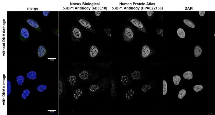
![Immunocytochemistry/Immunofluorescence: 53BP1 Antibody (6B3E10) [NBP2-25028] - Staining of human U-2 OS cells using anti-53BP1 antibody. Bottom images: human U-2 OS cells were treated with EPE to induce DNA-damage and showed increase in nuclear spots after the induction of DNA damage with EPE. Image from verified customer review.](http://images.novusbio.com/fullsize/53BP1-Antibody-6B3E10-Immunocytochemistry-Immunofluorescence-NBP2-25028-img0006.jpg "Immunocytochemistry/Immunofluorescence: 53BP1 Antibody (6B3E10) [NBP2-25028] - Staining of human U-2 OS cells using anti-53BP1 antibody. Bottom images: human U-2 OS cells were treated with EPE to induce DNA-damage and showed increase in nuclear spots after the induction of DNA damage with EPE. Image from verified customer review.")
| Immunogen | 53BP1 Antibody (6B3E10) was made to a partial recombinant human 53BP1 (between residues 500-800) expressed in E. coli [Uniprot: Q12888] |
| Localization | Nuclear |
| Marker | DNA Double Strand Break Marker |
| Predicted Species | Porcine (91%), Chimpanzee (99%), Orangutan (99%), Rabbit (92%), Feline (91%), Canine (93%), Primate (98%), Equine (91%). Backed by our 100% Guarantee. |
| Isotype | IgG1 |
| Clonality | Monoclonal |
| Host | Mouse |
| Gene | TP53BP1 |
| Purity | Protein A or G purified |
| Innovator's Reward | Test in a species/application not listed above to receive a full credit towards a future purchase. |
| Dilutions |
|
||||
| Theoretical MW | 213 kDa. Disclaimer note: The observed molecular weight of the protein may vary from the listed predicted molecular weight due to post translational modifications, post translation cleavages, relative charges, and other experimental factors. |
||||
| Control |
|
||||
| Reviewed Applications |
|
||||
| Publications |
|
| Storage | Store at 4C short term. Aliquot and store at -20C long term. Avoid freeze-thaw cycles. |
| Buffer | PBS |
| Preservative | 0.05% Sodium Azide |
| Concentration | 1.0 mg/ml |
| Purity | Protein A or G purified |


 - BSA Free")
| Images | Ratings | Applications | Species | Date | Details | ||||||||
|---|---|---|---|---|---|---|---|---|---|---|---|---|---|
Enlarge |
reviewed by:
Frida Danielsson |
ICC | Human | 12/18/2015 |
Summary
|
| Human Colon Transverse Whole Tissue Lysate (Adult Whole Normal) | |
| Human Uterus Whole Tissue Lysate (Adult Whole Tumor) |
Secondary Antibodies |
Isotype Controls |
Research Areas for 53BP1 Antibody (NBP2-25028)Find related products by research area.
|
|
The recent relationship of BRCA1 and 53BP1 The p53-binding protein 1 (53BP1) is a DNA damage response factor, which is recruited to nuclear structures at the site of DNA damage. DNA double-strand breaks (DSBs) are mutations that are detrimental to cell viability and genome stability, and m... Read full blog post. |
|
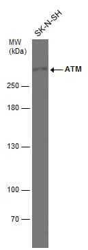
53BP1 - a marker for DNA Double Strand Break 53BP1 (p53 binding protein 1) was originally thought to be an enhancer for p53 transcriptional, but later studies have demonstrated that it is actually a substrate for ataxia telangiectasia mutated (ATM). 53BP1 is a classic late DNA damage response... Read full blog post. |
|
53BP1 - DNA damage is no fun The 53BP1 (p53 binding protein 1) was initially believed to be a p53 transcriptional enhancing partner, but it has now been established as an ataxia telangiectasia mutated (ATM) substrate. As a late DNA damage response (DDR) marker, 53BP1 appears duri... Read full blog post. |
|
53BP1, DNA Damage Response and Tumor Suppression 53BP1 (p53 binding protein 1) was originally thought to be a p53 transcriptional enhancing partner, but now has been shown to be an ataxia telangiectasia mutated (ATM) substrate. It is a late DNA damage response (DDR) marker, appearing in the telophas... Read full blog post. |
|
NUP153 & 53BP1: A Novel DNA Repair Pathway Mediating DNA damage is a crucial process, and one of the most important cellular guards against cancer. In response to DNA damage, sophisticated cellular machinery is recruited to repair the breaks, and if it fails, the cell is committed to death. De... Read full blog post. |
|
Blocking 53BP1 Expression Lessens Tumor Development in BRCA1-Defective Mice Our antibody database at Novus Biologicals provides research tools for the forefront of cancer research. Recently, a mouse study using 53BP1 and BRCA1 antibodies showed that deletion of 53BP1 greatly lessened the incidence of tumor development in mice... Read full blog post. |
The concentration calculator allows you to quickly calculate the volume, mass or concentration of your vial. Simply enter your mass, volume, or concentration values for your reagent and the calculator will determine the rest.


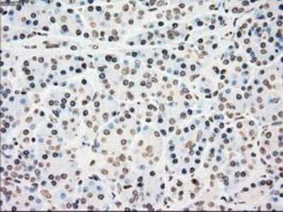
![Immunohistochemistry: 53BP1 Antibody (6B3E10) [NBP2-25028] - Analysis of paraffin-embedded colon cancer tissues using 53BP1 Antibody with DAB staining.](http://images.novusbio.com/fullsize/53BP1-Antibody-6B3E10-Immunohistochemistry-NBP2-25028-img0004.jpg "Immunohistochemistry: 53BP1 Antibody (6B3E10) [NBP2-25028] - Analysis of paraffin-embedded colon cancer tissues using 53BP1 Antibody with DAB staining.")
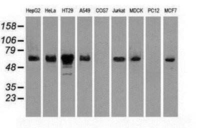
![Western Blot: 53BP1 Antibody (6B3E10) [NBP2-25028] - Western blot analysis using against HEK293 (1) and 53BP1 (AA: 574-773)-hIgGFc transfected HEK293 (2) cell lysate. The observed molecular weight is ~52 kDa and the theoretical molecular weight of the whole endogenous protein is 214 kDa.](http://images.novusbio.com/fullsize/53BP1-Antibody-6B3E10-Western-Blot-NBP2-25028-img0002.jpg "Western Blot: 53BP1 Antibody (6B3E10) [NBP2-25028] - Western blot analysis using against HEK293 (1) and 53BP1 (AA: 574-773)-hIgGFc transfected HEK293 (2) cell lysate. The observed molecular weight is ~52 kDa and the theoretical molecular weight of the whole endogenous protein is 214 kDa.")
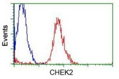
![Flow Cytometry: 53BP1 Antibody (6B3E10) [NBP2-25028] - Analysis of HepG2 cells using 53BP1 Antibody (green) and negative control (purple).](http://images.novusbio.com/fullsize/53BP1-Antibody-6B3E10-Flow-Cytometry-NBP2-25028-img0003.jpg "Flow Cytometry: 53BP1 Antibody (6B3E10) [NBP2-25028] - Analysis of HepG2 cells using 53BP1 Antibody (green) and negative control (purple).")
![Immunohistochemistry: 53BP1 Antibody (6B3E10) [NBP2-25028] - Analysis of paraffin-embedded endometrial cancer tissues using 53BP1 Antibody with DAB staining.](http://images.novusbio.com/fullsize/53BP1-Antibody-6B3E10-Immunohistochemistry-NBP2-25028-img0005.jpg "Immunohistochemistry: 53BP1 Antibody (6B3E10) [NBP2-25028] - Analysis of paraffin-embedded endometrial cancer tissues using 53BP1 Antibody with DAB staining.")
![Immunocytochemistry/Immunofluorescence: 53BP1 Antibody (6B3E10) [NBP2-25028] - Ntera2 cells were fixed in 4% paraformaldehyde for 10 minutes and permeabilized in 0.5% Triton X-100 in PBS for 5 minutes. The cells were incubated with anti-53BP1 Antibody [6B3E10] NBP2-25028 at 2 ug/ml overnight at 4C and detected with an anti-mouse DyLight 488 (Green) at a 1:1000 dilution for 60 minutes. Beta tubulin NB600-936 was used as a co-stain at a 1:1000 dilution and detected with an anti-rabbit DyLight 550 (Red) at a 1:1000 dilution. Nuclei were counterstained with DAPI (Blue). Cells were imaged using a 100X objective and digitally deconvolved.](http://images.novusbio.com/fullsize/53BP1-Antibody-6B3E10-Immunocytochemistry-Immunofluorescence-NBP2-25028-img0007.jpg "Immunocytochemistry/Immunofluorescence: 53BP1 Antibody (6B3E10) [NBP2-25028] - Ntera2 cells were fixed in 4% paraformaldehyde for 10 minutes and permeabilized in 0.5% Triton X-100 in PBS for 5 minutes. The cells were incubated with anti-53BP1 Antibody [6B3E10] NBP2-25028 at 2 ug/ml overnight at 4C and detected with an anti-mouse DyLight 488 (Green) at a 1:1000 dilution for 60 minutes. Beta tubulin NB600-936 was used as a co-stain at a 1:1000 dilution and detected with an anti-rabbit DyLight 550 (Red) at a 1:1000 dilution. Nuclei were counterstained with DAPI (Blue). Cells were imaged using a 100X objective and digitally deconvolved.")
![Flow Cytometry: 53BP1 Antibody (6B3E10) [NBP2-25028] - An intracellular stain was performed on HeLa cells with 53BP1 (6B3E10) Antibody NBP2-25028 (blue) and a matched isotype control MAB002 (orange). Cells were fixed with 4% PFA and then permeabilized with 0.1% saponin. Cells were incubated in an antibody dilution of 1.0 ug/mL for 30 minutes at room temperature, followed by Mouse IgG (H+L) Cross-Adsorbed Secondary Antibody, Dylight 550 (35503, Thermo Fisher).](http://images.novusbio.com/fullsize/53BP1-Antibody-6B3E10-Flow-Cytometry-NBP2-25028-img0011.jpg "Flow Cytometry: 53BP1 Antibody (6B3E10) [NBP2-25028] - An intracellular stain was performed on HeLa cells with 53BP1 (6B3E10) Antibody NBP2-25028 (blue) and a matched isotype control MAB002 (orange). Cells were fixed with 4% PFA and then permeabilized with 0.1% saponin. Cells were incubated in an antibody dilution of 1.0 ug/mL for 30 minutes at room temperature, followed by Mouse IgG (H+L) Cross-Adsorbed Secondary Antibody, Dylight 550 (35503, Thermo Fisher).")
![Western Blot: 53BP1 Antibody (6B3E10) [NBP2-25028] - Western blot analysis using 53BP1 Antibody against human 53BP1 recombinant protein. The expected molecular weight is 47.6 kDa which is demonstrated in this analysis, and the theoretical molecular weight of the whole endogenous protein is 214 kDa.](http://images.novusbio.com/fullsize/53BP1-Antibody-6B3E10-Western-Blot-NBP2-25028-img0001.jpg "Western Blot: 53BP1 Antibody (6B3E10) [NBP2-25028] - Western blot analysis using 53BP1 Antibody against human 53BP1 recombinant protein. The expected molecular weight is 47.6 kDa which is demonstrated in this analysis, and the theoretical molecular weight of the whole endogenous protein is 214 kDa.")
![Flow Cytometry: 53BP1 Antibody (6B3E10) [NBP2-25028] - An intracellular stain was performed on Ntera2 cells with 53BP1 Antibody [6B3E10] NBP2-25028 (blue) and a matched isotype control (orange). Cells were fixed with 4% PFA and then permeabilized with 0.1% saponin. Cells were incubated in an antibody dilution of 1.0 ug/mL for 30 minutes at room temperature, followed by Mouse IgG (H+L) Cross-Adsorbed Secondary Antibody, Dylight 550 (35503, Thermo Fisher).](http://images.novusbio.com/fullsize/53BP1-Antibody-6B3E10-Flow-Cytometry-NBP2-25028-img0010.jpg "Flow Cytometry: 53BP1 Antibody (6B3E10) [NBP2-25028] - An intracellular stain was performed on Ntera2 cells with 53BP1 Antibody [6B3E10] NBP2-25028 (blue) and a matched isotype control (orange). Cells were fixed with 4% PFA and then permeabilized with 0.1% saponin. Cells were incubated in an antibody dilution of 1.0 ug/mL for 30 minutes at room temperature, followed by Mouse IgG (H+L) Cross-Adsorbed Secondary Antibody, Dylight 550 (35503, Thermo Fisher).")


![Immunohistochemistry DDR1 Antibody [Unconjugated]](https://images.novusbio.com/images/antibody/DDR1_AF2396_Immunohistochemistry_7152.jpg)
![Simple Western DDR1 Antibody [Unconjugated]](https://images.novusbio.com/images/antibody/DDR1_AF2396_Immunohistochemistry_17635.jpg)
![Western Blot DDR1 Antibody [Unconjugated]](https://images.novusbio.com/images/antibody/DDR1_AF2396_Western_Blot_16723.jpg)