HAF016). A specific band was detected for N-Cadherin at approximately 130 kDa (as indicated). This experiment was conducted under reducing conditions and using
Immunoblot Buffer Group 1." class="big_lightbox">
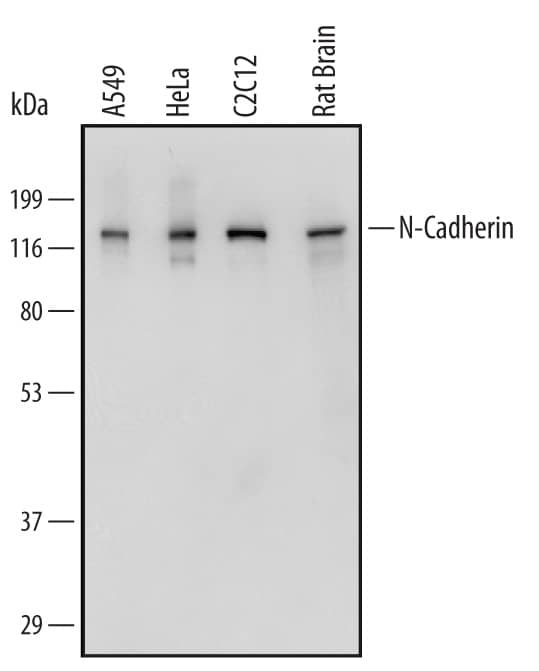
HAF016). A specific band was detected for N-Cadherin at approximately 130 kDa (as indicated). This experiment was conducted under reducing conditions and using
Immunoblot Buffer Group 1." alt="Western blot shows lysates of A549 human lung carcinoma cell line, HeLa human cervical epithelial carcinoma cell line, C2C12 mouse myoblast cell line, and rat brain tissue. PVDF Membrane was probed with 0.5 µg/mL of Sheep Anti-Human/Mouse/Rat N-Cadherin Antigen Affinity-purified Polyclonal Antibody (Catalog # AF6426) followed by HRP-conjugated Anti-Sheep IgG Secondary Antibody (Catalog #
HAF016). A specific band was detected for N-Cadherin at approximately 130 kDa (as indicated). This experiment was conducted under reducing conditions and using
Immunoblot Buffer Group 1." class="big_thumb" />
VC006). Before incubation with the primary antibody, tissue was subjected to heat-induced epitope retrieval using Antigen Retrieval Reagent-Basic (Catalog #
CTS013). Tissue was stained using DAB (brown) and counterstained with hematoxylin (blue). Specific staining was localized to intercalated disks between the myocardial cells. Staining was performed using our protocol for IHC Staining with VisUCyte HRP Polymer Detection Reagents." class="big_lightbox">
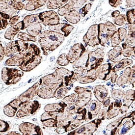
VC006). Before incubation with the primary antibody, tissue was subjected to heat-induced epitope retrieval using Antigen Retrieval Reagent-Basic (Catalog #
CTS013). Tissue was stained using DAB (brown) and counterstained with hematoxylin (blue). Specific staining was localized to intercalated disks between the myocardial cells. Staining was performed using our protocol for IHC Staining with VisUCyte HRP Polymer Detection Reagents." class="big_thumb" />
MAB13883) coated on a Clear Polystyrene Microplate (Catalog #
DY990). Sheep Anti-Human/Mouse/Rat N-Cadherin Antigen Affinity-purified Polyclonal Antibody (Catalog # AF6426) was biotinylated and incubated with the protein captured on the plate. Detection of the standard curve was achieved by incubating Streptavidin-HRP (Catalog #
DY998) followed by Substrate Solution (Catalog #
DY999) and stopping the enzymatic reaction with Stop Solution (Catalog #
DY994)." class="big_lightbox">
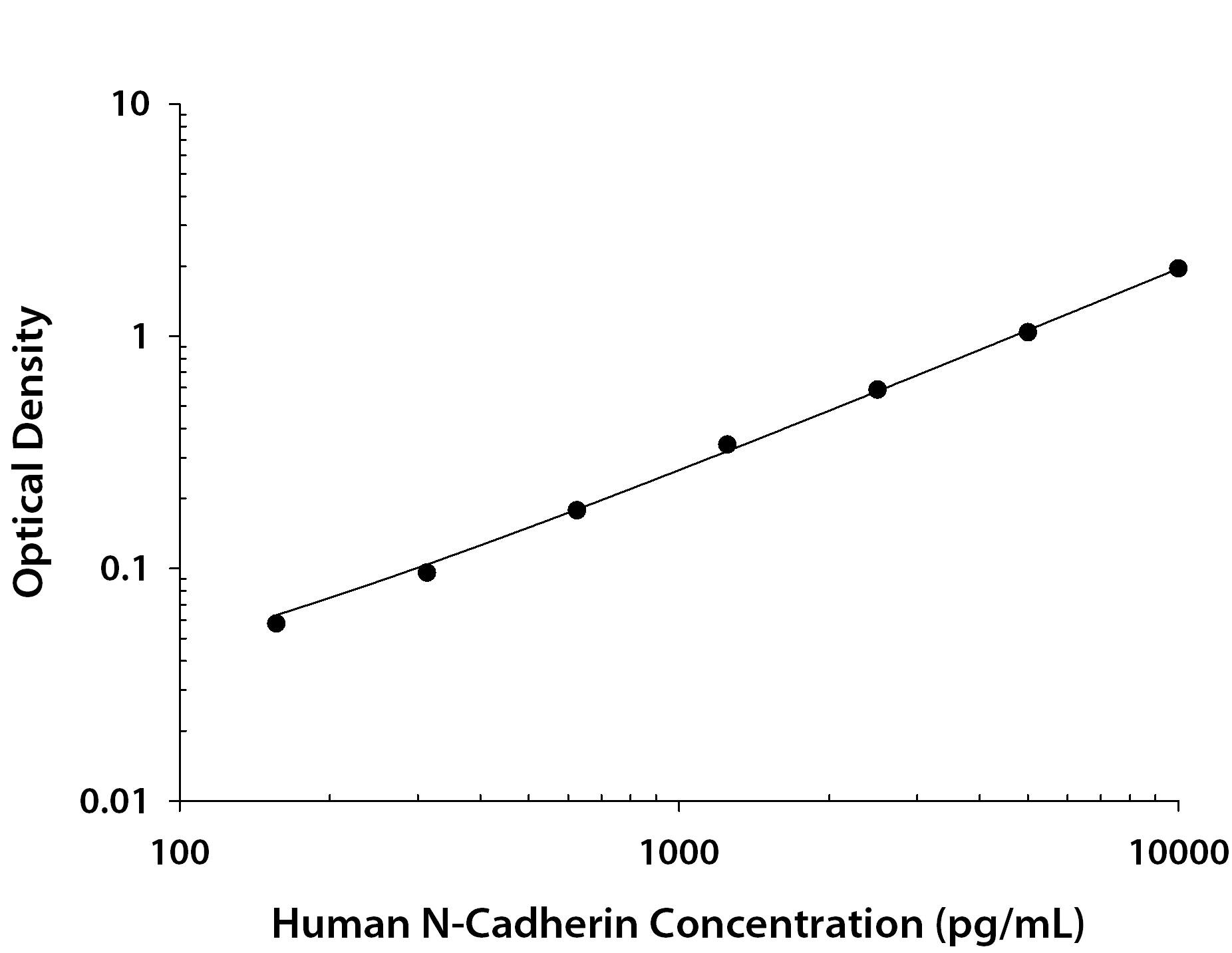
MAB13883) coated on a Clear Polystyrene Microplate (Catalog #
DY990). Sheep Anti-Human/Mouse/Rat N-Cadherin Antigen Affinity-purified Polyclonal Antibody (Catalog # AF6426) was biotinylated and incubated with the protein captured on the plate. Detection of the standard curve was achieved by incubating Streptavidin-HRP (Catalog #
DY998) followed by Substrate Solution (Catalog #
DY999) and stopping the enzymatic reaction with Stop Solution (Catalog #
DY994)." class="big_thumb" />
 HAF016). A specific band was detected for N-Cadherin at approximately 130 kDa (as indicated). This experiment was conducted under reducing conditions and using Immunoblot Buffer Group 1." alt="Western blot shows lysates of A549 human lung carcinoma cell line, HeLa human cervical epithelial carcinoma cell line, C2C12 mouse myoblast cell line, and rat brain tissue. PVDF Membrane was probed with 0.5 µg/mL of Sheep Anti-Human/Mouse/Rat N-Cadherin Antigen Affinity-purified Polyclonal Antibody (Catalog # AF6426) followed by HRP-conjugated Anti-Sheep IgG Secondary Antibody (Catalog # HAF016). A specific band was detected for N-Cadherin at approximately 130 kDa (as indicated). This experiment was conducted under reducing conditions and using Immunoblot Buffer Group 1." class="big_thumb" />


![Immunohistochemistry: Tyrosine Hydroxylase Antibody [NB300-109] - D409H GBA1 expression promotes reduction of dopaminergic fiber densities & motor deficit in the striatum of hA53T alpha -synuclein transgenic mice. a, The striatal TH-immunopositive fiber density was assessed in the striatum of the indicated genotypes at 6 months of age (n = 6 per each group). The scale bar is 250 μm. b, The optical densities of TH positive signals were quantified. c & d, Rotarod & Pole test were assessed at 6 months of age with the indicated genotypes (n = 7 per each group). e, the body weight of mice were measured with the indicated genotypes (n = 15 per each group). DATA are expressed as mean ± SEM Image collected & cropped by CiteAb from the following publication (//actaneurocomms.biomedcentral.com/articles/10.1186/s40478-018-0538-9), licensed under a CC-BY license. Not internally tested by Novus Biologicals.](http://images.novusbio.com/fullsize/nb300-109_rabbit-polyclonal-tyrosine-hydroxylase-antibody-310202415371995.jpg)
![Western Blot: Tyrosine Hydroxylase Antibody [NB300-109] - Dysregulation of RNF146-PARP1 pathways in PD pathogenesis. (C) Expression of PARsylated proteins, RNF146, TH & phosphorylated alpha -synuclein (pS129-a-syn) in postmortem striatum from PD patients & age-matched control subjects (Ctrl) determined by WBs using designated antibodies. Image collected & cropped by CiteAb from following publication (//www.oncotarget.com/lookup/doi/10.18632/oncotarget.21828), licensed under a CC-BY license. Not internally tested by Novus Biologicals.](http://images.novusbio.com/fullsize/nb300-109_rabbit-polyclonal-tyrosine-hydroxylase-antibody-310202415293344.jpg)
![Immunocytochemistry/ Immunofluorescence: Tyrosine Hydroxylase Antibody [NB300-109] - Image showing differentiation of neurospheres as evidenced morphologically (A) & through staining by Tyrosine Hydroxylase (B) to show the ability to derive dopaminergic neurons from the rat progenitor cells. Image collected & cropped by CiteAb from the following publication (//pubmed.ncbi.nlm.nih.gov/30042680), licensed under a CC-BY license. Not internally tested by Novus Biologicals.](http://images.novusbio.com/fullsize/nb300-109_rabbit-polyclonal-tyrosine-hydroxylase-antibody-310202415392574.jpg)
![Western Blot: Tyrosine Hydroxylase Antibody [NB300-109] - Immunoblots of tyrosine hydroxylase in the cricket & cockroach brain.Protein extracts (8 μl & 16 μl) from the male cricket, Gryllus bimaculatus & male cockroach, Periplaneta americana were electrophoresed, transferred to a membrane & reacted with anti-tyrosine hydroxylase (TH). The anti-TH recognized a single band of protein of ~ 66 kDa (arrow heads) in both the cricket (left two lanes) & cockroach (right two lanes). M, molecular marker. Image collected & cropped by CiteAb from the following publication (//dx.plos.org/10.1371/journal.pone.0160531), licensed under a CC-BY license. Not internally tested by Novus Biologicals.](http://images.novusbio.com/fullsize/nb300-109_rabbit-polyclonal-tyrosine-hydroxylase-antibody-31020241535660.jpg)
![Immunohistochemistry: Tyrosine Hydroxylase Antibody [NB300-109] - D409H GBA1 expression shortens lifespan & leads to dopaminergic degeneration in the hA53T alpha -synuclein transgenic mice. a, Survival was monitored from littermates with the following genotypes: hA53Ta-Syn/GBA1+/+ (n = 15), hA53T alpha -Syn/GBA1+/D409H(n = 16), hA53T alpha -Syn/GBA1D409H/D409H (n = 18) mice. GBA1 mutation induces the lethal phenotype & TH-positive neuronal loss in the SNpc of A53T mutant alpha -synuclein transgenic mice. b & c, The number of TH-positive neurons in the SNpc was counted using stereological analysis with the indicated genotypes at 6 months of age. The scale bar is 200 μm. DATA are expressed as mean ± SEM (n = 6 for the each group) Image collected & cropped by CiteAb from the following publication (//actaneurocomms.biomedcentral.com/articles/10.1186/s40478-018-0538-9), licensed under a CC-BY license. Not internally tested by Novus Biologicals.](http://images.novusbio.com/fullsize/nb300-109_rabbit-polyclonal-tyrosine-hydroxylase-antibody-310202415395928.jpg)
![Immunocytochemistry/ Immunofluorescence: Tyrosine Hydroxylase Antibody [NB300-109] - Tyrosine hydroxylase immunoreactivity in the deutocerebrum & medial antennal lobe tract.Tyrosine hydroxylase (TH) immunoreactivity is shown in magenta (A, D, G, J) & synapsin immunoreactivity in green (B, E, H, K). Corresponding merged images (C, F, I, L). A-C: Frontal sections through the antennal lobe (AL). Insets indicate single optical slice (< 2 μm thickness) of the three glomeruli, which are marked by asterisks in main panel B. TH-positive fibers innervate antennal lobe glomeruli, predominantly the peripheral areas (see arrowheads in inset of A). In the macroglomerular complex (MGC), TH-positive fibers innervate exclusively the medial side (arrows in A). D-F: Horizontal sections through the antennal lobe. A single TH-positive multiglomerular projection neuron (PN) is labeled, which projects its axon toward the medial antennal lobe tract (MALT, see also panel G). G-I: Frontal sections through the medial antennal lobe tract containing several TH-positive axons (arrowheads). J-L: Frontal sections through the lobus glomerulatus (LG) & the antennal mechanosensory & motor center (AMMC). TH-immunoreactive fibers are widely distributed in the AMMC. In the lobus glomerulatus, immunoreactive fibers predominantly innervate the ventral portion. D, dorsal; M, medial; ML, medial lobe; NO, noduli; P, posterior; PB, protocerebral bridge. Scale bars = 50 μm in inset of C; 100 μm in C, F, I, L. Image collected & cropped by CiteAb from the following publication (//dx.plos.org/10.1371/journal.pone.0160531), licensed under a CC-BY license. Not internally tested by Novus Biologicals.](http://images.novusbio.com/fullsize/nb300-109_rabbit-polyclonal-tyrosine-hydroxylase-antibody-310202416212328.jpg)
![Western Blot: Tyrosine Hydroxylase Antibody [NB300-109] - Specific knockdown of Hif1 alpha diminished phosphorylation of tyrosine hydroxylase & thereby reduced cellular dopamine content in PC12 cells. (A) Extrinsic hypoxia led to a time-dependent upregulation of Hif1 alpha & Hif2 alpha in PC12. (B) RNA interference using siRNA against Hif1 alpha repressed the expression of Hif1 alpha also under extrinsic hypoxia while Hif2 alpha remains unaffected. (C) Extrinsic hypoxia increased cellular dopamine content in PC12 control cells (transfection without siRNA). This effect was diminished by knockdown of Hif1 alpha . (D) Furthermore, phosphorylation of TH was decreased by knockdown of Hif1 alpha . Three independent experiments (n = 3–6). Mean ± SEM. ANOVA & Bonferroni post hoc test comparison vs. normoxia, * p < 0.05 or, ** p < 0.01, or vs. PC12 siRNA control 24 or 48 h hypoxia, # p < 0.05. Image collected & cropped by CiteAb from the following publication (//pubmed.ncbi.nlm.nih.gov/31035382), licensed under a CC-BY license. Not internally tested by Novus Biologicals.](http://images.novusbio.com/fullsize/nb300-109_rabbit-polyclonal-tyrosine-hydroxylase-antibody-310202416205183.jpg)
![Immunocytochemistry/ Immunofluorescence: Tyrosine Hydroxylase Antibody [NB300-109] - ErbB4 protein expression in mesencephalic nuclei.The overview on the mesencephalon (B) shows dense ErbB4 expression (mAb-10, green channel) in the dopaminergic nuclei SNc & VTA that is clearly above background levels, whereas expression in the red nucleus is weaker. Higher magnification shows abundant immunoreactivity on neuropil (C–E, C'–E'). At 100x magnification, a dense network of ErbB4-positive neuropil surrounding a neuron (asterisk) is evident in the VTA (E”). The red channel shows neurofilament H-immunofluorescence in panels (B–E) & TH-immunofluorescence in (F–G). Co-immunolabeling reveals that subsets of TH-positive dopaminergic somata express ErbB4 in the VTA (F) & SNc (G); coexpressing neurons are indicated by arrowheads (F–G), whereas lack of ErbB4 signal is indicated by arrows (G). The location of the overview image within the monkey brain is indicated by a red rectangle in (A). The location of the profiles through mesencephalic nuclei is indicated by yellow boxes in (A) & by rectangles in (B). Images were obtained with a confocal microscope. Scale bar = 3 mm (B), 400 µm (C–E), 175 µm (C'–E', F–G), 70 µm (E”). Abbreviations: cerebral peduncle (cp), oculomotor nerve (3n), substantia nigra pars reticulata (SNr). Image collected & cropped by CiteAb from the following publication (//dx.plos.org/10.1371/journal.pone.0027337), licensed under a CC0-1.0 license. Not internally tested by Novus Biologicals.](http://images.novusbio.com/fullsize/nb300-109_rabbit-polyclonal-tyrosine-hydroxylase-antibody-310202416205171.jpg)
![Western Blot: Tyrosine Hydroxylase Antibody [NB300-109] - Impact of extrinsic & intrinsic hypoxia on catecholamine biosynthesis. (A) Section of biosynthetic pathways of catecholamines. Squares highlighted the underlying enzymes (TH: tyrosine hydroxylase; DDC: DOPA decarboxylase; DBH: dopamine beta -hydroxylase; PNMT: phenylethanolamine N-methyltransferase). (B) Effect of extrinsic & intrinsic hypoxia on the protein expression & phosphorylation of TH at Ser40 in MTT cells. (C) Immunohistochemical staining showed an increased expression of TH (red) in the necrotic & hypoxic core of the spheroid (DAPI, blue). Scale bar: 200 µm. Catecholamine content of (D) MPC & (E) MTT cell spheroids in comparison to monolayer cultivation under normoxic or hypoxic conditions. Three independent experiments (n = 3–6). Mean ± SEM. ANOVA & Bonferroni post hoc test comparison vs. normoxia, ** p < 0.01, or vs. spheroid day 11, # p < 0.05 or, ## p < 0.01. Image collected & cropped by CiteAb from the following publication (//pubmed.ncbi.nlm.nih.gov/31035382), licensed under a CC-BY license. Not internally tested by Novus Biologicals.](http://images.novusbio.com/fullsize/nb300-109_rabbit-polyclonal-tyrosine-hydroxylase-antibody-31020241621826.jpg)
![Western Blot: Tyrosine Hydroxylase Antibody [NB300-109] - Validation of protein alterations & functional studies for pathways induced by genetic & toxic stress, & reversal of PD-related alterations following S-HSP90 inhibition. a Schematic illustration of key signaling proteins & pathways activated in response to genetic stress. Western blot confirms higher activity of these pathways in PD over WT mDA neurons, & shows that PU-H71 restores these to WT levels. Mean ± SEM, n = 3 individual values from the different experiments shown as points, t-test, ***p < 0.001; **p < 0.01; *p < 0.05; ns p > 0.05. b Heatmaps of proteins significantly enriched under toxic stress (10 µM CCCP; 20 nM rotenone) & their validation by western blot (Input, total levels & S-HSP90 bait isolate, chaperome network associated). Mean ± SEM, n = 3 individual values from the different experiments shown as points, t-test, ***p < 0.001; *p < 0.05. c Western blot & quantification of rotenone stress increase in the p-TH:TH ratio & its reduction by PU-H71 (200 nM). p-ERK/ERK, cell viability control; beta -actin, protein loading control. Mean ± SEM, n = 3 individual values from the different experiments shown as points, One-way ANOVA with Tukey’s post-hoc, ***p < 0.001; **p < 0.01. d Total intracellular dopamine levels in PD mDA neurons in conditions of toxic stress & under PU-H71 rescue; Mean ± SEM, n = 4–5 individual values from the different experiments shown as points, t-test, *p < 0.05. e PU-H71 treatment of rotenone- or CCCP-stressed mDA neurons significantly increases their viability, as measured by total ATP levels; means ± SEM, n = 3–8 from independent differentiations, t-test, ***p < 0.001; **p < 0.01; *p < 0.05 Image collected & cropped by CiteAb from the following publication (//pubmed.ncbi.nlm.nih.gov/30341316), licensed under a CC-BY license. Not internally tested by Novus Biologicals.](http://images.novusbio.com/fullsize/nb300-109_rabbit-polyclonal-tyrosine-hydroxylase-antibody-31020241620377.jpg)
![Immunocytochemistry/ Immunofluorescence: Tyrosine Hydroxylase Antibody [NB300-109] - ErbB4 protein expression in mesencephalic nuclei.The overview on the mesencephalon (B) shows dense ErbB4 expression (mAb-10, green channel) in the dopaminergic nuclei SNc & VTA that is clearly above background levels, whereas expression in the red nucleus is weaker. Higher magnification shows abundant immunoreactivity on neuropil (C–E, C'–E'). At 100x magnification, a dense network of ErbB4-positive neuropil surrounding a neuron (asterisk) is evident in the VTA (E”). The red channel shows neurofilament H-immunofluorescence in panels (B–E) & TH-immunofluorescence in (F–G). Co-immunolabeling reveals that subsets of TH-positive dopaminergic somata express ErbB4 in the VTA (F) & SNc (G); coexpressing neurons are indicated by arrowheads (F–G), whereas lack of ErbB4 signal is indicated by arrows (G). The location of the overview image within the monkey brain is indicated by a red rectangle in (A). The location of the profiles through mesencephalic nuclei is indicated by yellow boxes in (A) & by rectangles in (B). Images were obtained with a confocal microscope. Scale bar = 3 mm (B), 400 µm (C–E), 175 µm (C'–E', F–G), 70 µm (E”). Abbreviations: cerebral peduncle (cp), oculomotor nerve (3n), substantia nigra pars reticulata (SNr). Image collected & cropped by CiteAb from the following publication (//dx.plos.org/10.1371/journal.pone.0027337), licensed under a CC0-1.0 license. Not internally tested by Novus Biologicals.](http://images.novusbio.com/fullsize/nb300-109_rabbit-polyclonal-tyrosine-hydroxylase-antibody-310202416212334.jpg)
![Immunocytochemistry/ Immunofluorescence: Tyrosine Hydroxylase Antibody [NB300-109] - Tyrosine hydroxylase immunoreactivity in mushroom body.All panels show a right brain hemisphere, except panels M-O (posterior to top). Tyrosine hydroxylase (TH) immunoreactivity shown in magenta (A, D, G, J, M) & synapsin immunoreactivity (green: B, E, H, K, N). Corresponding merged images in C, F, I, L, O. A-C: Frontal sections through lateral protocerebrum. 2 groups of TH-positive cell bodies (DCa1 & 2) labeled. DCa2 appears to send axons ventrally (arrowhead). Superior lateral protocerebrum (SLP) as well as medial & lateral calyces (MCA & LCA) innervated by TH-positive fibers. D-F: Frontal sections through vertical lobe (VL) & parts of medial calyx (MCA), which receive many fine TH-positive fibers. G-I: Horizontal sections through a tip of vertical lobe & dorsal parts of calyces. Both calyces & vertical lobe innervated by fibrous TH-positive fibers. Note TH-immunoreactive fibers exclusively innervate synapsin-positive neuropil layers (NLs) of calyx where dendrites of Kenyon cells receive synaptic inputs from antennal-lobe projection neurons. J-L: Horizontal sections through calyces, ventral to panels G-I. Here, 3clusters of TH-positive cell bodies (DCa1, DCa2, & DSP1) visible. Immunoreactive fibers mainly invade anterior layers including gamma layer ( gamma ) in vertical lobe. M-O: Horizontal sections through distal portions of medial lobes (MLs), invaded by fine TH-positive fibers exhibiting moderate immunolabeling (arrows). in inferior medial protocerebrum (IMP) anterior to medial lobe, a pair of DIP1 clusters (triangles) is situated. D, dorsal; KFL, Kenyon fiber layer; LCA, lateral calyx; LO, lobula; M, medial; P, posterior; PED, pedunculus; SMP, superior medial protocerebrum. Scale bars = 100 μm. Image collected & cropped by CiteAb from following publication (//dx.plos.org/10.1371/journal.pone.0160531), licensed under a CC-BY license. Not internally tested by Novus Biologicals.](http://images.novusbio.com/fullsize/nb300-109_rabbit-polyclonal-tyrosine-hydroxylase-antibody-310202416212326.jpg)
![Immunohistochemistry: Tyrosine Hydroxylase Antibody [NB300-109] - Characterization of the nigrostriatal pathway of LRRK2 conditional transgenic mice. A, Representative TH immunohistochemistry of the midbrain coronal sections of 22-month-old LRRK2 GS & GSDA transgenic & age-matched littermate controls. B, C, Stereological assessment of TH-positive (B) & Nissl-positive (C) neurons in the SNpc (control, n = 9; GS, n = 9; GS/DA, n = 9). Data are the mean number of cells per region ± SEM, n = 9 mice per group. Statistical significance was determined by two-tailed unpaired Student’s t test. D, Representative images of TH immunostaining of nerve terminals in the striatum of LRRK2 conditional transgenic mice at 22 months of age. E, Quantitation of TH immunostaining in the striatum using ImageJ software (NIH; control, n = 7; GS, n = 7; GS/DA, n = 7). Differences between groups were assessed by two-way ANOVA. Bars represent the mean ± SEM (n ≥ 5 animals/genotype). n.s., Nonsignificant. Image collected & cropped by CiteAb from the following publication (//www.eneuro.org/lookup/doi/10.1523/ENEURO.0004-17.2017), licensed under a CC-BY license. Not internally tested by Novus Biologicals.](http://images.novusbio.com/fullsize/nb300-109_rabbit-polyclonal-tyrosine-hydroxylase-antibody-310202416205143.jpg)
![Immunocytochemistry/ Immunofluorescence: Tyrosine Hydroxylase Antibody [NB300-109] - Tyrosine hydroxylase (TH) immunoreactivity in mushroom body. Panels show a right brain hemisphere, except panels M-O (posterior top). Tyrosine hydroxylase immunoreactivity shown in magenta (A, D, G, J, M) & synapsin immunoreactivity in green (B, E, H, K, N). Corresponding merged images in C, F, I, L, O. A-C: Frontal sections through lateral protocerebrum. 2 groups of TH-positive cell bodies (DCa1 & 2) labeled. DCa2 appears to send axons ventrally (arrowhead). Superior lateral protocerebrum (SLP) as well as medial & lateral calyces (MCA & LCA) innervated by TH-positive fibers. D-F: Frontal sections through vertical lobe (VL) & parts of medial calyx (MCA), which receive many fine TH-positive fibers. G-I: Horizontal sections through tip of vertical lobe & dorsal parts of calyces. Both calyces & vertical lobe innervated by fibrous TH-positive fibers. Note TH-immunoreactive fibers exclusively innervate synapsin-positive neuropil layers (NLs) of calyx where dendrites of Kenyon cells receive synaptic inputs from antennal-lobe projection neurons. J-L: Horizontal sections through calyces, ventral to panels G-I. Here, 3clusters of TH-positive cell bodies (DCa1, DCa2, & DSP1) visible. Immunoreactive fibers mainly invade anterior layers including gamma layer ( gamma ) in vertical lobe. M-O: Horizontal sections through distal portions of medial lobes (MLs), invaded by fine TH-positive fibers exhibiting moderate immunolabeling (arrows). in inferior medial protocerebrum (IMP) anterior to medial lobe, pair of DIP1 clusters (triangles) situated. D, dorsal; KFL, Kenyon fiber layer; LCA, lateral calyx; LO, lobula; M, medial; P, posterior; PED, pedunculus; SMP, superior medial protocerebrum. Scale bars = 100 μm. Image collected & cropped by CiteAb from following publication (//dx.plos.org/10.1371/journal.pone.0160531), licensed under a CC-BY license. Not internally tested by Novus Biologicals.](http://images.novusbio.com/fullsize/nb300-109_rabbit-polyclonal-tyrosine-hydroxylase-antibody-310202416205113.jpg)


![Western blot analysis of whole brain tissue lysates using chicken pAb to microtubule associated protein 2 (MAP2), NB300-213, dilution 1:50,000 in green: [1] protein standard (red), [2] adult rat brain, [3] embryonic E20 rat brain, [4] adult mouse brain. Strong band observed at ~280 kDa mark corresponds to two major isoforms of MAP2 protein referred to as MAP2A and MAP2B. Smaller fragments of these isoforms are also detected if the antibody is used at higher concentrations.](http://images.novusbio.com/fullsize/MAP2-Antibody-Western-Blot-NB300-213-img0005.jpg)


![Immunocytochemistry/ Immunofluorescence: MAP2 Antibody [NB300-213] - SUMO1 conjugates are not localized to amyloid plaques. (a) Sagittal brain sections of 24‐week‐old KI/AD & WT/AD animals were stained using anti‐HA antibodies (red), 6E10 antibodies (green) that label amyloid beta 1–42 among other amyloid beta variants (epitope lies within amino acids 3–8 of amyloid beta) & anti‐MAP2 antibodies (blue). Sections of the hippocampal subiculum are shown. Images are representatives of three independent experiments. Scale bar, 25 μm. (b) Anti‐HA signal intensity in amyloid plaques & in cell nuclei was quantified using ImageJ (N = 3, ***significance between WT/AD & KI/AD, p = .0007 in Student's t test) Image collected & cropped by CiteAb from the following publication (//pubmed.ncbi.nlm.nih.gov/29633471), licensed under a CC-BY license. Not internally tested by Novus Biologicals.](http://images.novusbio.com/fullsize/nb300-213_chicken-map2-pab-310202415392532.jpg)
![Immunocytochemistry/ Immunofluorescence: MAP2 Antibody [NB300-213] - Oxytocin receptors (Oxtr) are expressed in mouse hippocampus.(a & b) Coronal sections of the hippocampus (a) & striatum (b) from OxtrVn/+ & Oxtr+/+ mice stained for Venus & DAPI (ac, anterior commisure; cx, cortex; hip, hippocampus; str, striatum). (c) Confocal images of rostral Oxtr+/+ & OxtrVn/+ brain sections including hippocampus, double-labeled for Venus (green) & NeuN (red). (d) Confocal images of Oxtr-expressing neurons in the CA1 subfields of the medial & caudal hippocampus from OxtrVn/+ sections, triple stained with antibodies directed against NeuN (red), MAP2 (magenta), & Venus (green). (e) Representative images of hippocampal mass cultures obtained from OxtrVn/+ mice at P0 mice & processed for immunofluorescence staining at 5, 10, & 14 DIV using antibodies to Venus (green) & GAD67 (red), & DAPI (blue). (f) Quantitative analysis of Venus-positive neurons with (GAD67+) & without colabeling for GAD67 (GAD67-) in hippocampal mass cultures obtained from OxtrVn/+ mice at P0 mice & processed for immunofluorescence staining at 5, 10, & 14 DIV. Data are shown as mean ± SEM.DOI://dx.doi.org/10.7554/eLife.22466.015 Image collected & cropped by CiteAb from the following publication (//pubmed.ncbi.nlm.nih.gov/28231043), licensed under a CC-BY license. Not internally tested by Novus Biologicals.](http://images.novusbio.com/fullsize/nb300-213_chicken-map2-pab-310202416212347.jpg)
![Immunocytochemistry/ Immunofluorescence: MAP2 Antibody [NB300-213] - SUMO1 conjugates remain nuclear during increased amyloid burden. (a) Western blot analysis of subcellular fractions of 36‐week‐old KI/WT & KI/AD mouse brain using anti‐HA antibody (upper two panels) & antibodies to GluN1 (a marker of the postsynaptic compartment) & Synaptophysin (a marker of the presynaptic compartment) to validate the fractionation procedure (lower two panels). H, homogenate; P1, nuclear pellet; S1, supernatant after P1 sedimentation; P2, crude synaptosomal pellet; S2, supernatant after P2 sedimentation; LP1, lysed synaptosomal membranes; LS1, supernatant after LP1 sedimentation; LP2, pellet after LS1 sedimentation, SPM, synaptic plasma membranes. Bracket indicates the anti‐HA signal representing SUMOylation, arrow indicates RanGAP1, stars indicate nonspecific signal detected by the anti‐HA antibody. (b) Brain sagittal sections of aged (36 weeks old) KI/AD (left panels) & WT/AD (right panels) mice were immunostained using antibodies directed against HA (red, labels HA‐HA conjugates), MAP2 (green, labels neuronal somata & dendrites), & Synapsin1 (Syn1, magenta, labels synapses). The images show triple‐labeled neurons of hippocampal subiculum (b), cortical layer 5 (c), & proximal apical dendrite from hippocampal CA3 (d). The white line shows the orientation of the line‐scan used to generate the image stack shown in the bottom side view. Scale bar: 10 μm. Note that the anti‐HA immunosignal is mainly located in neuronal nuclei, only background staining is observed in WT/AD mice. Little anti‐HA signal is observed along MAP2‐positive structures & does not colocalize with Synapsin1 Image collected & cropped by CiteAb from the following publication (//pubmed.ncbi.nlm.nih.gov/29633471), licensed under a CC-BY license. Not internally tested by Novus Biologicals.](http://images.novusbio.com/fullsize/nb300-213_chicken-map2-pab-310202416212317.jpg)
![Immunocytochemistry/ Immunofluorescence: MAP2 Antibody [NB300-213] - Validation of the CRISPR/Cas9 Alsin-/- cells in iPSC, NPC & iPSC-sMN.(A) Electrophoresis result of the PCR reaction using primers flanking exon three & within exon 3. Homozygous deletion was confirmed by the absence of a 2.8 kb. (B) Protein lysates from WT KOLF & Alsin-/- iPSC were loaded onto SDS-PAGE & immunoblotted with Alsin antibody. A band detected at ~184 kDa, but absent in Alsin-/-, corresponds to full-length Alsin. (C) WT KOLF, Alsin-/- iPSC, & neuroprogenitor cells (NPC) were immunostained with pluripotency markers Oct4 & Lin28, & neuroprogenitor markers Sox2 & Pax6, respectively. (D–E) WT KOLF & Alsin-/- iPSC-sMN were immunostained with motor neuron markers such as ChAT, HB9, & ISL1, nuclear dye DAPI, & cytoskeletal marker MAP2. (F) WT KOLF & Alsin-/- iPSC-sMN were immunostained with antibodies against Rab5 & Alsin, along with DAPI (nuclear) & phalloidin (actin) stains. Scale bars, 10 μm. Image collected & cropped by CiteAb from the following publication (//pubmed.ncbi.nlm.nih.gov/29469808), licensed under a CC-BY license. Not internally tested by Novus Biologicals.](http://images.novusbio.com/fullsize/nb300-213_chicken-map2-pab-310202416203714.jpg)
![Immunocytochemistry/ Immunofluorescence: MAP2 Antibody [NB300-213] - PEA decreases astrocyte activation in organotypic cultures of rat hippocampi & rescues neuronal CA3 damage caused by A beta challenge. A beta -challenged (1 μg/ml) slices of rat hippocampi treated for 24 hours w/ PEA (0.1 μM) in presence of selective PPAR gamma antagonist (GW9662, 9 nM) or selective PPAR alpha antagonist (MK886, 3 μM). (A) Nissl staining showing effect of treatment on morphology of organotypic hippocampal slices. Scale bar: 80 μm. A beta caused a marked neuronal loss, mainly in CA3 region of hippocampus, as highlighted by arrows. PEA able to reverse this effect. (B) Representative photomicrographs of CA3 region showing Results of immunofluorescence experiments aimed at investigating effect of treatments on astrocyte activation & neuronal loss, as determined by immunostaining for GFAP (green) & MAP2 (red) alone or merged, respectively. Nuclei stained w/ Hoechst (blue). Scale bar: 10 μm. Arrows in photomicrographs indicate astrocyte infiltration events & apoptotic condensation in nuclei of adjacent neurons. (C) Relative quantification of GFAP-positive cell number as a count of astrocyte proliferation. (D) Apoptotic events detected on MAP2-expressing cells as an indication of neuronal death. For (C) & (D), average value determined by counting cells in at least five microscopic fields for each treatment. Results are presented as means ± SEM of four separate experiments. At least four slices from each experimental group observed for each experiment. Statistical analysis performed using parametric one-way analysis of variance, & multiple comparisons performed using Bonferroni test. ***P < 0.001 & *P < 0.05 vs. control; ∘∘P < 0.01 & ∘P < 0.05 vs. A beta -challenge slices. Image collected & cropped by CiteAb from following publication (//jneuroinflammation.biomedcentral.com/articles/10.1186/1742-2094-9-49), licensed under a CC-BY license. Not internally tested by Novus Biologicals.](http://images.novusbio.com/fullsize/nb300-213_chicken-map2-pab-310202416203712.jpg)
![Immunocytochemistry/ Immunofluorescence: MAP2 Antibody [NB300-213] - PEA inhibits astroglial proliferation & reduces neuronal loss in mixed neuroglia co-cultures exposed to A beta . A beta -challenged (1 μg/ml) astrocyte/neuron mixed cultures were treated with PEA (0.1 μM) in the presence of the selective PPAR gamma antagonist (GW9662, 9 nM) or the selective PPAR alpha antagonist (MK886, 3 μM). After 24 hours of treatment, cells were processed for analyses. (A) Immunofluorescence photomicrographs showing the effect of the treatments on astrocyte proliferation & neuronal loss, as determined by immunostaining for GFAP (green) & MAP2 (red), respectively. Arrows indicate chromatin condensation in nuclei stained with Hoechst dye (blue) as markers of apoptotic events. Scale bar: 10 μm. (B) Relative quantification of GFAP-positive cell number as a count of astrocyte proliferation. (C) Apoptotic events detected on MAP2-expressing cells as an indication of neuronal death. For (B) & (C), the average value was determined by counting cells in at least nine microscopic fields for each treatment. Results are presented as means ± SEM of three separate experiments. Statistical analysis was performed using parametric one-way analysis of variance, & multiple comparisons were performed using the Bonferroni test. ***P < 0.001 vs. unstimulated cells, ∘∘P < 0.01 & ∘P < 0.05 vs. A beta -stimulated cultures. Image collected & cropped by CiteAb from the following publication (//jneuroinflammation.biomedcentral.com/articles/10.1186/1742-2094-9-49), licensed under a CC-BY license. Not internally tested by Novus Biologicals.](http://images.novusbio.com/fullsize/nb300-213_chicken-map2-pab-310202416205136.jpg)


![Analysis of whole brain tissue lysates using mouse mAb to FOX3/NeuN NBP1-92693, dilution 1:1,000 in green: [1] protein standard (red), [2] adult rat brain, [3] embryonic E20 rat brain, [4] adult mouse brain. Note the strong twin bands corresponding to the two alternate transcripts of FOX3/NeuN protein with apparent SDS-PAGE molecular weights of 46 and 48kDa. As with other FOX3/NeuN antibodies, an additional band at ~70kDa is revealed in some lysates.](http://images.novusbio.com/fullsize/RBFOX3-NeuN-Antibody-1B7-Western-Blot-NBP1-92693-img0020.jpg)


![Immunocytochemistry/ Immunofluorescence: RBFOX3/NeuN Antibody (1B7) [NBP1-92693] - NF kappa B localizes to both neuronal & glial nuclei in superior colliculus. Representative confocal images of NF kappa B in the superior colliculus (SC) of vehicle- & HE3286-treated rats with all nuclei indicated (DAPI). Sections were also labeled for astrocytes (A, GFAP), microglia (B, Iba1), or neurons using antibodies against either NeuN (C) or phosphorylated neurofilament heavy (pNFH; D). Representative nuclei from each cell class demonstrated NF kappa B localization (arrows). Insets (solid white, B) show region contained within dashed box with red channel removed to better visualize nuclear localization of NF kappa B. Scale: 10 μm. Image collected & cropped by CiteAb from the following publication (//journal.frontiersin.org/article/10.3389/fnins.2017.00045/full), licensed under a CC-BY license. Not internally tested by Novus Biologicals.](http://images.novusbio.com/fullsize/nbp1-92693_mouse-monoclonal-rbfox3-neun-antibody-1b7-310202415382419.jpg)
![Western Blot: Tyrosine Hydroxylase Antibody [NB300-110] - Analysis of rat caudate lysate showing specific immunolabeling of the ~60 kDa tyrosine hydroxylase protein.](http://images.novusbio.com/fullsize/Tyrosine-Hydroxylase-Antibody-Western-Blot-NB300-110-img0006.jpg "Western Blot: Tyrosine Hydroxylase Antibody [NB300-110] - Analysis of rat caudate lysate showing specific immunolabeling of the ~60 kDa tyrosine hydroxylase protein.")
![Western Blot: Tyrosine Hydroxylase Antibody [NB300-110] - Mice were injected with PBS (control group) or the dopaminergic neurotoxin MPTP. There is low concentration of TH in the striatum of mouse injected with MPTP. 30 ug total protein per lane. Antibody at 1:3000. Western blot image submitted by a verified customer review.](http://images.novusbio.com/fullsize/Tyrosine-Hydroxylase-Antibody-Western-Blot-NB300-110-img0009.jpg)

![Immunohistochemistry: Tyrosine Hydroxylase Antibody [NB300-110] - Mouse cryo-sections. Antibody at 1:500. Secondary antibody: dk-anti-sp Alexa Fluor488 at 1:500; 1 h. IHC image submitted by a verified customer review.](http://images.novusbio.com/fullsize/Tyrosine-Hydroxylase-Antibody-Immunohistochemistry-NB300-110-img0008.jpg)

![Immunohistochemistry-Paraffin: Tyrosine Hydroxylase Antibody [NB300-110] - Sagittal section of Pleurodeles waltl (amphibian) brain. The cell bodies of dopaminergic neurons in ventral tegmental area and dopaminergic fibers in striatum are stained with the TH antibody. IHC image submitted by a verified customer review.](http://images.novusbio.com/fullsize/Tyrosine-Hydroxylase-Antibody-Immunohistochemistry-Paraffin-NB300-110-img0011.jpg)
![Immunohistochemistry: Tyrosine Hydroxylase Antibody [NB300-110] - Immunohistochemistry analysis of gelatin section of mouse brain (substantia nigra pars compacta) using Tyrosine Hydroxylase antibody. IHC image submitted by a verified customer review.](http://images.novusbio.com/fullsize/Tyrosine-Hydroxylase-Antibody-Immunohistochemistry-NB300-110-img0003.jpg)
![Immunocytochemistry/ Immunofluorescence: Tyrosine Hydroxylase Antibody [NB300-110] - Activation of the DDR in mice transduced with AAV2/6 h-syn.a h-syn expression increases 53BP1 & gamma H2AX foci, & ATM phosphorylation in nigral dopaminergic neurons. b The DDR is not activated by viral delivery of GFP. Scale bar: 50 μm. (**p < 0.01; ***p < 0.001; Student’s t test). All bar graphs show mean + /− s.e.m Image collected & cropped by CiteAb from the following publication (//pubmed.ncbi.nlm.nih.gov/30050065), licensed under a CC-BY license. Not internally tested by Novus Biologicals.](http://images.novusbio.com/fullsize/nb300-110_sheep-polyclonal-tyrosine-hydroxylase-antibody-31020241534314.jpg)
![Immunocytochemistry/ Immunofluorescence: Tyrosine Hydroxylase Antibody [NB300-110] - Activation of the DDR in mice injected with alpha -syn PFF in the striatum.Augmented levels 53BP1 & gamma H2AX foci, & ATM phosphorylation in dopamine neurons of the substantia nigra. Scale bar: 50 μm. (**p < 0.01; ***p < 0.001; Student’s t test). All bar graphs show mean + /− s.e.m Image collected & cropped by CiteAb from the following publication (//pubmed.ncbi.nlm.nih.gov/30050065), licensed under a CC-BY license. Not internally tested by Novus Biologicals.](http://images.novusbio.com/fullsize/nb300-110_sheep-polyclonal-tyrosine-hydroxylase-antibody-31020241528453.jpg)
![Immunocytochemistry/ Immunofluorescence: Tyrosine Hydroxylase Antibody [NB300-110] - Cytoplasmic Wlds protects dopaminergic neurons from MPP+ toxicity. (A) Dissociated dopaminergic cultures from both WT & cyto Wlds mice were co-stained with TH & Wlds antibodies to confirm the subcellular localization of Wlds. (B) Cultures were treated with 2 μm MPP+ for 48 hours prior to fixing & staining. (C) Quantification of TH+ cell bodies & (D) TH+ neurites shows that cytoplasmic WldS protected both cell bodies & neurites against MPP+. Data are normalized to control cultures & denote the mean ± SEM of representative determinations made in three separate cultures. *p < 0.05. Image collected & cropped by CiteAb from the following publication (//pubmed.ncbi.nlm.nih.gov/22315973), licensed under a CC-BY license. Not internally tested by Novus Biologicals.](http://images.novusbio.com/fullsize/nb300-110_sheep-polyclonal-tyrosine-hydroxylase-antibody-310202415382449.jpg)
![Immunocytochemistry/ Immunofluorescence: Tyrosine Hydroxylase Antibody [NB300-110] - Activation of the DDR in mice transduced with AAV2/6 h-syn.a h-syn expression increases 53BP1 & gamma H2AX foci, & ATM phosphorylation in nigral dopaminergic neurons. b The DDR is not activated by viral delivery of GFP. Scale bar: 50 μm. (**p < 0.01; ***p < 0.001; Student’s t test). All bar graphs show mean + /− s.e.m Image collected & cropped by CiteAb from the following publication (//pubmed.ncbi.nlm.nih.gov/30050065), licensed under a CC-BY license. Not internally tested by Novus Biologicals.](http://images.novusbio.com/fullsize/nb300-110_sheep-polyclonal-tyrosine-hydroxylase-antibody-31020241535634.jpg)
![Western Blot: Tyrosine Hydroxylase Antibody [NB300-110] - Rab10 is a mediator of GCase activity by LRRK2 in fibroblasts & DA neurons.Western blot analysis of fibroblasts, from healthy controls or from patients with the LRRK2 G2019S mutation treated with lentivirus encoding Rab8 & Rab10 shRNA, probed for Rab8, Rab10, & GAPDH (loading control) a. Examination of relative lysosomal GCase activity in fibroblasts upon Rab8 & Rab10 knock-down b–e. Western blot analysis of fibroblasts from 3 patients with LRRK2 G2019S & from 3 healthy controls were probed for phospho-Rab10 (p-Rab10), Rab10, and GAPDH (loading control). The data is presented as the average p-Rab10 signal for the 3 G2019S samples relative to the 3 controls f. Western blot analysis of fibroblasts from 3 controls treated with MLi-2 were probed for phospho-Rab10 (p-Rab10), Rab10, GCase, & tubulin (loading control). Data is presented as the ratio of p-Rab10 to total Rab10 for the treated relative to untreated cells g. Representative western blots of lysates from LRRK2 G2019S & R1441C DA neurons relative to the corresponding isogenic controls h or relative to MLi-2 treated neurons i were probed for p-Rab10, Rab10 & beta -3-tubulin (loading control). The data are presented as the mean ± SEM, n = 3; *p < 0.05, **p < 0.01, using one-way ANOVA followed by Tukey’s multiple comparison post hoc testb–e, or paired two-tailed t-test f, g. Source data are provided as a Source Data file. Image collected & cropped by CiteAb from the following publication (//pubmed.ncbi.nlm.nih.gov/31804465), licensed under a CC-BY license. Not internally tested by Novus Biologicals.](http://images.novusbio.com/fullsize/nb300-110_sheep-polyclonal-tyrosine-hydroxylase-antibody-31020241620519.jpg)
![Western Blot: Tyrosine Hydroxylase Antibody [NB300-110] - LRRK2 kinase inhibitors rescue PD-related pathophysiologic phenotypes in LRRK2 & GBA1 mutant neurons. Measurement of insoluble oxidized dopamine by near-IR fluorescence from LRRK2 G2019S.a & R1441C c mutant DA neurons treated with 6166 or MLi-2. Treated cultures were also subjected to western blot analysis of phospho-S129 aSyn (P-S129), total aSyn, & tyrosine hydrolase (TH) with beta -3-tubulin used as a loading control b, d. Measurement of relative levels of insoluble oxidized dopamine by near-IR fluorescence from DA neurons containing GBA1 E326K e or N370S h mutations. Treated cultures were also subjected to western blot analysis of P-S129, total aSyn, & TH with beta -3-tubulin used as a loading control f, i. Representative images from additional DA neurons containing GBA1 E326K g or N370S j mutations treated with MLi-2 & stained with antibodies targeted to P-S129 & beta -3-tubulin, scale bars, 50 µm. The data are presented as the mean ± SEM, n = 3; *p < 0.05, **p < 0.01 relative to untreated, one-way ANOVA followed by Tukey’s multiple comparison post hoc test. Source data are provided as a Source Data file. Image collected & cropped by CiteAb from the following publication (//pubmed.ncbi.nlm.nih.gov/31804465), licensed under a CC-BY license. Not internally tested by Novus Biologicals.](http://images.novusbio.com/fullsize/nb300-110_sheep-polyclonal-tyrosine-hydroxylase-antibody-310202416212362.jpg)
![Immunocytochemistry/ Immunofluorescence: Tyrosine Hydroxylase Antibody [NB300-110] - Comparison of Mouse & Human Dopaminergic Neuronal Development(A) WNT1 compartments marking lateral population of the floor plate in human & mouse tissue (scale bar, 100 μm).(B) Bar plot of cell types of the human & mouse dopaminergic lineage, showing the expression of key genes. Bars show average mRNA expression, scaled to the absolute molecule counts indicated on the right axis. Error bars show SEM.(C) Validation of mNbM by in situ hybridization for Igfbpl1 & Nhlh1 (scale bar, 50 μm).(D) Neuroblasts in human & mouse ventral midbrain (scale bar, 100 μm; magnification, 20 μm).(E) Selected genes showing similar (left) or distinct (right) expression in mouse & human ventral midbrain. Blue, expressed above baseline in mouse (>99.8% posterior probability); green, expressed above baseline in human (>99.8% posterior probability); gray, not expressed above baseline.(F) Validation of LMO3 expression by immunohistochemistry in a subset of TH+ neurons in the E18.5 mouse ventral midbrain (scale bar left, 100 μm; right, 20 μm).(G) Validation of BNC2 expression by immunohistochemistry in TH+ neurons in the E18.5 mouse ventral midbrain (scale bar left, 100 μm; right, 20 μm). Image collected & cropped by CiteAb from the following publication (//linkinghub.elsevier.com/retrieve/pii/S0092867416313095), licensed under a CC-BY license. Not internally tested by Novus Biologicals.](http://images.novusbio.com/fullsize/nb300-110_sheep-polyclonal-tyrosine-hydroxylase-antibody-310202416205145.jpg)
![Immunocytochemistry/ Immunofluorescence: Tyrosine Hydroxylase Antibody [NB300-110] - Developmental song exposure & social context affect cFOS expression in TH neurons of the caudal VTA. Sagittal drawings (A,C) & photomicrographs of tyrosine hydroxylase (TH; green label) expression (B,D) in the ventral tegmental area (VTA) & periaqueductal gray (PAG; panels A,B), & the substantia nigra pars compacta (SNc) & locus coeruleus (LC; panels C,D). (E) Examples of co-localized expression of TH (green) & cFOS (red) in the caudal VTA of a normally-reared bird that heard silence (left panel), UD song (middle panel; UD song) & FD song (FD; right panel). White arrows indicate examples of colocalized expression. (F) Percent of TH neurons co-localized with cFOS in the caudal VTA (cVTA), rostral VTA (rVTA) & SNc. Box-and-whisker plots for normally-reared (yellow colors) & song-naïve (green colors) hearing UD (UD; lighter colors) or female-directed (FD; darker colors) songs. Each box spans the interquartile range, horizontal white lines indicate the median & whiskers show the minima & maxima. Levels of cFOS expression in TH neurons for silence controls are plotted as the mean (gray dashed line) +/− standard error (gray boxes). *Indicates a significant difference at p < 0.05 for all comparisons within a brain area. Image collected & cropped by CiteAb from the following publication (//pubmed.ncbi.nlm.nih.gov/30082796), licensed under a CC-BY license. Not internally tested by Novus Biologicals.](http://images.novusbio.com/fullsize/nb300-110_sheep-polyclonal-tyrosine-hydroxylase-antibody-310202416212353.jpg)
![Immunocytochemistry/ Immunofluorescence: Tyrosine Hydroxylase Antibody [NB300-110] - Comparison of Mouse & Human Dopaminergic Neuronal Development(A) WNT1 compartments marking lateral population of the floor plate in human & mouse tissue (scale bar, 100 μm).(B) Bar plot of cell types of the human & mouse dopaminergic lineage, showing the expression of key genes. Bars show average mRNA expression, scaled to the absolute molecule counts indicated on the right axis. Error bars show SEM.(C) Validation of mNbM by in situ hybridization for Igfbpl1 & Nhlh1 (scale bar, 50 μm).(D) Neuroblasts in human & mouse ventral midbrain (scale bar, 100 μm; magnification, 20 μm).(E) Selected genes showing similar (left) or distinct (right) expression in mouse & human ventral midbrain. Blue, expressed above baseline in mouse (>99.8% posterior probability); green, expressed above baseline in human (>99.8% posterior probability); gray, not expressed above baseline.(F) Validation of LMO3 expression by immunohistochemistry in a subset of TH+ neurons in the E18.5 mouse ventral midbrain (scale bar left, 100 μm; right, 20 μm).(G) Validation of BNC2 expression by immunohistochemistry in TH+ neurons in the E18.5 mouse ventral midbrain (scale bar left, 100 μm; right, 20 μm). Image collected & cropped by CiteAb from the following publication (//linkinghub.elsevier.com/retrieve/pii/S0092867416313095), licensed under a CC-BY license. Not internally tested by Novus Biologicals.](http://images.novusbio.com/fullsize/nb300-110_sheep-polyclonal-tyrosine-hydroxylase-antibody-31020241621840.jpg)
![Immunocytochemistry/ Immunofluorescence: Tyrosine Hydroxylase Antibody [NB300-110] - Conversion of hNES cells into hPRogFPM & their differentiation into midbrain dopaminergic neurons. a Schematic representation of the conversion & differentiation protocols. b, cRT-qPCR analysis at day 8, showing the expression of midbrain-hindbrain TFs, such as OTX2, GBX2, LMX1A, & FOXA2 (b), as well as the dopaminergic neuron markers, NR4A2, TH, SLC18A22, & SLC6A3 (c). d Immunocytochemistry analysis of the presence of OTX2 & TH in control unconverted NES cultures, compared with NES cells converted with SAF+Dkk1 & differentiated until day 8. e, f Percentage of OTX2+ & TH+ cells in the conditions in d. P = 0.02673 (e), P = 0.03233 (f), n = 3. g–i Expression of the key midbrain TFs, LMX1A, NR4A2, & PBX1, in TH+cells derived from SAI2-NES cells after conversion & differentiation. j, k TH+ cells express the mature neuronal marker, MAP2 (j), & some acquire mature neuronal morphologies, with long processes & varicosities at day 8 (k). Scale 50μm. Box plots (b, c, e, f): Center line, median; hinges, 25% & 75% quartiles; whiskers, 1.5 interquartile range. Statistics: (b, c) ANOVA, followed by pair-wise t-test with Bonferroni correction for multiple testing. (e, f). Two sample t-test; *P ≤ 0,05; **P ≤ 0,01; ***P ≤ 0,001. N = 3 (GBX2, FOXA2, TH, SLC6A3), n = 4 (LMX1A, OTX2, NR4A2, SLC18A2) Image collected & cropped by CiteAb from the following publication (//pubmed.ncbi.nlm.nih.gov/29968757), licensed under a CC-BY license. Not internally tested by Novus Biologicals.](http://images.novusbio.com/fullsize/nb300-110_sheep-polyclonal-tyrosine-hydroxylase-antibody-31020241621231.jpg)
![Immunocytochemistry/ Immunofluorescence: Tyrosine Hydroxylase Antibody [NB300-110] - Conversion of hNES cells into hPRogFPM & their differentiation into midbrain dopaminergic neurons. a Schematic representation of the conversion & differentiation protocols. b, cRT-qPCR analysis at day 8, showing the expression of midbrain-hindbrain TFs, such as OTX2, GBX2, LMX1A, & FOXA2 (b), as well as the dopaminergic neuron markers, NR4A2, TH, SLC18A22, & SLC6A3 (c). d Immunocytochemistry analysis of the presence of OTX2 & TH in control unconverted NES cultures, compared with NES cells converted with SAF+Dkk1 & differentiated until day 8. e, f Percentage of OTX2+ & TH+ cells in the conditions in d. P = 0.02673 (e), P = 0.03233 (f), n = 3. g–i Expression of the key midbrain TFs, LMX1A, NR4A2, & PBX1, in TH+cells derived from SAI2-NES cells after conversion & differentiation. j, k TH+ cells express the mature neuronal marker, MAP2 (j), & some acquire mature neuronal morphologies, with long processes & varicosities at day 8 (k). Scale 50μm. Box plots (b, c, e, f): Center line, median; hinges, 25% & 75% quartiles; whiskers, 1.5 interquartile range. Statistics: (b, c) ANOVA, followed by pair-wise t-test with Bonferroni correction for multiple testing. (e, f). Two sample t-test; *P ≤ 0,05; **P ≤ 0,01; ***P ≤ 0,001. N = 3 (GBX2, FOXA2, TH, SLC6A3), n = 4 (LMX1A, OTX2, NR4A2, SLC18A2) Image collected & cropped by CiteAb from the following publication (//pubmed.ncbi.nlm.nih.gov/29968757), licensed under a CC-BY license. Not internally tested by Novus Biologicals.](http://images.novusbio.com/fullsize/nb300-110_sheep-polyclonal-tyrosine-hydroxylase-antibody-31020241621841.jpg)
![Immunocytochemistry/ Immunofluorescence: Tyrosine Hydroxylase Antibody [NB300-110] - Comparison of Mouse & Human Dopaminergic Neuronal Development(A) WNT1 compartments marking lateral population of the floor plate in human & mouse tissue (scale bar, 100 μm).(B) Bar plot of cell types of the human & mouse dopaminergic lineage, showing the expression of key genes. Bars show average mRNA expression, scaled to the absolute molecule counts indicated on the right axis. Error bars show SEM.(C) Validation of mNbM by in situ hybridization for Igfbpl1 & Nhlh1 (scale bar, 50 μm).(D) Neuroblasts in human & mouse ventral midbrain (scale bar, 100 μm; magnification, 20 μm).(E) Selected genes showing similar (left) or distinct (right) expression in mouse & human ventral midbrain. Blue, expressed above baseline in mouse (>99.8% posterior probability); green, expressed above baseline in human (>99.8% posterior probability); gray, not expressed above baseline.(F) Validation of LMO3 expression by immunohistochemistry in a subset of TH+ neurons in the E18.5 mouse ventral midbrain (scale bar left, 100 μm; right, 20 μm).(G) Validation of BNC2 expression by immunohistochemistry in TH+ neurons in the E18.5 mouse ventral midbrain (scale bar left, 100 μm; right, 20 μm). Image collected & cropped by CiteAb from the following publication (//linkinghub.elsevier.com/retrieve/pii/S0092867416313095), licensed under a CC-BY license. Not internally tested by Novus Biologicals.](http://images.novusbio.com/fullsize/nb300-110_sheep-polyclonal-tyrosine-hydroxylase-antibody-31020241621827.jpg)
![Immunocytochemistry/ Immunofluorescence: Tyrosine Hydroxylase Antibody [NB300-110] - alpha -Synuclein overexpression or intracerebral seeding impacts the dopaminergic system.a–c Intranigral injection of AAV2/6 serotype expressing human alpha -syn (h-syn) results in increased protein expression paralleled by reduction in tyrosine hydroxylase (TH) levels. (d) h-syn is also expressed in dopaminergic cell bodies (arrows). e Unbiased stereological counts demonstrate a reduction in nigral dopaminergic cell bodies. f, g Intracranial injection of alpha -syn pre-formed fibrils (PFF) causes striatal dopaminergic denervation as evidenced by a reduction in TH immunoreactivity. h Increased levels of ser129-phosphosynuclein in the substantia nigra (arrows) indicate alpha -syn stress in PFF injected animals. i Unbiased stereological counts showing a decrease in dopaminergic cell bodies in the substantia nigra. Scale bars: 1 mm in a, b 50 μm in d. (**p < 0.01; ***p < 0.001; Student’s t test). All bar graphs show mean + /− s.e.m Image collected & cropped by CiteAb from the following publication (//pubmed.ncbi.nlm.nih.gov/30050065), licensed under a CC-BY license. Not internally tested by Novus Biologicals.](http://images.novusbio.com/fullsize/nb300-110_sheep-polyclonal-tyrosine-hydroxylase-antibody-310202416212352.jpg)
![Immunohistochemistry-Paraffin: GAP-43 Antibody [NB300-143] - Review image from confirmed customer on mouse E15.5 paraffin sections.](http://images.novusbio.com/fullsize/GAP-43-Antibody-Immunohistochemistry-Paraffin-NB300-143-img0019.jpg "Immunohistochemistry-Paraffin: GAP-43 Antibody [NB300-143] - Review image from confirmed customer on mouse E15.5 paraffin sections.")
![Western Blot: GAP-43 Antibody [NB300-143] - Different tissue and cell lysates using rabbit pAb to GAP43, dilution 1:20,000 in green: [1] protein standard (red), [2] rat brain, [3] rat spinal cord, [4] mouse brain, [5] mouse spinal cord, [6] SH-SY5Y cells, [7] C6 cells. Single band at 43 kDa mark corresponds to GAP43 protein. The GAP43 protein is detected only in the lysates of neuronal origin. C6 cells are a rat glioma cell line and do not express GAP43 protein](http://images.novusbio.com/fullsize/GAP-43-Antibody-Western-Blot-NB300-143-img0022.jpg)


![Immunocytochemistry/Immunofluorescence: GAP-43 Antibody [NB300-143] - Cortical neuron-glial cell culture from E20 rat stained with rabbit pAb to GAP43, dilution 1:2,000 in green, and costained with mouse mAb to vimentin, dilution 1:2,000, in red. Blue: DAPI staining of nuclear DNA. GAP43 antibody labels protein expressed in the axonal membrane of neuronal cells, while vimentin antibody stains intermediate filaments in fibroblasts and other non-neuronal cells.](http://images.novusbio.com/fullsize/GAP-43-Antibody-Immunocytochemistry-Immunofluorescence-NB300-143-img0021.jpg)
![Immunocytochemistry/Immunofluorescence: GAP-43 Antibody [NB300-143] - Rat E18 mixed neuron/glia cultures with rabbit GAP43 (red) and 5B10, mouse monoclonal to MAP-tau (green).](http://images.novusbio.com/fullsize/GAP-43-Antibody-Immunocytochemistry-Immunofluorescence-NB300-143-img0018.jpg)
![Western Blot: GAP-43 Antibody [NB300-143] - Western blots of homogenate of cow cerebellum stained with RPCA-GAP-43 . A prominent band running at ~43kDa represents the full length GAP-43.](http://images.novusbio.com/fullsize/GAP-43-Antibody-Western-Blot-NB300-143-img0020.jpg)
![Immunocytochemistry/Immunofluorescence: GAP-43 Antibody [NB300-143] - Immunofluorescence of GAP-43 (green), a molecular marker of neurite outgrowth, demonstrates intense staining in overexpressing wild-type PS-1 (E) PC-12 cells. (Teo, et al, 2005)](http://images.novusbio.com/fullsize/GAP-43-Antibody-Immunocytochemistry-Immunofluorescence-NB300-143-img0015.jpg)
![Immunocytochemistry/Immunofluorescence: GAP-43 Antibody [NB300-143] - Mixed neuron-glial cultures stained with RPCA-GAP43 (red), blue is DNA staining.](http://images.novusbio.com/fullsize/GAP-43-Antibody-Immunocytochemistry-Immunofluorescence-NB300-143-img0017.jpg)
![Western Blot: GAP-43 Antibody [NB300-143] - Silencing TFAP4 induces neuroblastoma cell differentiation. a Morphology of neuroblastoma cells after silencing TFAP4. Cells were grown in culture for 8 days, with 1 μg/ml doxycycline (shRNA induced) or without doxycycline. b Neurites # per field, & neurite length (pixels) were quantified with or without doxycycline. Mean ± std dev. *, P < 0.05; **, P < 0.01, ****, P < 0.0001. cGAP43 expression level (qRT-PCR) & protein level (western blot) in neuroblastoma cells. Gene expression was measured by quantitative PCR 4 days after induction of shRNA against TFAP4. Protein samples for western blot were collected from MYCN-amplified cells that were cultured with or without doxycycline (1 μg/ml) for 6 days Image collected & cropped by CiteAb from the following publication (//pubmed.ncbi.nlm.nih.gov/29880876), licensed under a CC-BY license. Not internally tested by Novus Biologicals.](http://images.novusbio.com/fullsize/nb300-143_rabbit-polyclonal-gap-43-antibody-310202415384199.jpg)
![Immunocytochemistry/ Immunofluorescence: GAP-43 Antibody [NB300-143] - LAMP1 increases in SDC-null corneas 14 days after trephine wound. Representative 3D images rotated to generate cross-sectional views show the localization of GAP43 (green), beta III tubulin (red), & LAMP1 (blue) in WT (top) & SDC1-null (bottom) corneas. These images show staining within a 0.5-μm cross-sections. Data indicate that axon fragments are being degraded (arrows). Representative INTs (*) & SBNs (#) have been highlighted & enlarged 3-fold. When LAMP1 expression within apical & basal cell layers 14 days after trephine injury is quantified & compared with unwounded controls (Fig. 2B), LAMP1 increases over 3-fold in SDC1-null corneas but remains the same in WT corneas. Scale bar: 10 μm. Image collected & cropped by CiteAb from the following publication (//pubmed.ncbi.nlm.nih.gov/28973369), licensed under a CC-BY license. Not internally tested by Novus Biologicals.](http://images.novusbio.com/fullsize/nb300-143_rabbit-polyclonal-gap-43-antibody-310202415334931.jpg)
![Western Blot: GAP-43 Antibody [NB300-143] - Obesity & diabetes led to white adipose tissue neuropathy.The BTBR ob/ob (MUT) model of obesity & diabetes was compared to BTBR +/+ wild-type (WT) for body weight measurements (a), & adiposity (b). Body & tissue weight data were analyzed by two-tailed Student’s t-test. Von Frey tactile allodynia analysis was performed on MUT & WT animals to determine onset of peripheral neuropathy (c). Von Frey data was analyzed by ANOVA with Sidak’s post hoc test. For (a-c), all males; WT N = 8, 12–20 weeks old; MUT N = 6, 12–24 weeks old. Protein levels of PGP9.5 (d), as well as TH (e) in inguinal scWAT of the MUT & WT mice were measured by western blotting. For (e), lane 5 was excluded from analyses due to uneven resolution of housekeeper. For (d-e), all males; WT N = 5, 12–20 weeks old & MUT N = 4, 12–20 weeks old. Protein expression of PGP9.5 (f), TH (g), PSD95 (h), & GAP43 (i) in inguinal scWAT of 12–25 weeks old WT, 12 week old MUT & 24–28 week old MUT was measured by western blotting. All males, N = 3 per group. Western blot data were normalized to either beta -Tubulin or Cyclophilin B, band intensity were quantified in Image J, & analyzed by two-tailed Student’s t-test. Error bars are SEMs. Image collected & cropped by CiteAb from the following publication (//pubmed.ncbi.nlm.nih.gov/31509546), licensed under a CC-BY license. Not internally tested by Novus Biologicals.](http://images.novusbio.com/fullsize/nb300-143_rabbit-polyclonal-gap-43-antibody-310202416212375.jpg)
![Immunocytochemistry/ Immunofluorescence: GAP-43 Antibody [NB300-143] - Intraepithelial nerve terminals extension into the apical most cell layers increases in SDC1-null corneas after trephine injury. Representative 3D images rotated to generate cross-sectional views show the localization of beta III tubulin (red) & GAP43 (green) at 4, 14, & 28 days after trephine wounding in WT & SDC1-null corneas. Punctate GAP43 is increased within the epithelium in SDC1-null compared with WT corneas at 14 days (*). beta III tubulin+ & GAP43+ INTs in the apical most cell layers were quantified as a function of time after trephine injury & normalized relative to control (presented in Fig. 2A). Asterisks within bars indicate differences that are significant relative to unwounded controls; asterisks between bars indicate significant differences between time points. In WT corneas, the numbers of beta III tubulin+ & GAP43+ INTs extending apically decrease relative to controls at 28 days for beta III tubulin+ INTs & at 4 & 28 days for GAP43+ INTS, in SDC1-null corneas, both beta III tubulin+ & GAP43+ INTs increase at all time points assessed. Scale bar: 25 μm. Image collected & cropped by CiteAb from the following publication (//pubmed.ncbi.nlm.nih.gov/28973369), licensed under a CC-BY license. Not internally tested by Novus Biologicals.](http://images.novusbio.com/fullsize/nb300-143_rabbit-polyclonal-gap-43-antibody-310202416212312.jpg)
![Immunocytochemistry/ Immunofluorescence: GAP-43 Antibody [NB300-143] - Differences in INTs & LAMP1 localization in WT & SDC1-null adult corneas. Representative high-resolution confocal 3D images rotated as indicated in the cartoon to the left were used to generate cross sectional views to show the localization of beta III tubulin (red) & GAP43 (green) in intraepithelial nerve terminals in control WT & SDC1-null corneas. The images shown in (A) project through 135 μm of tissue, images in (B) project through 0.5 μm of tissue. (A) Intraepithelial corneal nerves are shown to be linear, arise from the subbasal nerves (SBNs), & project apically. Quantitation of beta III tubulin & GAP43 within apical & basal layers of the corneal epithelium reveals that WT corneas have significantly more INTs in both basal & apical cell layers compared with SDC1-null corneas. Although SDC1-null corneas have fewer INTs, they are equally capable of extending toward the apical most cell layers. (B) In the 0.5-μm cross-sectional slices through the tissue, INTs appear discontinuous. In addition to beta III tubulin & GAP43, LAMP1 is shown in blue. LAMP1 is a lysosomal marker; co-localization of GAP43 and/or beta III tubulin within corneal epithelial cell lysosomes indicate that axon fragments have been phagocytozed & are being degraded (arrows). The sites indicated by the asterisks have been digitally enlarged 3-fold. LAMP1 was quantified in WT & SDC1-null corneas. Data show that cells within the apical layers express more LAMP1 compared with basal cells in both genotypes of mice. In addition, WT corneas express 2- to 3-fold more LAMP1 in apical & basal layers compared with SDC1-null corneas. Scale bar: 10 μm. Image collected & cropped by CiteAb from the following publication (//pubmed.ncbi.nlm.nih.gov/28973369), licensed under a CC-BY license. Not internally tested by Novus Biologicals.](http://images.novusbio.com/fullsize/nb300-143_rabbit-polyclonal-gap-43-antibody-31020241620512.jpg)
![Immunohistochemistry-Paraffin: RBFOX3/NeuN Antibody [NBP1-77686] - IHC analysis of formalin fixed paraffin embedded tissue section from hippocampus of mouse brain with NeuN antibody at 1:200 dilution. The antibody generated a strong nuclear with weak cytoplasmic staining in NeuN reactive cells.](http://images.novusbio.com/fullsize/RBFOX3-NeuN-Antibody-Immunohistochemistry-Paraffin-NBP1-77686-img0007.jpg "Immunohistochemistry-Paraffin: RBFOX3/NeuN Antibody [NBP1-77686] - IHC analysis of formalin fixed paraffin embedded tissue section from hippocampus of mouse brain with NeuN antibody at 1:200 dilution. The antibody generated a strong nuclear with weak cytoplasmic staining in NeuN reactive cells.")

![Immunocytochemistry/Immunofluorescence: RBFOX3/NeuN Antibody [NBP1-77686] - Neuro2a cells were fixed in 4% paraformaldehyde for 10 minutes and permeabilized in 0.5% Triton X-100 in PBS for 5 minutes. The cells were incubated with anti-RBFOX3/NeuN Antibody NBP1-77686 at 1 ug/ml overnight at 4C and detected with an anti-rabbit Dylight 488 (Green) at a 1:1000 dilution for 60 minutes. Nuclei were counterstained with DAPI (Blue). Cells were imaged using a 40X objective.](http://images.novusbio.com/fullsize/RBFOX3-NeuN-Antibody-Immunocytochemistry-Immunofluorescence-NBP1-77686-img0013.jpg)
![Western Blot: RBFOX3/NeuN Antibody [NBP1-77686] - Western blot image submitted by a verified customer review.](http://images.novusbio.com/fullsize/RBFOX3-NeuN-Antibody-Western-Blot-NBP1-77686-img0012.jpg)
![Flow Cytometry: RBFOX3/NeuN Antibody [NBP1-77686] - An intracellular stain was performed on Hek293 cells with NBP1-77686AF647 (blue) and a matched isotype control (orange). Cells were fixed with 4% PFA and then permeabilized with 0.1% saponin. Cells were incubated in an antibody dilution of 2.5 ug/mL for 30 minutes at room temperature. Both antibodies were conjugated to Alexa Fluor 647.](http://images.novusbio.com/fullsize/RBFOX3-NeuN-Antibody-Flow-Cytometry-NBP1-77686-img0010.jpg)
![Immunocytochemistry/Immunofluorescence: RBFOX3/NeuN Antibody [NBP1-77686] - NeuN antibody was tested in U2OS cells with DyLight 488 (green). Nuclei and alpha-tubulin were counterstained with DAPI (blue) and DyLight 550 (red).](http://images.novusbio.com/fullsize/RBFOX3-NeuN-Antibody-Immunocytochemistry-Immunofluorescence-NBP1-77686-img0005.jpg)
![Immunohistochemistry: RBFOX3/NeuN Antibody [NBP1-77686] - Analysis of NeuN in mouse brain using DAB with hematoxylin counterstain.](http://images.novusbio.com/fullsize/RBFOX3-NeuN-Antibody-Immunohistochemistry-NBP1-77686-img0008.jpg)
![Flow Cytometry: RBFOX3/NeuN Antibody [NBP1-77686] - An intracellular stain was performed on U2OS cells with RBFOX3/NeuN Antibody NBP1-77686AF647 (blue) and a matched isotype control (orange). Cells were fixed with 4% PFA and then permeabilized with 0.1% saponin. Cells were incubated in an antibody dilution of 5 ug/mL for 30 minutes at room temperature. Both antibodies were conjugated to Alexa Fluor 647.](http://images.novusbio.com/fullsize/RBFOX3-NeuN-Antibody-Flow-Cytometry-NBP1-77686-img0009.jpg)


![Immunocytochemistry/ Immunofluorescence: RBFOX3/NeuN Antibody - BSA Free [NBP1-77686] - Double immunofluorescence indicates that NF-kappa B & CCL5 were co-localized with microglia, astrocytes, & neurons in the ipsilateral L4–5 spinal cord on day 7 after CCI.NF-kappa B (green) colocalizes with Iba-1, GFAP, & NeuN (red) in laminae II–III of the superficial dorsal horn (A). CCL5 (red) co-localizes with Iba-1, GFAP, & NeuN (green) in the medial superficial dorsal horn (laminae II–III) (B). NF-kappa B (green) co-localizes with CCL5 (red) in laminae II–IV of the superficial dorsal horn following CCI on day 7 in the sham & CCI groups in the ipsilateral L4–5 spinal cord (C). Two single stained images (yellow) were merged. Scale = 100 μm. Image collected & cropped by CiteAb from the following publication (//dx.plos.org/10.1371/journal.pone.0115120), licensed under a CC-BY license. Not internally tested by Novus Biologicals.](http://images.novusbio.com/fullsize/nbp1-77686_rabbit-polyclonal-rbfox3-neun-antibody-31020241538241.jpg)
![Immunocytochemistry/ Immunofluorescence: RBFOX3/NeuN Antibody - BSA Free [NBP1-77686] - (A) Representative Western blots depicting phosphorylated JAK3 (pJAK3), total JAK3, & corresponding beta -actin levels in the ipsilateral (CXI) versus the contralateral (CXC) cortex at 24 h in sham & mice subjected to permanent middle cerebral artery occlusion (pMCAO). (B,C) Quantified pJAK3 & JAK3 values normalized to beta -actin in the cortex of sham & mice subjected to stroke. Both pJAK3 & JAK3 are significantly increased in the ipsilateral cortex of stroked animals (*P < 0.05 & **P < 0.01 with respect to sham ipsilateral/contralateral & stroke contralateral; sham, n = 5; stroke, n = 13). Data were analyzed using a two-way ANOVA (ipsilateral/contralateral or sham/stroke) with Bonferroni post-test. (D) Diagram showing peri-infarct brain region from which images were taken (n = 3). Scale bar is 10 µm, & magnification is 60×. (E) JAK3 colocalized with microglia/macrophages labeled with Iba-1. (F) JAK3 colocalized with neurons labeled with NeuN. (G) JAK3 colocalized with endothelial cells labeled with CD31. (H) JAK3 is not colocalized with astrocytes labeled with GFAP. Image collected & cropped by CiteAb from the following publication (//pubmed.ncbi.nlm.nih.gov/28790974), licensed under a CC-BY license. Not internally tested by Novus Biologicals.](http://images.novusbio.com/fullsize/nbp1-77686_rabbit-polyclonal-rbfox3-neun-antibody-310202416175267.jpg)


![Western Blot: N-Cadherin Antibody (13A9) [NBP1-48309] - Analysis of N-Cadherin expression in HeLa whole cell lysate.](http://images.novusbio.com/fullsize/N-Cadherin-Antibody-13A9-Western-Blot-NBP1-48309-img0005.jpg)

![Flow (Intracellular): N-Cadherin Antibody (13A9) [NBP1-48309] - An intracellular stain was performed on HeLa with NBP1-48309 and a matched isotype control. Cells were fixed with 4% PFA and then permeablized with 0.1% saponin. Cells were incubated in an antibody dilution of 1 ug/mL for 30 minutes at room temperature, followed by Mouse F(ab)2 IgG (H+L) PE-conjugated Antibody.](http://images.novusbio.com/fullsize/N-Cadherin-Antibody-13A9-Flow-Intracellular-NBP1-48309-img0010.jpg)
![Simple Western: N-Cadherin Antibody (13A9) [NBP1-48309] - Simple Western lane view shows a specific band for N Cadherin in 1.0 mg/mL of HeLa lysate. This experiment was performed under reducing conditions using the 12-230 kDa separation system.](http://images.novusbio.com/fullsize/N-Cadherin-Antibody-13A9-Simple-Western-NBP1-48309-img0008.jpg)

![Immunocytochemistry/Immunofluorescence: N-Cadherin Antibody (13A9) [NBP1-48309] - Adult mouse neural stem cells stained for N-Cadherin. Antibody at 1:100. ICC/IF image submitted by a verified customer review.](http://images.novusbio.com/fullsize/N-Cadherin-Antibody-13A9-Immunocytochemistry-Immunofluorescence-NBP1-48309-img0027.jpg)

![Flow Cytometry: N-Cadherin Antibody (13A9) [NBP1-48309] - An intracellular stain was performed on U-251 MG cells with N-Cadherin Antibody (13A9) NBP1-48309AF594 (blue) and a matched isotype control (orange). Cells were fixed with 4% PFA and then permeabilized with 0.1% saponin. Cells were incubated in an antibody dilution of 2.5 ug/mL for 30 minutes at room temperature. Both antibodies were conjugated to Alexa Fluor 594.](http://images.novusbio.com/fullsize/N-Cadherin-Antibody-13A9-Flow-Cytometry-NBP1-48309-img0030.jpg)
![Immunocytochemistry/Immunofluorescence: N-Cadherin Antibody (13A9) [NBP1-48309] - HEK 293 cells were fixed for 10 minutes using 10% formalin and then permeabilized for 5 minutes using 1X PBS + 0.5% Triton X-100. The cells were incubated with anti- at 5 ug/mL overnight at 4C and detected with an anti-mouse DyLight 488 (Green) at a 1:500 dilution. Actin was detected with Phalloidin 568 (Red) at a 1:200 dilution. Nuclei were counterstained with DAPI (Blue). Cells were imaged using a 40X objective.](http://images.novusbio.com/fullsize/N-Cadherin-Antibody-13A9-Immunocytochemistry-Immunofluorescence-NBP1-48309-img0021.jpg)
![Immunohistochemistry: N-Cadherin Antibody (13A9) [NBP1-48309] - IHC analysis of N Cadherin in mouse liver using DAB with hematoxylin counterstain.](http://images.novusbio.com/fullsize/N-Cadherin-Antibody-13A9-Immunohistochemistry-NBP1-48309-img0007.jpg)
![Immunohistochemistry-Paraffin: N-Cadherin Antibody (13A9) [NBP1-48309] - Analysis of a FFPE tissue section of human brain using 1:200 dilution of N-Cadherin antibody. The staining was developed using HRP labeled anti-mouse secondary antibody and DAB reagent, and nuclei of cells were counter-stained with hematoxylin.](http://images.novusbio.com/fullsize/N-Cadherin-Antibody-13A9-Immunohistochemistry-Paraffin-NBP1-48309-img0028.jpg)
![Flow Cytometry: N-Cadherin Antibody (13A9) [NBP1-48309] - An intracellular stain was performed on HeLa cells with NBP1-48309AF488 and a matched isotype control (orange). Cells were fixed with 4% PFA and then permeabilized with 0.1% saponin. Cells were incubated in an antibody dilution of 10 ug/mL for 30 minutes at room temperature. Both antibodies were conjugated to Alexa Fluor 488.](http://images.novusbio.com/fullsize/N-Cadherin-Antibody-13A9-Flow-Cytometry-NBP1-48309-img0016.jpg)


 NL010) and counterstained with DAPI (blue). Specific staining was localized to cell surfaces. View our protocol for
  VC006). Before incubation with the primary antibody, tissue was subjected to heat-induced epitope retrieval using Antigen Retrieval Reagent-Basic (Catalog #
  MAB13883) coated on a Clear Polystyrene Microplate (Catalog #
  5-001-A, open histogram), followed by Fluorescein-conjugated Anti-Sheep IgG Secondary Antibody (Catalog #

![Immunohistochemistry-Paraffin: NrCAM Antibody (MM0495-8E25) [NBP2-11826] - 10% Buffer formalin fixed and parffine embedded human placetal tissue section (4um) is subjected IHC staining using NBP2-11826. Tissue section was pretreated in citric buffer (pH 6.0) with microwave for antige retrieval before IHC is applied.](http://images.novusbio.com/fullsize/NrCAM-Antibody-MM0495-8E25-Immunohistochemistry-Paraffin-NBP2-11826-img0001.jpg "<b>Biological Strategies Validation. </b>Immunohistochemistry-Paraffin: NrCAM Antibody (MM0495-8E25) [NBP2-11826] - 10% Buffer formalin fixed and parffine embedded human placetal tissue section (4um) is subjected IHC staining using NBP2-11826. Tissue section was pretreated in citric buffer (pH 6.0) with microwave for antige retrieval before IHC is applied.")
![Immunohistochemistry-Paraffin: NrCAM Antibody [NBP1-88365] - Staining of human pancreas shows membranous positivity in exocrine glandular cells.](http://images.novusbio.com/fullsize/NrCAM-Antibody-Immunohistochemistry-Paraffin-NBP1-88365-img0011.jpg "Immunohistochemistry-Paraffin: NrCAM Antibody [NBP1-88365] - Staining of human pancreas shows membranous positivity in exocrine glandular cells.")
![Immunohistochemistry-Paraffin: NrCAM Antibody [NBP1-88365] - Staining of human cerebellum shows positivity in neuropil.](http://images.novusbio.com/fullsize/NrCAM-Antibody-Immunohistochemistry-Paraffin-NBP1-88365-img0009.jpg)
![Immunohistochemistry-Paraffin: NrCAM Antibody [NBP1-88365] - Staining of human cerebral cortex shows positivity in neuropil.](http://images.novusbio.com/fullsize/NrCAM-Antibody-Immunohistochemistry-Paraffin-NBP1-88365-img0010.jpg)


