 MAB002, open histogram), followed by Phycoerythrin-conjugated Anti-Mouse IgG F(ab')2Secondary Antibody (Catalog # F0102B). To facilitate intracellular staining, cells were fixed with paraformaldehyde and permeabilized with ice-cold methanol." title="Jurkat human acute T cell leukemia cell line was stained with Human T‑bet/TBX21 Monoclonal Antibody (Catalog # MAB5385, filled histogram) or isotype control antibody (Catalog # MAB002, open histogram), followed by Phycoerythrin-conjugated Anti-Mouse IgG F(ab')2Secondary Antibody (Catalog # F0102B). To facilitate intracellular staining, cells were fixed with paraformaldehyde and permeabilized with ice-cold methanol." />
| Immunogen | E. coli-derived recombinant human T-bet Glu326-Asn535 Accession # Q9UL17 |
| Specificity | Detects human T-bet. |
| Source | N/A |
| Isotype | IgG1 |
| Clonality | Monoclonal |
| Host | Mouse |
| Gene | TBX21 |
| Purity Statement | Protein A or G purified from hybridoma culture supernatant |
| Innovator's Reward | Test in a species/application not listed above to receive a full credit towards a future purchase. |
| Dilutions |
|
|
| Publications |
|
| Storage | Use a manual defrost freezer and avoid repeated freeze-thaw cycles.
|
| Buffer | Lyophilized from a 0.2 μm filtered solution in PBS with Trehalose. *Small pack size (SP) is supplied either lyophilized or as a 0.2 µm filtered solution in PBS. |
| Preservative | No Preservative |
| Concentration | LYOPH |
| Reconstitution Instructions | Reconstitute at 0.5 mg/mL in sterile PBS. |
T-box expressed in T cells (T-bet), also known as T-box transcription factor TBX21, is a 62 kDa member of the T-box family of transcription factors and the Tbr1 subfamily. Human T-bet is 535 amino acids in length and contains a T-box DNA binding domain (aa 136-327). Human T-bet shares 88% aa sequence identity with mouse T-bet. T-bet is a nuclear protein highly apparent in Th1-cells. Northern blot analysis revealed that it is also expressed in lung, thymus and spleen. Functionally, T-bet controls the expression of the Th1 cytokine, IFN gamma , and initiates Th1 lineage development from naïve Th precursor cells by both activating Th1 genetic programs and by repressing the opposing Th2 programs.


![Western Blot STAT1 [p Tyr701] Antibody [Unconjugated]](https://images.novusbio.com/images/antibody/STAT1_AF2894_Western_Blot_5226.jpg)
![Intracellular Staining by Flow Cytometry STAT1 [p Tyr701] Antibody [Unconjugated]](https://images.novusbio.com/images/antibody/STAT1_AF2894_Flow_Cytometry_8189.jpg)
![Western Blot STAT1 [p Tyr701] Antibody [Unconjugated]](https://images.novusbio.com/images/af2894_human-mouse-phospho-stat1-y701-affinity-purified-pab-western-blot-121220258554112.jpg)
Secondary Antibodies |
Isotype Controls |
The concentration calculator allows you to quickly calculate the volume, mass or concentration of your vial. Simply enter your mass, volume, or concentration values for your reagent and the calculator will determine the rest.


 HAF007). A specific band was detected for T-bet/TBX21 at approximately 55 kDa (as indicated). This experiment was conducted under reducing conditions and using
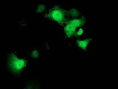
  MAB002) followed by Fluorescein-conjugated Anti-Mouse IgG Secondary Antibody (Catalog #
 ![Bioactivity IL-4 [Unconjugated]](https://images.novusbio.com/images/protein/6507-ilcf_recombinant-human-il-4-cho-expressed-protein-cf-bioactivity-272020133214.jpg)
![SEC-MALS IFN-gamma [Unconjugated]](https://images.novusbio.com/images/485-mi_recombinant-mouse-ifn-gamma-protein-sec-mals-1612202583245.jpg)
![Bioactivity IFN-gamma [Unconjugated]](https://images.novusbio.com/images/protein/IFN-gamma_485-MI_455.jpg)
![SDS-Page IFN-gamma [Unconjugated]](https://images.novusbio.com/images/protein/IFN-gamma_485-MI_407.jpg)
![Western Blot GATA-3 Antibody [Unconjugated]](https://images.novusbio.com/images/antibody/GATA-3_AF2605_Western_Blot_5199.jpg)
![Immunocytochemistry GATA-3 Antibody [Unconjugated]](https://images.novusbio.com/images/antibody/GATA-3_AF2605_Immunocytochemistry_6486.jpg)
![Immunohistochemistry GATA-3 Antibody [Unconjugated]](https://images.novusbio.com/images/antibody/GATA-3_AF2605_Immunohistochemistry_7156.jpg)
![N/A IL-10 [Biotin]](https://images.novusbio.com/images/elisa/DATA_IL10_DY417_ELISA_2014.jpg)
![Bioactivity IL-2 [Unconjugated]](https://images.novusbio.com/images/202-il_recombinant-human-il-2-protein-bioactivity-174202314946.jpg)
![N/A IL-17/IL-17A [Biotin]](https://images.novusbio.com/images/elisa/DATA_IL17_DY421_ELISA_2448.jpg)
![N/A IL-5 [HRP]](https://images.novusbio.com/images/elisa/IL-5_M5000_ELISA_237.jpg)
![N/A IL-5 [HRP]](https://images.novusbio.com/images/elisa/DATA_IL5_M5000_ELISA_935.jpg)
![N/A IL-13 [Biotin]](https://images.novusbio.com/images/elisa/DATA_IL13_DY413_ELISA_2010.jpg)
![SDS-Page TNF-alpha [Unconjugated]](https://images.novusbio.com/images/protein/TNF-alpha_210-TA_256.jpg)
![Bioactivity TNF-alpha [Unconjugated]](https://images.novusbio.com/images/protein/TNFalpha_210TA_1658.jpg)
![SEC-MALS TNF-alpha [Unconjugated]](https://images.novusbio.com/images/210-ta_recombinant-human-tnf-alpha-protein-sec-mals-35202312244..jpg)
![N/A IL-6 [HRP]](https://images.novusbio.com/images/elisa/DATA_IL6_M6000_ELISA_936.jpg)
![N/A IL-6 [HRP]](https://images.novusbio.com/images/elisa/IL-6_M6000_ELISA_415.jpg)
![N/A IL-6 [HRP]](https://images.novusbio.com/images/m6000b_mouse-il-6-quantikine-elisa-kit-1752025024034.jpg)
![Intracellular Staining by Flow Cytometry IL-17D Antibody (312724) [Unconjugated]](https://images.novusbio.com/images/antibody/IL-17D_MAB2274_Flow_Cytometry_8369.jpg)
![Flow Cytometry IL-17D Antibody (312724) [Unconjugated]](https://images.novusbio.com/images/mab2274_mouse-il-17d-mab-clone-312724-flow-cytometry-1212202593071.jpg)
![Flow Cytometry IL-17D Antibody (312724) [Unconjugated]](https://images.novusbio.com/images/mab2274_mouse-il-17d-mab-clone-312724-flow-cytometry-12122025932377.jpg)
![Bioactivity CTLA-4 [Unconjugated]](https://images.novusbio.com/images/protein/CTLA4_7268CT_2293.jpg)


