 or isotype control antibody (Catalog # MAB002, open histogram) followed by Allophycocyanin-conjugated Anti-Mouse IgG Secondary Antibody (Catalog # F0101B). To facilitate intracellular staining, cells were fixed with Flow Cytometry Fixation Buffer (Catalog # FC004) and permeabilized with Flow Cytometry Permeabilization/Wash Buffer I (Catalog # FC005). View our protocol for Staining Intracellular Molecules.")
| Reactivity | HuSpecies Glossary |
| Applications | ELISA, IHC, CyTOF-ready, Flow |
| Clone | 122511 |
| Clonality | Monoclonal |
| Host | Mouse |
| Conjugate | Unconjugated |
| Immunogen | Mouse myeloma cell line NS0-derived recombinant human SPARC Ala18-Ile303 Accession # P09486 |
| Specificity | Detects human SPARC/Osteonectin in direct ELISAs. In direct ELISAs, no cross-reactivity with recombinant mouse SPARC/Osteonectin is observed. |
| Source | N/A |
| Isotype | IgG1 |
| Clonality | Monoclonal |
| Host | Mouse |
| Gene | SPARC |
| Purity Statement | Protein A or G purified from hybridoma culture supernatant |
| Innovator's Reward | Test in a species/application not listed above to receive a full credit towards a future purchase. |
| Dilutions |
|
|
| Publications |
|
| Storage | Use a manual defrost freezer and avoid repeated freeze-thaw cycles.
|
| Buffer | Lyophilized from a 0.2 μm filtered solution in PBS with Trehalose. See Certificate of Analysis for details. *Small pack size (-SP) is supplied either lyophilized or as a 0.2 µm filtered solution in PBS. |
| Preservative | No Preservative |
| Reconstitution Instructions | Reconstitute at 0.5 mg/mL in sterile PBS. For liquid material, refer to CoA for concentration. |
SPARC, an acronym for “secreted protein, acidic and rich in cysteine”, is also known as osteonectin or BM-40 (1-5). It is the founding member of a family of secreted matricellular proteins with similar domain structure. The 286 amino acid (aa), 43 kDa protein contains an N-terminal acidic region that binds calcium, a follistatin domain that contains Kazal-like sequences, and a C-terminal extracellular calcium (EC) binding domain with two EF-hand motifs (1-5). Crystal structure modeling shows that residues implicated in cell binding, inhibition of cell spreading, and disassembly of focal adhesions cluster on one face of SPARC, while a collagen binding epitope and an N-glycosylation site are opposite this face (6). SPARC is produced by fibroblasts, capillary endothelial cells, platelets and macrophages, especially in areas of tissue morphogenesis and remodeling (3, 7). SPARC shows context-specific effects, but generally inhibits adhesion, spreading and proliferation, and promotes collagen matrix formation (3-5). For endothelial cells, SPARC disrupts focal adhesions and binds and sequesters PDGF and VEGF (3-5). SPARC is abundantly expressed in bone, where it promotes osteoblast differentiation and inhibits adipogenesis (5, 8). SPARC is potentially cleaved by metalloproteinases, producing an angiogenic peptide that includes the copper-binding sequence KGHK (7). Paradoxically, SPARC is highly expressed in many tumor types undergoing an endothelial to mesenchymal transition; its expression, however, mainly decreases the likelihood of metastasis and confers sensitivity to chemotherapy and radiation (4, 9-11). Stabilin-1, which is expressed on alternately activated macrophages, is the first SPARC receptor to be identified. It binds the SPARC EC domain and mediates endocytosis for degradation (12). Mature human SPARC shows 92%, 92%, 97%, 99%, 96% and 85% aa identity with mouse, rat, canine, bovine, porcine and chick SPARC, respectively.
![Western Blot Osteopontin/OPN Antibody [Unconjugated]](https://images.novusbio.com/images/af808_mouse-osteopontin-opn-affinity-purified-polyclonal-ab-western-blot-121220259133019.jpg)
![Western Blot Osteopontin/OPN Antibody [Unconjugated]](https://images.novusbio.com/images/af808_mouse-osteopontin-opn-affinity-purified-polyclonal-ab-western-blot-12122025932150.jpg)
![Western Blot Osteopontin/OPN Antibody [Unconjugated]](https://images.novusbio.com/images/af808_mouse-osteopontin-opn-affinity-purified-polyclonal-ab-western-blot-121220259224819.jpg)


![SPARC Antibody (122511) [Unconjugated]](/sites/all/modules/enterprise-tech/et_datasheets/images/novus_guarantee.png "SPARC Antibody (122511) [Unconjugated]")
Secondary Antibodies |
Isotype Controls |
|
Caspase 8 - a key mediator of apoptosis Programmed cell death via apoptosis is a key controlled physiological process instigated by the cell death receptor family, their ligands, and the caspase cysteine protease family. All caspases exist in a precursor form that contains a prodomain, a... Read full blog post. |
|
GAPDH: More than a housekeeping gene GAPDH is a 146kD tetramer glycolytic pathway metabolic enzyme composed of four 30-40 kDa subunits. It is responsible for reversibly phosphorylating its substrate glyceraldehyde 3-phosphate within the glycolytic pathway. Apart from its role in gl... Read full blog post. |
|
GAPDH (Glyceraldehyde 3-Phosphate Dehydrogenase) GAPDH is a 146 kD tetramer glycolytic pathway metabolic enzyme responsible for reversibly phosphorylating glyceraldehyde 3-phosphate. It may have other possible functions in transcriptional activation. GAPDH is highly expressed due to this housekeepin... Read full blog post. |
|
FLICE, FLICE, baby Cell death via apoptosis is a fundamental cellular function triggered by the cell death receptor family and their ligands which signal through downstream adaptor molecules and the caspase protease family. All caspases exist in a precursor form compose... Read full blog post. |
The concentration calculator allows you to quickly calculate the volume, mass or concentration of your vial. Simply enter your mass, volume, or concentration values for your reagent and the calculator will determine the rest.


 or isotype control antibody (Catalog # MAB002, open histogram), followed by Allophycocyanin-conjugated Anti-Mouse IgG Secondary Antibody (Catalog # F0101B). To facilitate intracellular staining, cells were fixed with Flow Cytometry Fixation Buffer (Catalog # FC004) and permeabilized with Flow Cytometry Permeabilization/Wash Buffer I (Catalog # FC005). View our protocol for Staining Intracellular Molecules.")
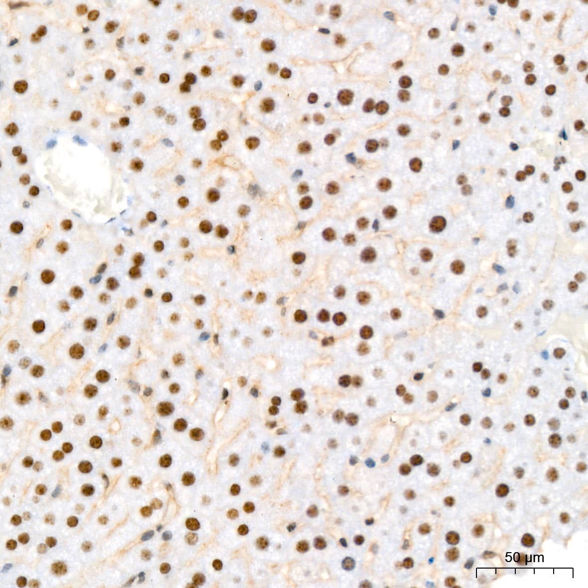
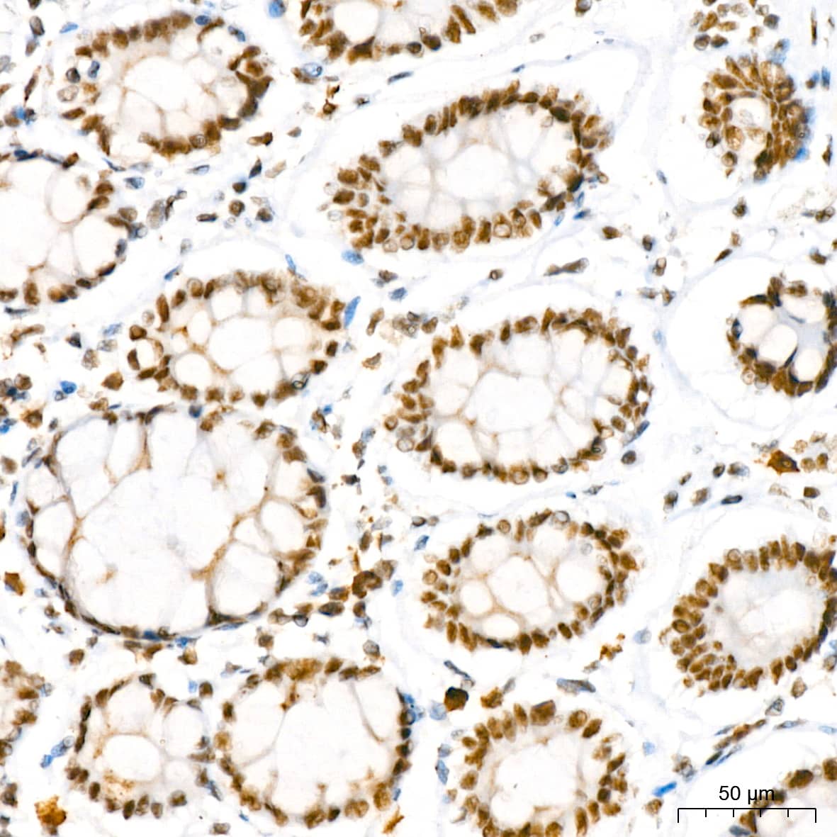
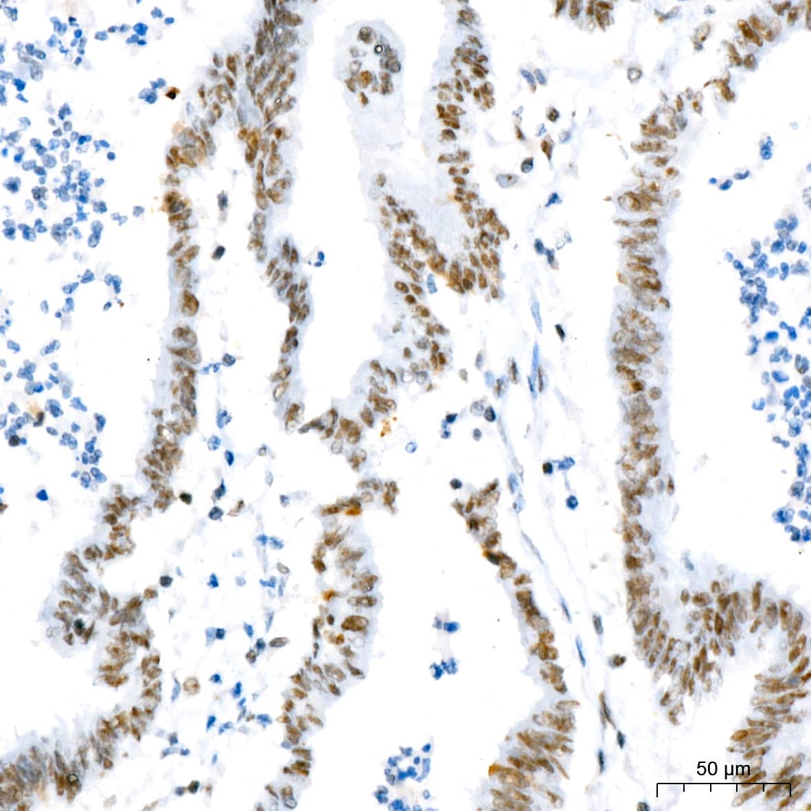
 at 25 µg/mL overnight at 4 °C. Tissue was stained using the Anti-Mouse HRP-DAB Cell & Tissue Staining Kit (brown; Catalog # CTS002) and counterstained with hematoxylin (blue). View our protocol for Chromogenic IHC Staining of Paraffin-embedded Tissue Sections.")
 coated on a Clear Polystyrene Microplate (Catalog # DY990). Goat Anti-Human SPARC Antigen Affinity-purified Polyclonal Antibody (Catalog # AF941) was biotinylated and incubated with the protein captured on the plate. Detection of the standard curve was achieved by incubating Streptavidin-HRP (Catalog # DY998) followed by Substrate Solution (Catalog # DY999) and stopping the enzymatic reaction with Stop Solution (Catalog # DY994).")


![Bioactivity IBSP/Sialoprotein II [Unconjugated]](https://images.novusbio.com/images/4014-sp_recombinant-human-ibsp-sialoprotein-ii-protein-cf-bioactivity-4420249391..jpg)
![N/A Thrombospondin-1 [HRP]](https://images.novusbio.com/images/elisa/Thrombospondin-1_DTSP10_ELISA_199.jpg)
![N/A Thrombospondin-1 [HRP]](https://images.novusbio.com/images/dtsp10_human-thrombospondin-1-quantikine-elisa-kit-23120261145.jpg)


![N/A VEGF [HRP]](https://images.novusbio.com/images/elisa/VEGF_DVE00_ELISA_208.jpg)
![N/A VEGF [HRP]](https://images.novusbio.com/images/elisa/DATA_VEGF_DVE00_ELISA_871.jpg)
![N/A VEGF [HRP]](https://images.novusbio.com/images/elisa/DATA_VEGF_DVE00_ELISA_872.jpg)
![N/A MMP-2 [HRP]](https://images.novusbio.com/images/elisa/DATA_MMP2_MMP200_ELISA_1003.jpg)
![N/A MMP-2 [HRP]](https://images.novusbio.com/images/elisa/MMP2_MMP200_ELISA_474.jpg)
![N/A SLPI [HRP]](https://images.novusbio.com/images/elisa/SLPI_DPI00_ELISA_160.jpg)
![N/A SLPI [HRP]](https://images.novusbio.com/images/elisa/DATA_SLPI_DPI00_ELISA_792.jpg)
![Western Blot RUNX2/CBFA1 Antibody (232902) [Unconjugated]](https://images.novusbio.com/images/mab2006_human-runx2-cbfa1-mab-clone-232902-western-blot-1212202597422.jpg)
![Western Blot RUNX2/CBFA1 Antibody (232902) [Unconjugated]](https://images.novusbio.com/images/mab2006_human-runx2-cbfa1-mab-clone-232902-western-blot-121220259172010.jpg)
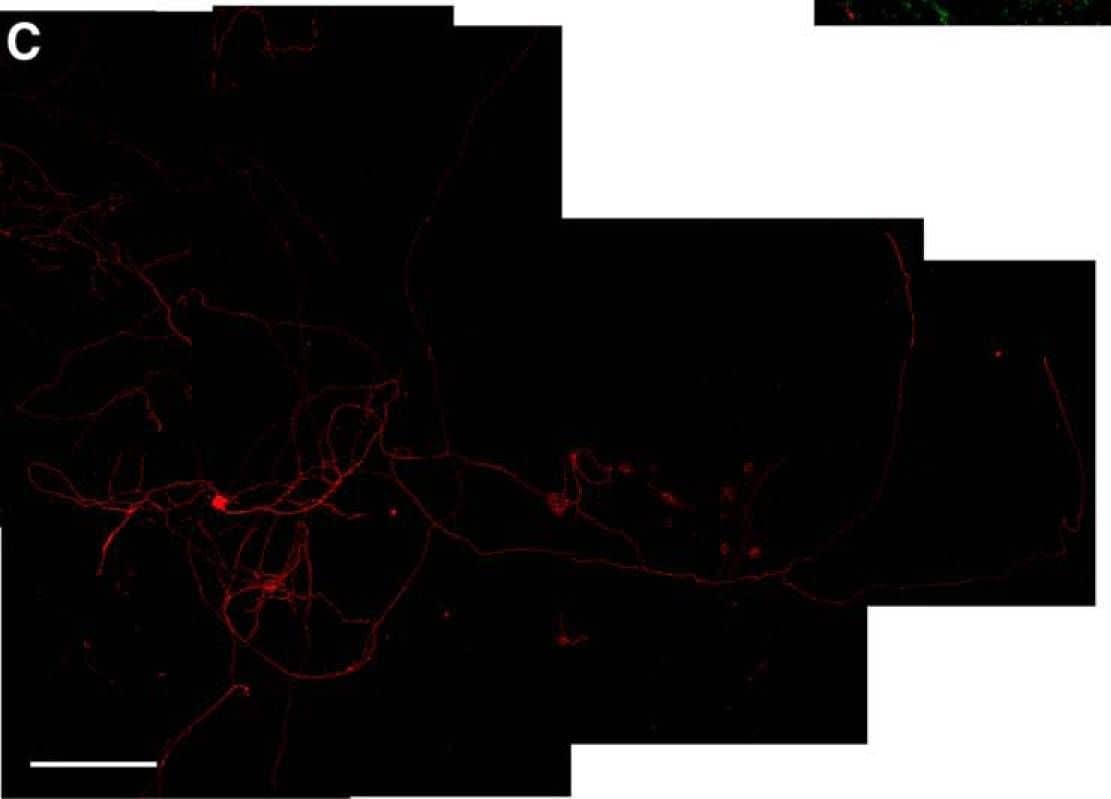
![Immunocytochemistry RUNX2/CBFA1 Antibody (232902) [Unconjugated]](https://images.novusbio.com/images/antibody/RUNX2_MAB2006_Immunocytochemistry_13309.jpg)
![N/A CCL27/CTACK [HRP]](https://images.novusbio.com/images/elisa/DATA_CCL27_DCC270_ELISA_615.jpg)
![N/A CCL27/CTACK [HRP]](https://images.novusbio.com/images/elisa/CCL27_DCC270_ELISA_54.jpg)


