MAB002, open histogram) followed by Allophycocyanin-conjugated Anti-Mouse IgG Secondary Antibody (Catalog #
F0101B). To facilitate intracellular staining, cells were fixed with Flow Cytometry Fixation Buffer (Catalog #
FC004) and permeabilized with Flow Cytometry Permeabilization/Wash Buffer I (Catalog #
FC005). View our protocol for
Staining Intracellular Molecules." class="big_lightbox">
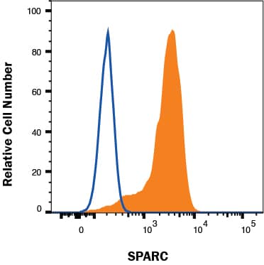
MAB002, open histogram) followed by Allophycocyanin-conjugated Anti-Mouse IgG Secondary Antibody (Catalog #
F0101B). To facilitate intracellular staining, cells were fixed with Flow Cytometry Fixation Buffer (Catalog #
FC004) and permeabilized with Flow Cytometry Permeabilization/Wash Buffer I (Catalog #
FC005). View our protocol for
Staining Intracellular Molecules." alt="MG-63 cells were stained with Mouse Anti-Human SPARC Monoclonal Antibody (Catalog # MAB941, filled histogram) or isotype control antibody (Catalog #
MAB002, open histogram) followed by Allophycocyanin-conjugated Anti-Mouse IgG Secondary Antibody (Catalog #
F0101B). To facilitate intracellular staining, cells were fixed with Flow Cytometry Fixation Buffer (Catalog #
FC004) and permeabilized with Flow Cytometry Permeabilization/Wash Buffer I (Catalog #
FC005). View our protocol for
Staining Intracellular Molecules." class="big_thumb" />
MAB002, open histogram), followed by Allophycocyanin-conjugated Anti-Mouse IgG Secondary Antibody (Catalog #
F0101B). To facilitate intracellular staining, cells were fixed with Flow Cytometry Fixation Buffer (Catalog #
FC004) and permeabilized with Flow Cytometry Permeabilization/Wash Buffer I (Catalog #
FC005). View our protocol for
Staining Intracellular Molecules." class="big_lightbox">
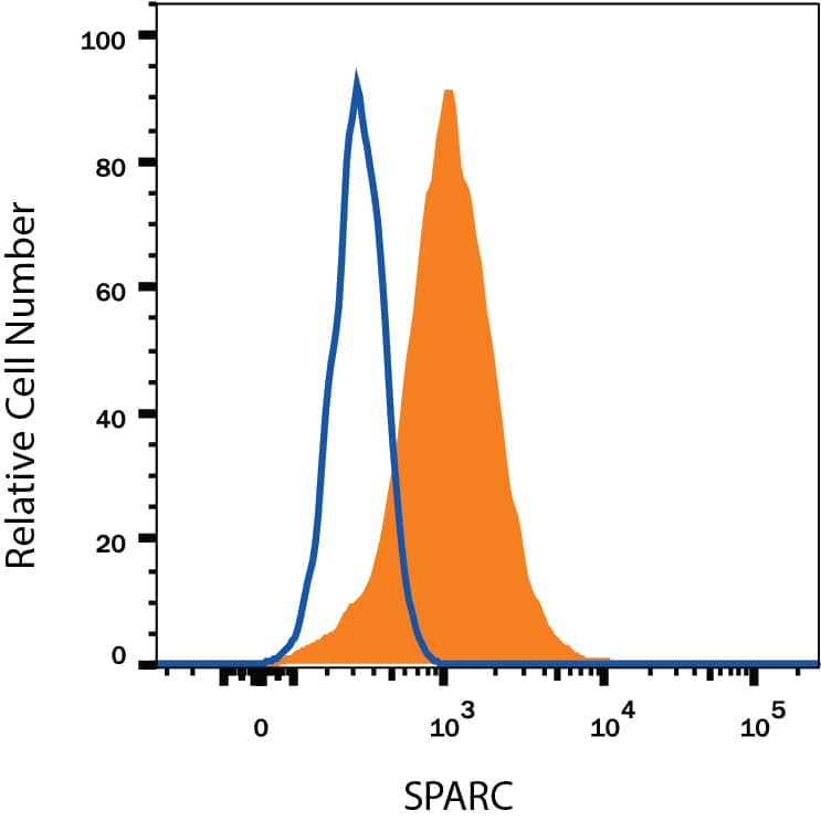
MAB002, open histogram), followed by Allophycocyanin-conjugated Anti-Mouse IgG Secondary Antibody (Catalog #
F0101B). To facilitate intracellular staining, cells were fixed with Flow Cytometry Fixation Buffer (Catalog #
FC004) and permeabilized with Flow Cytometry Permeabilization/Wash Buffer I (Catalog #
FC005). View our protocol for
Staining Intracellular Molecules." class="big_thumb" />
DY990). Goat Anti-Human SPARC Antigen Affinity-purified Polyclonal Antibody (Catalog #
AF941) was biotinylated and incubated with the protein captured on the plate. Detection of the standard curve was achieved by incubating Streptavidin-HRP (Catalog #
DY998) followed by Substrate Solution (Catalog #
DY999) and stopping the enzymatic reaction with Stop Solution (Catalog #
DY994)." class="big_lightbox">
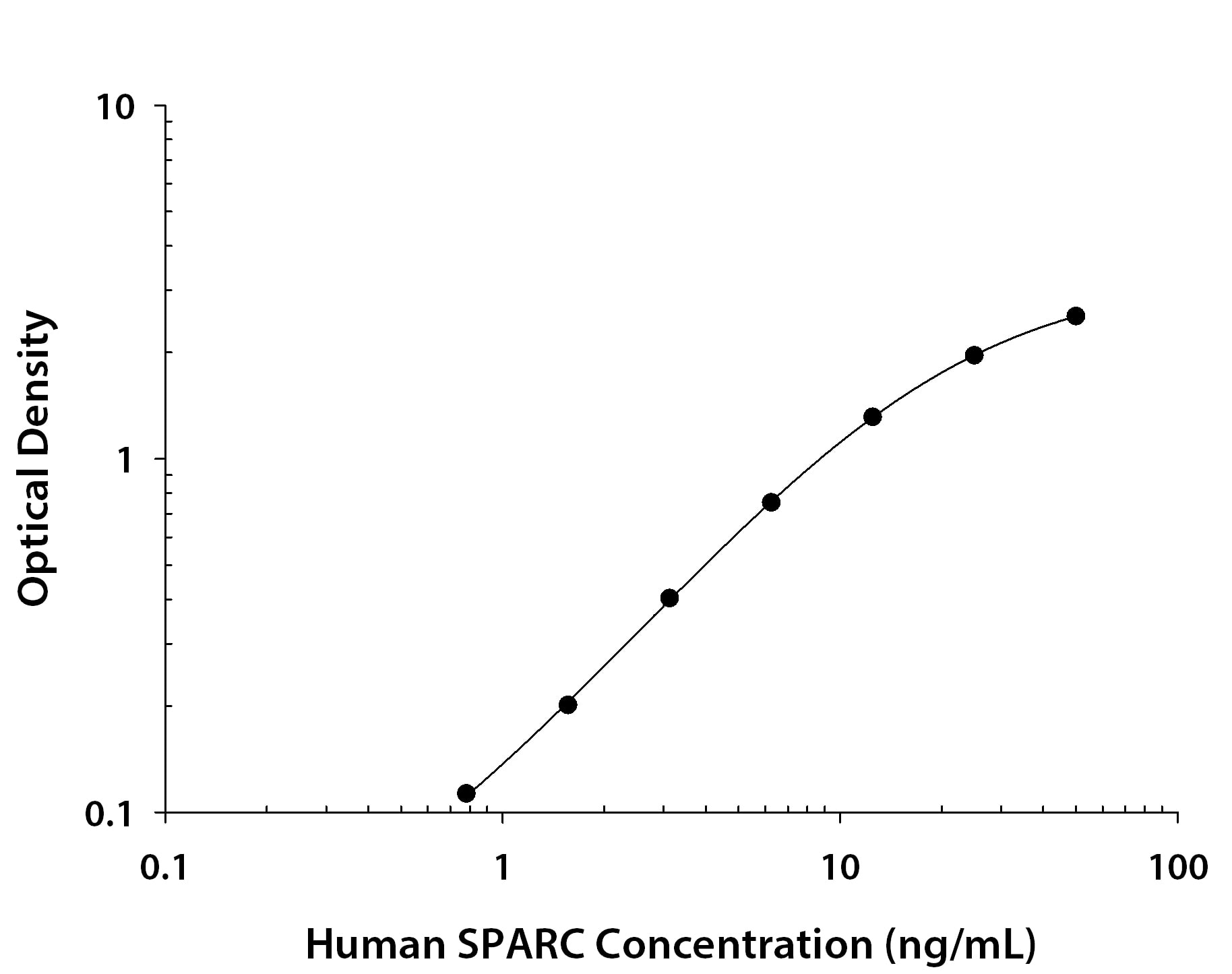
DY990). Goat Anti-Human SPARC Antigen Affinity-purified Polyclonal Antibody (Catalog #
AF941) was biotinylated and incubated with the protein captured on the plate. Detection of the standard curve was achieved by incubating Streptavidin-HRP (Catalog #
DY998) followed by Substrate Solution (Catalog #
DY999) and stopping the enzymatic reaction with Stop Solution (Catalog #
DY994)." class="big_thumb" />
941-SP) was serially diluted and captured by Mouse Anti-Human SPARC Monoclonal Antibody (Catalog # MAB941) coated on a Clear Polystyrene Microplate (Catalog #
DY990). Goat Anti-Human SPARC Antigen Affinity-purified Polyclonal Antibody (Catalog #
AF941) was biotinylated and incubated with the protein captured on the plate. Detection of the standard curve was achieved by incubating Streptavidin-HRP (Catalog #
DY998)" class="big_lightbox">

941-SP) was serially diluted and captured by Mouse Anti-Human SPARC Monoclonal Antibody (Catalog # MAB941) coated on a Clear Polystyrene Microplate (Catalog #
DY990). Goat Anti-Human SPARC Antigen Affinity-purified Polyclonal Antibody (Catalog #
AF941) was biotinylated and incubated with the protein captured on the plate. Detection of the standard curve was achieved by incubating Streptavidin-HRP (Catalog #
DY998)" class="big_thumb" />
 HAF017). A specific band was detected for SPARC at approximately 35-37 kDa (as indicated). This experiment was conducted under reducing conditions and using Immunoblot Buffer Group 1." alt="Western blot shows lysates of C2C12 mouse myoblast cell line and mouse placenta tissue. PVDF membrane was probed with 0.2 µg/mL of Goat Anti-Mouse SPARC Antigen Affinity-purified Polyclonal Antibody (Catalog # AF942) followed by HRP-conjugated Anti-Goat IgG Secondary Antibody (Catalog # HAF017). A specific band was detected for SPARC at approximately 35-37 kDa (as indicated). This experiment was conducted under reducing conditions and using Immunoblot Buffer Group 1." class="big_thumb" />
 AB-108-C, open histogram) followed by Allophycocyanin-conjugated Anti-Goat IgG Secondary Antibody (Catalog # F0108). To facilitate intracellular staining, cells were fixed with Flow Cytometry Fixation Buffer (Catalog # FC004) and permeabilized with Flow Cytometry Permeabilization/Wash Buffer I (Catalog # FC005). View our protocol for Staining Intracellular Molecules." alt="MG-63 cells were stained with Goat Anti-Human SPARC Antigen Affinity-purified Polyclonal Antibody (Catalog # AF941, filled histogram) or isotype control antibody (Catalog # AB-108-C, open histogram) followed by Allophycocyanin-conjugated Anti-Goat IgG Secondary Antibody (Catalog # F0108). To facilitate intracellular staining, cells were fixed with Flow Cytometry Fixation Buffer (Catalog # FC004) and permeabilized with Flow Cytometry Permeabilization/Wash Buffer I (Catalog # FC005). View our protocol for Staining Intracellular Molecules." class="big_thumb" />
 HAF005). A specific band was detected for SPARC at approximately 35-37 kDa (as indicated). This experiment was conducted under reducing conditions and using Immunoblot Buffer Group 1." alt="Western blot shows lysates of C2C12 mouse myoblast cell line and mouse placenta tissue. PVDF membrane was probed with 2 µg/mL of Rat Anti-Mouse SPARC Monoclonal Antibody (Catalog # MAB942) followed by HRP-conjugated Anti-Rat IgG Secondary Antibody (Catalog # HAF005). A specific band was detected for SPARC at approximately 35-37 kDa (as indicated). This experiment was conducted under reducing conditions and using Immunoblot Buffer Group 1." class="big_thumb" />
 MAB002, open histogram) followed by Allophycocyanin-conjugated Anti-Mouse IgG Secondary Antibody (Catalog # F0101B). To facilitate intracellular staining, cells were fixed with Flow Cytometry Fixation Buffer (Catalog # FC004) and permeabilized with Flow Cytometry Permeabilization/Wash Buffer I (Catalog # FC005). View our protocol for Staining Intracellular Molecules." alt="MG-63 cells were stained with Mouse Anti-Human SPARC Monoclonal Antibody (Catalog # MAB941, filled histogram) or isotype control antibody (Catalog # MAB002, open histogram) followed by Allophycocyanin-conjugated Anti-Mouse IgG Secondary Antibody (Catalog # F0101B). To facilitate intracellular staining, cells were fixed with Flow Cytometry Fixation Buffer (Catalog # FC004) and permeabilized with Flow Cytometry Permeabilization/Wash Buffer I (Catalog # FC005). View our protocol for Staining Intracellular Molecules." class="big_thumb" />


 CTS008) and counterstained with hematoxylin (blue). Specific staining was localized to developing cartilage. View our protocol for
  NL001) and counterstained with DAPI (blue). Specific staining was localized to endoplasmic reticuli. View our protocol for
  AB-108-C, open histogram), followed by Phycoerythrin-conjugated anti-Goat IgG (
 HAF019). A specific band was detected for SPARC at approximately 43 kDa (as indicated). This experiment was conducted under reducing conditions and using
  CTS008) and counterstained with hematoxylin (blue). View our protocol for
  HAF109). This experiment was conducted under reducing conditions and using the 12-230 kDa separation system." class="big_thumb" />
 MAB941) coated on a Clear Polystyrene Microplate (


 941-SP) was serially diluted and captured by Mouse Anti-Human SPARC Monoclonal Antibody (Catalog #
  CTS017) and counterstained with hematoxylin (blue). View our protocol for
  MAB002, open histogram), followed by Allophycocyanin-conjugated Anti-Mouse IgG Secondary Antibody (Catalog #
  CTS002) and counterstained with hematoxylin (blue). View our protocol for
  DY990). Goat Anti-Human SPARC Antigen Affinity-purified Polyclonal Antibody (Catalog #
  941-SP) was serially diluted and captured by Mouse Anti-Human SPARC Monoclonal Antibody (Catalog # MAB941) coated on a Clear Polystyrene Microplate (Catalog #
 ![Western Blot: SPARC Antibody [NBP1-80972] - Analysis in human cell lines U2OS and HeLa. Corresponding RNA-seq data are presented for the same cell lines. Loading control: Anti-HSP90B1.](http://images.novusbio.com/fullsize/SPARC-Antibody-Western-Blot-NBP1-80972-img0010.jpg "<b>Orthogonal Strategies Validation. </b>Western Blot: SPARC Antibody [NBP1-80972] - Analysis in human cell lines U2OS and HeLa. Corresponding RNA-seq data are presented for the same cell lines. Loading control: Anti-HSP90B1.")
![<b>Independent Antibodies Validation and Orthogonal Strategies Validation.</b>Western Blot: SPARC Antibody [NBP1-80972] - Analysis using Anti-SPARC antibody NBP1-80972 (A) shows similar pattern to independent antibody NBP1-80971 (B).](http://images.novusbio.com/fullsize/SPARC-Antibody-Western-Blot-NBP1-80972-img0013.jpg)
![Immunohistochemistry-Paraffin: SPARC Antibody [NBP1-80972] - Staining of human placenta shows strong positivity in endothelial cells and fibroblasts.](http://images.novusbio.com/fullsize/SPARC-Antibody-Immunohistochemistry-Paraffin-NBP1-80972-img0011.jpg)
![Western Blot: SPARC Antibody [NBP1-80971] - Analysis in human cell lines U-251MG and MCF-7. Corresponding RNA-seq data are presented for the same cell lines. Loading control: Anti-GAPDH.](http://images.novusbio.com/fullsize/SPARC-Antibody-Western-Blot-NBP1-80971-img0007.jpg "<b>Orthogonal Strategies Validation. </b>Western Blot: SPARC Antibody [NBP1-80971] - Analysis in human cell lines U-251MG and MCF-7. Corresponding RNA-seq data are presented for the same cell lines. Loading control: Anti-GAPDH.")
![<b>Independent Antibodies Validation and Orthogonal Strategies Validation.</b>Western Blot: SPARC Antibody [NBP1-80971] - Analysis using Anti-SPARC antibody NBP1-80971 (A) shows similar pattern to independent antibody NBP1-80972 (B).](http://images.novusbio.com/fullsize/SPARC-Antibody-Western-Blot-NBP1-80971-img0010.jpg)
![Western Blot: SPARC Antibody [NBP1-80971] - Analysis in control (vector only transfected HEK293T lysate) and SPARC over-expression lysate (Co-expressed with a C-terminal myc-DDK tag (3.1 kDa) in mammalian HEK293T cells).](http://images.novusbio.com/fullsize/SPARC-Antibody-Western-Blot-NBP1-80971-img0009.jpg)


![Western Blot: SPARC Antibody [NBP2-27358] - Analysis of human A375 lysate probed with SPARC antibody. Goat anti-rabbit IgG HRP secondary antibody and PicoTect ECL substrate solution were used for this test.](http://images.novusbio.com/fullsize/SPARC-Antibody-Western-Blot-NBP2-27358-img0001.jpg "Western Blot: SPARC Antibody [NBP2-27358] - Analysis of human A375 lysate probed with SPARC antibody. Goat anti-rabbit IgG HRP secondary antibody and PicoTect ECL substrate solution were used for this test.")
![Immunohistochemistry-Paraffin: SPARC Antibody [NBP2-27358] - FFPE human placenta tissue stained with SPARC antibody, peroxidase-conjugate and DAB chromogen.](http://images.novusbio.com/fullsize/SPARC-Antibody-Immunohistochemistry-Paraffin-NBP2-27358-img0002.jpg)
![Western Blot: SPARC Antibody (001) [NBP2-90612] - Anti-SPARC rabbit monoclonal antibody at 1:500 dilution. Lane A: A549 Whole Cell Lysate. Lysates/proteins at 30 ug per lane. Secondary Goat Anti-Rabbit IgG H&L (Dylight 800) at 1/10000 dilution. Developed using the Odyssey technique. Performed under reducing conditions. Predicted band size: 35 kDa. Observed band size: 40 kDa](http://images.novusbio.com/fullsize/SPARC-Antibody-001-Western-Blot-NBP2-90612-img0003.jpg "Western Blot: SPARC Antibody (001) [NBP2-90612] - Anti-SPARC rabbit monoclonal antibody at 1:500 dilution. Lane A: A549 Whole Cell Lysate. Lysates/proteins at 30 ug per lane. Secondary Goat Anti-Rabbit IgG H&L (Dylight 800) at 1/10000 dilution. Developed using the Odyssey technique. Performed under reducing conditions. Predicted band size: 35 kDa. Observed band size: 40 kDa")
![Immunoprecipitation: SPARC Antibody (001) [NBP2-90612] - Mouse SPARC was immunoprecipitated using: Lane A: 0.5 mg A549 Whole Cell Lysate. Lane B: 0.5 mg 293T Whole Cell Lysate. 0.5 ul anti-Mouse SPARC rabbit monoclonal antibody and 15 ul of 50 % Protein G agarose. Primary antibody: Anti-Mouse SPARC rabbit monoclonal antibody, at 1:500 dilution. Secondary antibody: Dylight 800-labeled antibody to rabbit IgG (H+L), at 1:5000 dilution. Developed using the Odyssey technique. Performed under reducing conditions. Predicted band size: 35 kDa. Observed band size: 38 kDa](http://images.novusbio.com/fullsize/SPARC-Antibody-001-Immunoprecipitation-NBP2-90612-img0004.jpg)
![Immunohistochemistry: SPARC Antibody (PD00-57) [NBP3-32974] - Immunohistochemical analysis of paraffin-embedded human malignant melanoma tissue with Rabbit anti-SPARC antibody (NBP3-32974) at 1/500 dilution.<br/><br/> The section was pre-treated using heat mediated antigen retrieval with Tris-EDTA buffer (pH 9.0) for 20 minutes. The tissues were blocked in 1% BSA for 20 minutes at room temperature, washed with ddH2O and PBS, and then probed with the primary antibody (NBP3-32974) at 1/500 dilution for 1 hour at room temperature. The detection was performed using an HRP conjugated compact polymer system. DAB was used as the chromogen. Tissues were counterstained with hematoxylin and mounted with DPX.](http://images.novusbio.com/fullsize/nbp3-32974_rabbit-sparc-mab-pd00-57-298202414333618.jpg "Immunohistochemistry: SPARC Antibody (PD00-57) [NBP3-32974] - Immunohistochemical analysis of paraffin-embedded human malignant melanoma tissue with Rabbit anti-SPARC antibody (NBP3-32974) at 1/500 dilution.<br/><br/> The section was pre-treated using heat mediated antigen retrieval with Tris-EDTA buffer (pH 9.0) for 20 minutes. The tissues were blocked in 1% BSA for 20 minutes at room temperature, washed with ddH2O and PBS, and then probed with the primary antibody (NBP3-32974) at 1/500 dilution for 1 hour at room temperature. The detection was performed using an HRP conjugated compact polymer system. DAB was used as the chromogen. Tissues were counterstained with hematoxylin and mounted with DPX.")
![Western blot analysis on different lysates with NBP3-32974 at 1/5,000 dilution. Lane 1: A375 cell lysate Lane 2: SH-SY5Y cell lysate Lysates/proteins at 20 µg/Lane. Predicted band size: 35 kDa Observed band size: 40 kDa Exposure time: Lane 1: 59 seconds; Lane 2: 3 minutes; 4-20% SDS-PAGE gel. Proteins were transferred to a PVDF membrane and blocked with 5% NFDM/TBST for 1 hour at room temperature. NBP3-32974 at 1/5,000 dilution was used in primary antibody dilution at 4℃ overnight. Goat Anti-Rabbit IgG [HRP] Secondary Antibody at 1/50,000 dilution was used for 1 hour at room temperature.](http://images.novusbio.com/fullsize/nbp3-32974_-western-blot-639204594551263433.jpg)


