After August 17, 2026, Novus Biologicals products and services will no longer be available on this website; you will access all products and services on rndsystems.com. Create your R&D Systems online account today.
Western Blot: S100A9 Antibody [NB110-89726] - Cells were transfected with the pCMV6-ENTRY S100A9 cDNA or the pCMV6-ENTRY control for 48 hrs and lysed. Equivalent amounts of cell lysates (5 ug per lane) were separated by ...read more
Immunocytochemistry/ Immunofluorescence: S100A9 Antibody [NB110-89726] - S100A9 antibody was tested in A431 cells with FITC (green). Nuclei and alpha-tubulin were counterstained with Dapi (blue) and Dylight 550 (red).
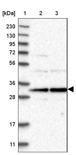
Biological Strategies: Western Blot: S100A9 Antibody [NB110-89726] - Analysis of S100A9 Antibody in DMSO treated HL60 whole cell lysates.
Simple Western: S100A9 Antibody [NB110-89726] - Simple Western lane view shows a specific band for S100A9 in 0.5 mg/ml of Human PBMC's lysate. This experiment was performed under reducing conditions using the 12-230 ...read more
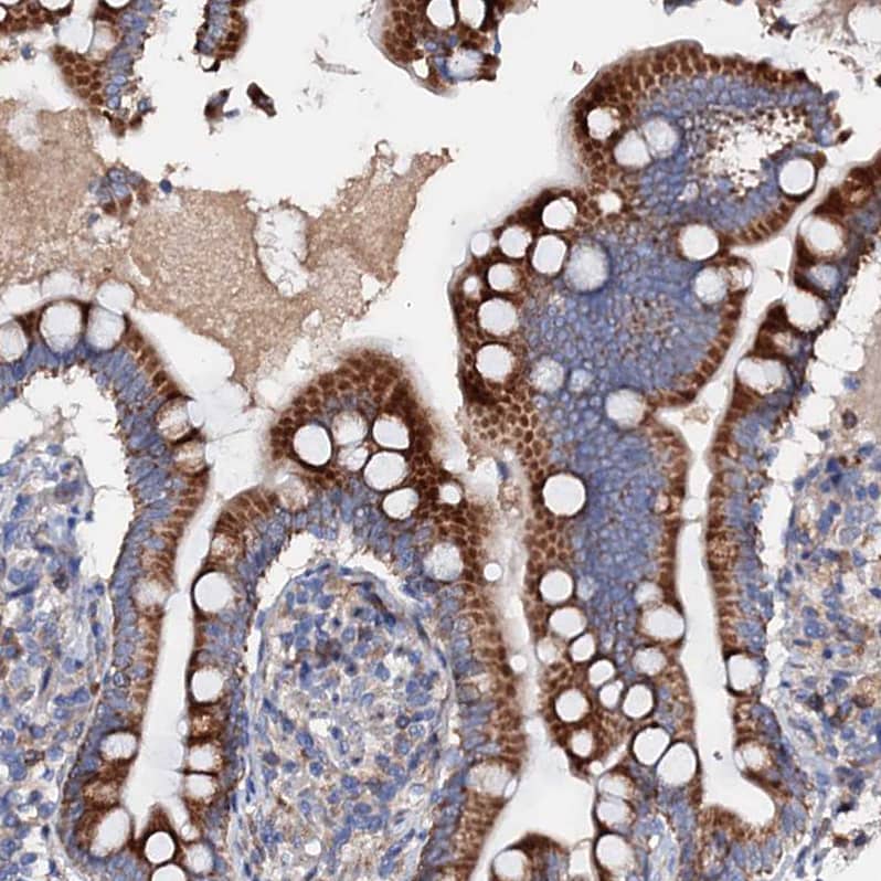
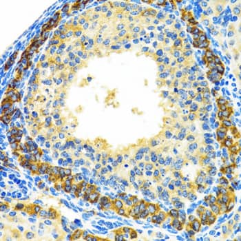
Immunohistochemistry: S100A9 Antibody [NB110-89726] - Key molecules of specific signaling pathways are assayed by immunohistochemistry in the colorectum of mice. Immunohistochemistry (200× magnification) of (A) S100a9, ...read more
Immunohistochemistry: S100A9 Antibody [NB110-89726] - Key molecules of specific signaling pathways are assayed by immunohistochemistry in the colorectum of mice. Immunohistochemistry (200× magnification) of (A) S100a9, ...read more
Immunohistochemistry: S100A9 Antibody [NB110-89726] - Key molecules of specific signaling pathways are assayed by immunohistochemistry in the colorectum of mice. Immunohistochemistry (200× magnification) of (A) S100a9, ...read more
Immunohistochemistry: S100A9 Antibody [NB110-89726] - Key molecules of specific signaling pathways are assayed by immunohistochemistry in the colorectum of mice. Immunohistochemistry (200× magnification) of (A) S100a9, ...read more
Immunohistochemistry: S100A9 Antibody [NB110-89726] - Key molecules of specific signaling pathways are assayed by immunohistochemistry in the colorectum of mice. Immunohistochemistry (200× magnification) of (A) S100a9, ...read more
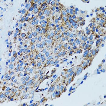
Immunohistochemistry: S100A9 Antibody [NB110-89726] - Effects of anti-S100a9 Ab administration on the azoxymethane (AOM)/dextran sulfate sodium (DSS)-induced colitis-associated cancer development. (A) Experimental ...read more
Immunohistochemistry: S100A9 Antibody [NB110-89726] - Effects of anti-S100a9 Ab on the frequency of neutrophils, macrophages, & dendritic cells (DCs) in the colon of the dextran sulfate sodium (DSS) mouse model. (A) ...read more
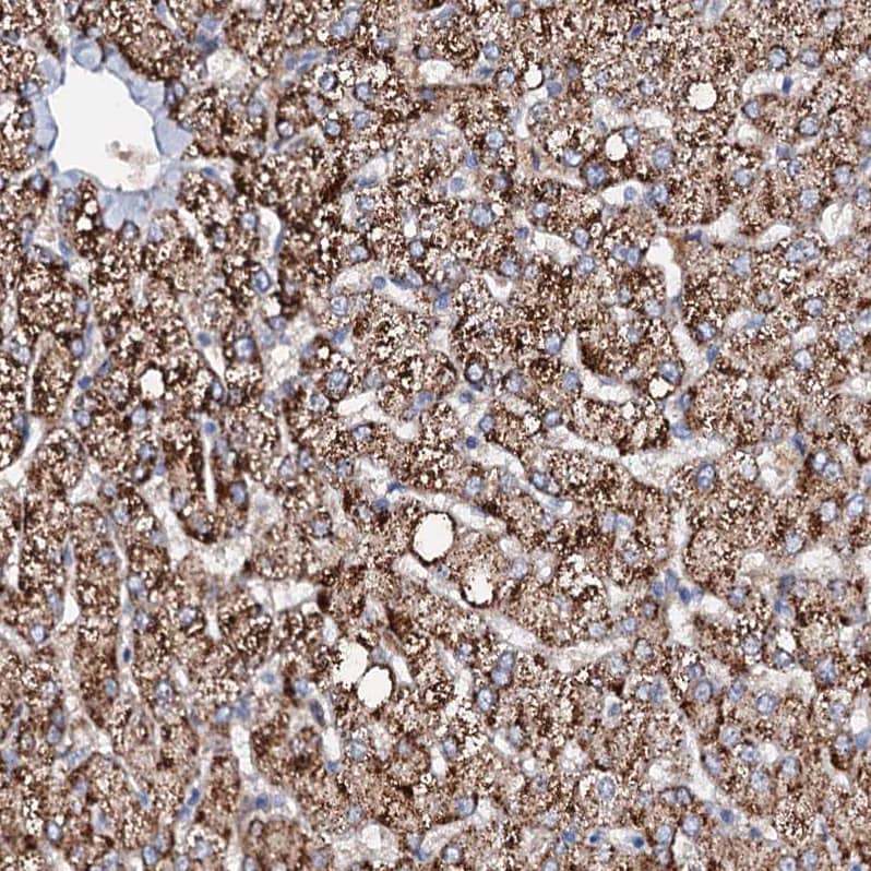
Immunohistochemistry: S100A9 Antibody [NB110-89726] - Anti-S100a9 Ab ameliorates inflammatory response of dextran sulfate sodium (DSS)-induced colitis in mice. (A) 6 days after DSS treatment, representative ...read more
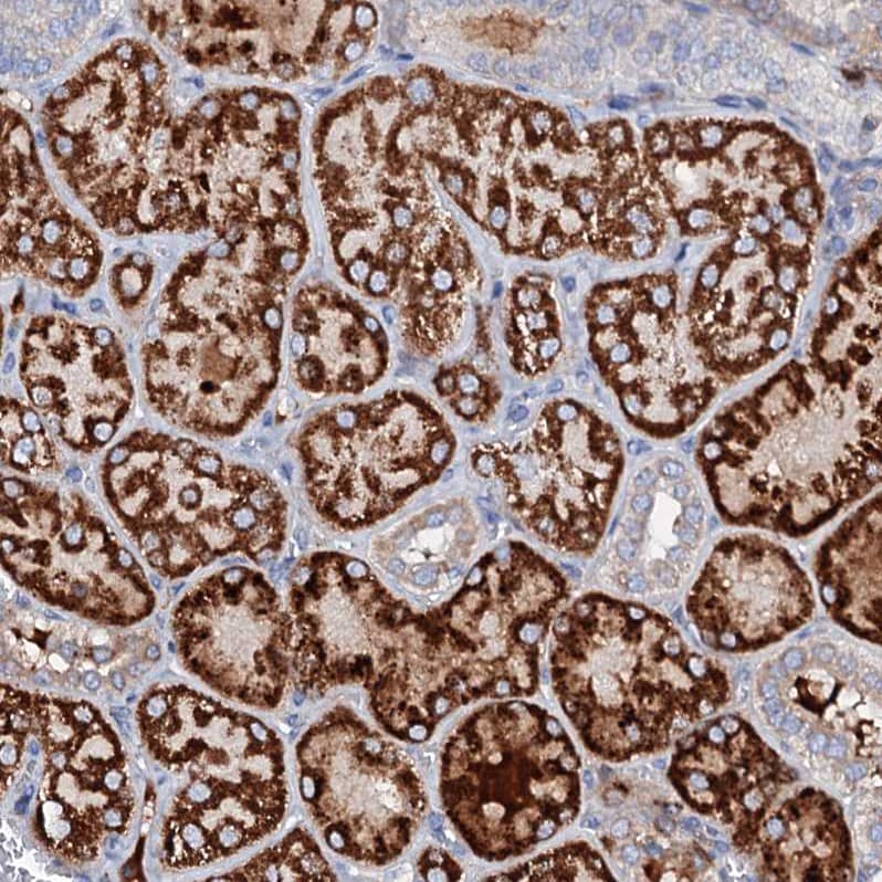
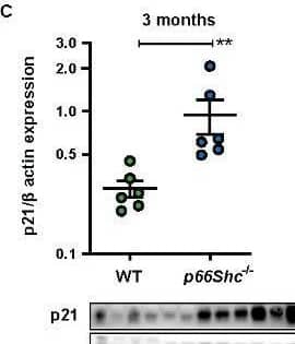
Immunohistochemistry: S100A9 Antibody [NB110-89726] - Host CP (S100A8/A9) is elevated in H. pylori infected stomach tissue.A) s100a8/s100a9 transcript abundance in RNA extracted from C57BL/6 mice infected with H. pylori ...read more
Immunohistochemistry: S100A9 Antibody [NB110-89726] - Anti-S100a9 Ab ameliorates inflammatory response of dextran sulfate sodium (DSS)-induced colitis in mice. (A) 6 days after DSS treatment, representative ...read more
Immunohistochemistry: S100A9 Antibody [NB110-89726] - Anti-S100a9 Ab ameliorates inflammatory response of dextran sulfate sodium (DSS)-induced colitis in mice. (A) 6 days after DSS treatment, representative ...read more
Immunohistochemistry reported in scientific literature (PMID 25792748)
Immunohistochemistry-Paraffin reported in scientific literature (PMID 21653680)
Simple Western 1:100
Western Blot 1:1000
Application Notes
In Western blot, a band is seen at approx. 16 kDa.
In Simple Western only 10 - 15 uL of the recommended dilution is used per data point. See Simple Western Antibody Database for Simple Western validation: Tested in Human PBMC's lysate 0.5 mg/mL, separated by Size, antibody dilution of 1:100. Separated by Size-Wes, Sally Sue/Peggy Sue. The observed molecular weight of the protein may vary from the listed predicted molecular weight due to post translational modifications, post translation cleavages, relative charges, and other experimental factors.
Theoretical MW
16 kDa. Disclaimer note: The observed molecular weight of the protein may vary from the listed predicted molecular weight due to post translational modifications, post translation cleavages, relative charges, and other experimental factors.
Store at 4C short term. Aliquot and store at -20C long term. Avoid freeze-thaw cycles.
Buffer
Whole antisera
Preservative
0.1% Sodium Azide
Purity
Unpurified
Alternate Names for S100A9 Antibody - BSA Free
CAGBMigration inhibitory factor-related protein 14
Calgranulin B
calgranulin-B
Calprotectin L1H subunit
CFAGMRP-14,60B8AG
CGLB
L1AG
LIAG
MAC387
MIF
MRP-14
MRP14Leukocyte L1 complex heavy chain
NIF
P14
protein S100-A9
S100 calcium binding protein A9 (calgranulin B)
S100 calcium binding protein A9
S100 calcium-binding protein A9 (calgranulin B)
S100 calcium-binding protein A9
S100A9
Background
S100A9 (S100 calcium-binding protein A9) is a member of S-100 family (family named so as its members are 100% solubile in ammonium sulfate at normal pH) and it exist as a homodimer or as heterodimer/heterotetramer with S100A8. S100A9 is a calcium binding protein with several post-translation modifications as well as multiple ligands, and when bound to calcium, it binds to arachidonic acid, cytoskeletal elements (e.g. keratin intermediate filaments), RAGE (receptor for advanced glycation endproducts), TLR4, CD36, MMPs, fibronectin, and heparin sulfate glycosaminoglycans for serving diverse biological functions. Localization of S100A9 varies according to cell type/disease state, however, it predominantly localize to cytoplasm, from where it translocates to cytoskeleton/cell membrane upon elevation of intracellular calcium levels. During neutrophil activation or endothelial adhesion of monocytes, it gets secreted via a microtubule-mediated alternative pathway. S100A9 is expressed in normal cell types such as myeloid cells, but its expression levels are increased in conditions of inflammatory diseases (RA, SLE, cystic fibrosis, inflammatory bowel disease, Crohn's disease, Sjogren's syndrome, and progressive systemic sclerosis) as well as in several types of cancers including gastric, esophageal, colon, pancreatic, bladder, ovarian, thyroid, breast and skin cancers.
Limitations
This product is for research use only and is not approved for use in humans or in clinical diagnosis. Primary Antibodies are guaranteed for 1 year from date of receipt.
Yoshikawa T, Takeichi T, Hirabayashi T et al. IL-17 axis is a significant driver of skin inflammation in Card14 mutant pityriasis rubra pilaris model mice Research Square 2023-02-02 (IHC, Mouse)
Langle Y, Sandes E, Belgorosky D. La expresion de S100A9, vinculada con el oxidontrico, es un marcador de mal pronostico enpacientes con cancer de vejiga, siendo su inhibicionun posible blanco terapeutico. Rev. Arg. de Urol. 2014-03-01
Turchi R, Tortolici F, Benvenuto M et al. Low Sulfur Amino Acid, High Polyunsaturated Fatty Acid Diet Inhibits Breast Cancer Growth International Journal of Molecular Sciences 2022-12-23 (IHC-Fr, Mouse)
FAQs for S100A9 Antibody (NB110-89726). (Showing 1 - 3 of 3 FAQs).
Have any of your S100A9 antibodies been tested for use in neutralization, or blocking assays, on human samples?
Unfortunately at this time we have not tested, or received customer feedback on our S100A9 antibodies for use in blocking or neutralizing assays. If you were planning on testing any of our S100A9 antibodies for neutralizing or blocking capabilities, we would recommend our Innovator’s Reward Program. Under the terms of this program we would be happy to provide a credit for a free vial in exchange for new data on this previously untested or reported application. Please submit this data in the form of an online review. Additional <a href="http://www.novusbio.com/support/innovators-reward.html" target="_self">Innovators Reward Program</a> information can be found using this link.
We would like to stain paraffin embedded human intestinal tissue for Calprotectin. One question will be which cells express the majority of Calprotectin - invading neutrophils vs epithelium for example. The antibody list you provide is quite extensive. Which antibody would you recommend for our purpose? Which tissue would you recommend as positive control.
Nine of our antibodies to Calprotectin have been validated for IHC-P with human tissue, and you can see all of these products. A number of our IHC images were generated using human spleen samples, and as such this may be a good choice of a positive control tissue. We do sell a human spleen slide product, which is suitable for IHC-P. Unfortunately I do not have an in-depth knowledge of Calprotectin, however the image shown for our antibody with catalogue number NBP1-02826 demonstrates clear staining of neutrophils. The following paper also suggests that the protein is abundant in neutrophils: PMID 11435495.
We would like to know detail information about immunocytochemistry and immunofluorescence. Could you provide protocol and other image for our reference?
For our ICC testing of this product, our lab tested in A431 cells. This image is visible on our datasheet here: Datasheet. The lab followed the following protocol: Protocol, and used the antibody at a 1:100 dilution.
The concentration calculator allows you to quickly calculate the volume, mass or concentration of your vial. Simply enter your mass, volume, or concentration values for your reagent and the calculator will determine the rest.
=
÷
Review this Product
Be the first to review our S100A9 Antibody - BSA Free and receive a gift card or discount.
![Western Blot: S100A9 Antibody [NB110-89726] - Cells were transfected with the pCMV6-ENTRY S100A9 cDNA or the pCMV6-ENTRY control for 48 hrs and lysed. Equivalent amounts of cell lysates (5 ug per lane) were separated by SDS-PAGE and immunoblotted with anti-S100A9.](http://images.novusbio.com/fullsize/S100A9-Antibody-Western-Blot-NB110-89726-img0007.jpg "Western Blot: S100A9 Antibody [NB110-89726] - Cells were transfected with the pCMV6-ENTRY S100A9 cDNA or the pCMV6-ENTRY control for 48 hrs and lysed. Equivalent amounts of cell lysates (5 ug per lane) were separated by SDS-PAGE and immunoblotted with anti-S100A9.")


![Immunocytochemistry/Immunofluorescence: S100A9 Antibody [NB110-89726] - S100A9 antibody was tested in A431 cells with FITC (green). Nuclei and alpha-tubulin were counterstained with Dapi (blue) and Dylight 550 (red).](http://images.novusbio.com/fullsize/S100A9-Antibody-Immunocytochemistry-Immunofluorescence-NB110-89726-img0008.jpg "Immunocytochemistry/Immunofluorescence: S100A9 Antibody [NB110-89726] - S100A9 antibody was tested in A431 cells with FITC (green). Nuclei and alpha-tubulin were counterstained with Dapi (blue) and Dylight 550 (red).")

![Western Blot: S100A9 Antibody [NB110-89726] - Analysis of S100A9 Antibody in DMSO treated HL60 whole cell lysates.](http://images.novusbio.com/fullsize/S100A9-Antibody-Western-Blot-NB110-89726-img0006.jpg "Western Blot: S100A9 Antibody [NB110-89726] - Analysis of S100A9 Antibody in DMSO treated HL60 whole cell lysates.")
![Simple Western: S100A9 Antibody [NB110-89726] - Simple Western lane view shows a specific band for S100A9 in 0.5 mg/ml of Human PBMC's lysate. This experiment was performed under reducing conditions using the 12-230 kDa separation system.](http://images.novusbio.com/fullsize/S100A9-Antibody-Simple-Western-NB110-89726-img0009.jpg "Simple Western: S100A9 Antibody [NB110-89726] - Simple Western lane view shows a specific band for S100A9 in 0.5 mg/ml of Human PBMC's lysate. This experiment was performed under reducing conditions using the 12-230 kDa separation system.")
![Immunohistochemistry: S100A9 Antibody [NB110-89726] - Key molecules of specific signaling pathways are assayed by immunohistochemistry in the colorectum of mice. Immunohistochemistry (200× magnification) of (A) S100a9, (B) beta -catenin, (C) c-Myc, (D) p-Akt, (E) p-Smad2, & (F) Cxcl5 in normal control, IgG Ab, & anti-S100a9 Ab-treated colorectal tissues of the colitis-associated cancer mouse (n = 4). Scale bar, 50 µm. Staining scores were determined by semi-quantitative optical analysis. Image collected & cropped by CiteAb from the following publication (//pubmed.ncbi.nlm.nih.gov/29326691), licensed under a CC-BY license. Not internally tested by Novus Biologicals.](http://images.novusbio.com/fullsize/nb110-89726_rabbit-polyclonal-s100a9-antibody-310202415291935.jpg "Immunohistochemistry: S100A9 Antibody [NB110-89726] - Key molecules of specific signaling pathways are assayed by immunohistochemistry in the colorectum of mice. Immunohistochemistry (200× magnification) of (A) S100a9, (B) beta -catenin, (C) c-Myc, (D) p-Akt, (E) p-Smad2, & (F) Cxcl5 in normal control, IgG Ab, & anti-S100a9 Ab-treated colorectal tissues of the colitis-associated cancer mouse (n = 4). Scale bar, 50 µm. Staining scores were determined by semi-quantitative optical analysis. Image collected & cropped by CiteAb from the following publication (//pubmed.ncbi.nlm.nih.gov/29326691), licensed under a CC-BY license. Not internally tested by Novus Biologicals.")
![Immunohistochemistry: S100A9 Antibody [NB110-89726] - Key molecules of specific signaling pathways are assayed by immunohistochemistry in the colorectum of mice. Immunohistochemistry (200× magnification) of (A) S100a9, (B) beta -catenin, (C) c-Myc, (D) p-Akt, (E) p-Smad2, & (F) Cxcl5 in normal control, IgG Ab, & anti-S100a9 Ab-treated colorectal tissues of the colitis-associated cancer mouse (n = 4). Scale bar, 50 µm. Staining scores were determined by semi-quantitative optical analysis. Image collected & cropped by CiteAb from the following publication (//pubmed.ncbi.nlm.nih.gov/29326691), licensed under a CC-BY license. Not internally tested by Novus Biologicals.](http://images.novusbio.com/fullsize/nb110-89726_rabbit-polyclonal-s100a9-antibody-310202415371968.jpg "Immunohistochemistry: S100A9 Antibody [NB110-89726] - Key molecules of specific signaling pathways are assayed by immunohistochemistry in the colorectum of mice. Immunohistochemistry (200× magnification) of (A) S100a9, (B) beta -catenin, (C) c-Myc, (D) p-Akt, (E) p-Smad2, & (F) Cxcl5 in normal control, IgG Ab, & anti-S100a9 Ab-treated colorectal tissues of the colitis-associated cancer mouse (n = 4). Scale bar, 50 µm. Staining scores were determined by semi-quantitative optical analysis. Image collected & cropped by CiteAb from the following publication (//pubmed.ncbi.nlm.nih.gov/29326691), licensed under a CC-BY license. Not internally tested by Novus Biologicals.")
![Immunohistochemistry: S100A9 Antibody [NB110-89726] - Key molecules of specific signaling pathways are assayed by immunohistochemistry in the colorectum of mice. Immunohistochemistry (200× magnification) of (A) S100a9, (B) beta -catenin, (C) c-Myc, (D) p-Akt, (E) p-Smad2, & (F) Cxcl5 in normal control, IgG Ab, & anti-S100a9 Ab-treated colorectal tissues of the colitis-associated cancer mouse (n = 4). Scale bar, 50 µm. Staining scores were determined by semi-quantitative optical analysis. Image collected & cropped by CiteAb from the following publication (//pubmed.ncbi.nlm.nih.gov/29326691), licensed under a CC-BY license. Not internally tested by Novus Biologicals.](http://images.novusbio.com/fullsize/nb110-89726_rabbit-polyclonal-s100a9-antibody-310202415395938.jpg "Immunohistochemistry: S100A9 Antibody [NB110-89726] - Key molecules of specific signaling pathways are assayed by immunohistochemistry in the colorectum of mice. Immunohistochemistry (200× magnification) of (A) S100a9, (B) beta -catenin, (C) c-Myc, (D) p-Akt, (E) p-Smad2, & (F) Cxcl5 in normal control, IgG Ab, & anti-S100a9 Ab-treated colorectal tissues of the colitis-associated cancer mouse (n = 4). Scale bar, 50 µm. Staining scores were determined by semi-quantitative optical analysis. Image collected & cropped by CiteAb from the following publication (//pubmed.ncbi.nlm.nih.gov/29326691), licensed under a CC-BY license. Not internally tested by Novus Biologicals.")
![Immunohistochemistry: S100A9 Antibody [NB110-89726] - Key molecules of specific signaling pathways are assayed by immunohistochemistry in the colorectum of mice. Immunohistochemistry (200× magnification) of (A) S100a9, (B) beta -catenin, (C) c-Myc, (D) p-Akt, (E) p-Smad2, & (F) Cxcl5 in normal control, IgG Ab, & anti-S100a9 Ab-treated colorectal tissues of the colitis-associated cancer mouse (n = 4). Scale bar, 50 µm. Staining scores were determined by semi-quantitative optical analysis. Image collected & cropped by CiteAb from the following publication (//pubmed.ncbi.nlm.nih.gov/29326691), licensed under a CC-BY license. Not internally tested by Novus Biologicals.](http://images.novusbio.com/fullsize/nb110-89726_rabbit-polyclonal-s100a9-antibody-31020241539727.jpg "Immunohistochemistry: S100A9 Antibody [NB110-89726] - Key molecules of specific signaling pathways are assayed by immunohistochemistry in the colorectum of mice. Immunohistochemistry (200× magnification) of (A) S100a9, (B) beta -catenin, (C) c-Myc, (D) p-Akt, (E) p-Smad2, & (F) Cxcl5 in normal control, IgG Ab, & anti-S100a9 Ab-treated colorectal tissues of the colitis-associated cancer mouse (n = 4). Scale bar, 50 µm. Staining scores were determined by semi-quantitative optical analysis. Image collected & cropped by CiteAb from the following publication (//pubmed.ncbi.nlm.nih.gov/29326691), licensed under a CC-BY license. Not internally tested by Novus Biologicals.")
![Immunohistochemistry: S100A9 Antibody [NB110-89726] - Key molecules of specific signaling pathways are assayed by immunohistochemistry in the colorectum of mice. Immunohistochemistry (200× magnification) of (A) S100a9, (B) beta -catenin, (C) c-Myc, (D) p-Akt, (E) p-Smad2, & (F) Cxcl5 in normal control, IgG Ab, & anti-S100a9 Ab-treated colorectal tissues of the colitis-associated cancer mouse (n = 4). Scale bar, 50 µm. Staining scores were determined by semi-quantitative optical analysis. Image collected & cropped by CiteAb from the following publication (//pubmed.ncbi.nlm.nih.gov/29326691), licensed under a CC-BY license. Not internally tested by Novus Biologicals.](http://images.novusbio.com/fullsize/nb110-89726_rabbit-polyclonal-s100a9-antibody-31020241529199.jpg "Immunohistochemistry: S100A9 Antibody [NB110-89726] - Key molecules of specific signaling pathways are assayed by immunohistochemistry in the colorectum of mice. Immunohistochemistry (200× magnification) of (A) S100a9, (B) beta -catenin, (C) c-Myc, (D) p-Akt, (E) p-Smad2, & (F) Cxcl5 in normal control, IgG Ab, & anti-S100a9 Ab-treated colorectal tissues of the colitis-associated cancer mouse (n = 4). Scale bar, 50 µm. Staining scores were determined by semi-quantitative optical analysis. Image collected & cropped by CiteAb from the following publication (//pubmed.ncbi.nlm.nih.gov/29326691), licensed under a CC-BY license. Not internally tested by Novus Biologicals.")
![Immunohistochemistry: S100A9 Antibody [NB110-89726] - Effects of anti-S100a9 Ab administration on the azoxymethane (AOM)/dextran sulfate sodium (DSS)-induced colitis-associated cancer development. (A) Experimental procedure of the control group & the AOM/DSS group treated with IgG Ab or anti-S100a9 Ab. (B) DAI of the IgG Ab or anti-S100a9 Ab-treated AOM/DSS mice & normal controls. (C) General observation of the colorectums in mice at the end of the 13th & 18th week. (D) Colon length, tumor rate, & number of macroscopic neoplasms were statisticed at 13 & 18 weeks, individually. n = 5–9 per group. Results were representative of the three experiments performed. (E) Histopathological examination of colon sections was shown under the Pannoramic Viewer (H&E staining, upper panels: original magnification 10×, scale bar: 2000 µm; lower panels: original magnification 200×, scale bar: 100 µm). Image collected & cropped by CiteAb from the following publication (//pubmed.ncbi.nlm.nih.gov/29326691), licensed under a CC-BY license. Not internally tested by Novus Biologicals.](http://images.novusbio.com/fullsize/nb110-89726_rabbit-polyclonal-s100a9-antibody-310202416205192.jpg "Immunohistochemistry: S100A9 Antibody [NB110-89726] - Effects of anti-S100a9 Ab administration on the azoxymethane (AOM)/dextran sulfate sodium (DSS)-induced colitis-associated cancer development. (A) Experimental procedure of the control group & the AOM/DSS group treated with IgG Ab or anti-S100a9 Ab. (B) DAI of the IgG Ab or anti-S100a9 Ab-treated AOM/DSS mice & normal controls. (C) General observation of the colorectums in mice at the end of the 13th & 18th week. (D) Colon length, tumor rate, & number of macroscopic neoplasms were statisticed at 13 & 18 weeks, individually. n = 5–9 per group. Results were representative of the three experiments performed. (E) Histopathological examination of colon sections was shown under the Pannoramic Viewer (H&E staining, upper panels: original magnification 10×, scale bar: 2000 µm; lower panels: original magnification 200×, scale bar: 100 µm). Image collected & cropped by CiteAb from the following publication (//pubmed.ncbi.nlm.nih.gov/29326691), licensed under a CC-BY license. Not internally tested by Novus Biologicals.")
![Immunohistochemistry: S100A9 Antibody [NB110-89726] - Effects of anti-S100a9 Ab on the frequency of neutrophils, macrophages, & dendritic cells (DCs) in the colon of the dextran sulfate sodium (DSS) mouse model. (A) Colon lamina propria cells were isolated from normal control & IgG Ab or anti-S100a9 Ab-treated DSS mice at day 6 post-DSS colitis induction. Frequencies of neutrophils, macrophages, & DCs in the colon were determined by flow cytometry. Cells were gated on CD45+CD3−CD4−CD11b+Ly6G+, CD45+CD3−CD4−CD11b+F4/80+, & CD45+CD3−CD4−CD11b+CD11c+ populations respectively. Representative flow cytometric figures were shown. The percentage of cells was presented as the mean ± SEM of four to six individual mice per group. *p < 0.05 in a one-way analysis of variance followed by Bonferroni correction. Data were representative of three independent experiments. (B) Immunohistochemical staining of myeloperoxidase (MPO), CD68, & CD11c proteins in the normal control & IgG Ab or anti-S100a9 Ab-treated colitis mice at day 6 (left panels: original magnification 40×, scale bar: 200 µm; right panels: original magnification 200×, scale bar: 50 µm). Staining scores were counted. One-way analysis of variance followed by Bonferroni correction. Results were representative of the three experiments performed. Error bars represent SD. (C) Expression of S100a9, Tnf alpha , Il1 beta , Il6, Il17a, Ifn gamma , Il12a, Il23a, Il4, & Il10 mRNA, as assessed by quantitative real-time PCR in normal control & IgG Ab, or anti-S100a9 Ab-treated colitis tissues. Image collected & cropped by CiteAb from the following publication (//pubmed.ncbi.nlm.nih.gov/29326691), licensed under a CC-BY license. Not internally tested by Novus Biologicals.](http://images.novusbio.com/fullsize/nb110-89726_rabbit-polyclonal-s100a9-antibody-310202416205114.jpg "Immunohistochemistry: S100A9 Antibody [NB110-89726] - Effects of anti-S100a9 Ab on the frequency of neutrophils, macrophages, & dendritic cells (DCs) in the colon of the dextran sulfate sodium (DSS) mouse model. (A) Colon lamina propria cells were isolated from normal control & IgG Ab or anti-S100a9 Ab-treated DSS mice at day 6 post-DSS colitis induction. Frequencies of neutrophils, macrophages, & DCs in the colon were determined by flow cytometry. Cells were gated on CD45+CD3−CD4−CD11b+Ly6G+, CD45+CD3−CD4−CD11b+F4/80+, & CD45+CD3−CD4−CD11b+CD11c+ populations respectively. Representative flow cytometric figures were shown. The percentage of cells was presented as the mean ± SEM of four to six individual mice per group. *p < 0.05 in a one-way analysis of variance followed by Bonferroni correction. Data were representative of three independent experiments. (B) Immunohistochemical staining of myeloperoxidase (MPO), CD68, & CD11c proteins in the normal control & IgG Ab or anti-S100a9 Ab-treated colitis mice at day 6 (left panels: original magnification 40×, scale bar: 200 µm; right panels: original magnification 200×, scale bar: 50 µm). Staining scores were counted. One-way analysis of variance followed by Bonferroni correction. Results were representative of the three experiments performed. Error bars represent SD. (C) Expression of S100a9, Tnf alpha , Il1 beta , Il6, Il17a, Ifn gamma , Il12a, Il23a, Il4, & Il10 mRNA, as assessed by quantitative real-time PCR in normal control & IgG Ab, or anti-S100a9 Ab-treated colitis tissues. Image collected & cropped by CiteAb from the following publication (//pubmed.ncbi.nlm.nih.gov/29326691), licensed under a CC-BY license. Not internally tested by Novus Biologicals.")
![Immunohistochemistry: S100A9 Antibody [NB110-89726] - Anti-S100a9 Ab ameliorates inflammatory response of dextran sulfate sodium (DSS)-induced colitis in mice. (A) 6 days after DSS treatment, representative H&E-stained colon sections were shown (upper panels: original magnification 40×, scale bar: 200 µm; lower panels: original magnification 100×, scale bar: 100 µm). (B) Colon inflammation, ulceration, & crypt damage were scored individually, & composite total score was scored. n = 5 per group. (C) Isolated lymphoid follicles (ILFs) area was measured at day 6. Representative TUNEL staining (D) & ethynyl-2′-deoxyuridine (EdU) staining (E) of normal mice & DSS-induced mice, which were treated with IgG Ab or anti-S100a9 Ab on day 6. The percent of positive cells was measured. At least six fields were counted per mouse. Scale bar, 100 µm. Image collected & cropped by CiteAb from the following publication (//pubmed.ncbi.nlm.nih.gov/29326691), licensed under a CC-BY license. Not internally tested by Novus Biologicals.](http://images.novusbio.com/fullsize/nb110-89726_rabbit-polyclonal-s100a9-antibody-31020241621842.jpg "Immunohistochemistry: S100A9 Antibody [NB110-89726] - Anti-S100a9 Ab ameliorates inflammatory response of dextran sulfate sodium (DSS)-induced colitis in mice. (A) 6 days after DSS treatment, representative H&E-stained colon sections were shown (upper panels: original magnification 40×, scale bar: 200 µm; lower panels: original magnification 100×, scale bar: 100 µm). (B) Colon inflammation, ulceration, & crypt damage were scored individually, & composite total score was scored. n = 5 per group. (C) Isolated lymphoid follicles (ILFs) area was measured at day 6. Representative TUNEL staining (D) & ethynyl-2′-deoxyuridine (EdU) staining (E) of normal mice & DSS-induced mice, which were treated with IgG Ab or anti-S100a9 Ab on day 6. The percent of positive cells was measured. At least six fields were counted per mouse. Scale bar, 100 µm. Image collected & cropped by CiteAb from the following publication (//pubmed.ncbi.nlm.nih.gov/29326691), licensed under a CC-BY license. Not internally tested by Novus Biologicals.")
![Immunohistochemistry: S100A9 Antibody [NB110-89726] - Host CP (S100A8/A9) is elevated in H. pylori infected stomach tissue.A) s100a8/s100a9 transcript abundance in RNA extracted from C57BL/6 mice infected with H. pylori PMSS1 or SS1 for 1, 2, or 3 months relative to uninfected animals as determined by real-time RT-PCR analysis. Points indicate mean relative units of transcript abundance +/− SEM (levels of s100a8 in PMSS1-infected mice compared to uninfected mice; 1 mo p = 0.0511; 2 mo p = 0.0432; 3 mo p = 0.0127; levels of s100a8 in SS1-infected mice compared to uninfected mice at 2 mo p = 0.0623 Student's t test). (B) Inflammation scores of H. pylori infected mice at 1, 2, & 3 months post infection. (C) s100a8/s100a9 transcript abundance in RNA extracted from gastric biopsies derived from human patients, which were either H. pylori-positive or H. pylori-negative (s100a8 p = 0.15; s100a9 *p = 0.05). Bars indicate mean relative units of transcript abundance +/− SEM. Each experimental group represents≥5 individuals (mice or human samples). D) Gastric samples derived from H. pylori PMSS1-infected WT mice or SS1-infected WT mice at 2 months post-infection were analyzed via immunohistochemistry using a polyclonal antibody to S100A9 (scale bars are 50 microns). E) Real-time RT-PCR was performed on gastric tissue to quantify s100a8 & s100a9 transcript abundance from WT (C57BL/6 mice) & IL-17RA-/- mice infected with PMSS1. Data represent relative units of transcript abundance +/− SEM in WT mice & IL-17RA-/- mice, *p = 0.0169 & p = 0.0143, respectively. Image collected & cropped by CiteAb from the following publication (//pubmed.ncbi.nlm.nih.gov/25330071), licensed under a CC0-1.0 license. Not internally tested by Novus Biologicals.](http://images.novusbio.com/fullsize/nb110-89726_rabbit-polyclonal-s100a9-antibody-310202416205130.jpg "Immunohistochemistry: S100A9 Antibody [NB110-89726] - Host CP (S100A8/A9) is elevated in H. pylori infected stomach tissue.A) s100a8/s100a9 transcript abundance in RNA extracted from C57BL/6 mice infected with H. pylori PMSS1 or SS1 for 1, 2, or 3 months relative to uninfected animals as determined by real-time RT-PCR analysis. Points indicate mean relative units of transcript abundance +/− SEM (levels of s100a8 in PMSS1-infected mice compared to uninfected mice; 1 mo p = 0.0511; 2 mo p = 0.0432; 3 mo p = 0.0127; levels of s100a8 in SS1-infected mice compared to uninfected mice at 2 mo p = 0.0623 Student's t test). (B) Inflammation scores of H. pylori infected mice at 1, 2, & 3 months post infection. (C) s100a8/s100a9 transcript abundance in RNA extracted from gastric biopsies derived from human patients, which were either H. pylori-positive or H. pylori-negative (s100a8 p = 0.15; s100a9 *p = 0.05). Bars indicate mean relative units of transcript abundance +/− SEM. Each experimental group represents≥5 individuals (mice or human samples). D) Gastric samples derived from H. pylori PMSS1-infected WT mice or SS1-infected WT mice at 2 months post-infection were analyzed via immunohistochemistry using a polyclonal antibody to S100A9 (scale bars are 50 microns). E) Real-time RT-PCR was performed on gastric tissue to quantify s100a8 & s100a9 transcript abundance from WT (C57BL/6 mice) & IL-17RA-/- mice infected with PMSS1. Data represent relative units of transcript abundance +/− SEM in WT mice & IL-17RA-/- mice, *p = 0.0169 & p = 0.0143, respectively. Image collected & cropped by CiteAb from the following publication (//pubmed.ncbi.nlm.nih.gov/25330071), licensed under a CC0-1.0 license. Not internally tested by Novus Biologicals.")
![Immunohistochemistry: S100A9 Antibody [NB110-89726] - Anti-S100a9 Ab ameliorates inflammatory response of dextran sulfate sodium (DSS)-induced colitis in mice. (A) 6 days after DSS treatment, representative H&E-stained colon sections were shown (upper panels: original magnification 40×, scale bar: 200 µm; lower panels: original magnification 100×, scale bar: 100 µm). (B) Colon inflammation, ulceration, & crypt damage were scored individually, & composite total score was scored. n = 5 per group. (C) Isolated lymphoid follicles (ILFs) area was measured at day 6. Representative TUNEL staining (D) & ethynyl-2′-deoxyuridine (EdU) staining (E) of normal mice & DSS-induced mice, which were treated with IgG Ab or anti-S100a9 Ab on day 6. The percent of positive cells was measured. At least six fields were counted per mouse. Scale bar, 100 µm. Image collected & cropped by CiteAb from the following publication (//pubmed.ncbi.nlm.nih.gov/29326691), licensed under a CC-BY license. Not internally tested by Novus Biologicals.](http://images.novusbio.com/fullsize/nb110-89726_rabbit-polyclonal-s100a9-antibody-310202416205132.jpg "Immunohistochemistry: S100A9 Antibody [NB110-89726] - Anti-S100a9 Ab ameliorates inflammatory response of dextran sulfate sodium (DSS)-induced colitis in mice. (A) 6 days after DSS treatment, representative H&E-stained colon sections were shown (upper panels: original magnification 40×, scale bar: 200 µm; lower panels: original magnification 100×, scale bar: 100 µm). (B) Colon inflammation, ulceration, & crypt damage were scored individually, & composite total score was scored. n = 5 per group. (C) Isolated lymphoid follicles (ILFs) area was measured at day 6. Representative TUNEL staining (D) & ethynyl-2′-deoxyuridine (EdU) staining (E) of normal mice & DSS-induced mice, which were treated with IgG Ab or anti-S100a9 Ab on day 6. The percent of positive cells was measured. At least six fields were counted per mouse. Scale bar, 100 µm. Image collected & cropped by CiteAb from the following publication (//pubmed.ncbi.nlm.nih.gov/29326691), licensed under a CC-BY license. Not internally tested by Novus Biologicals.")
![Immunohistochemistry: S100A9 Antibody [NB110-89726] - Anti-S100a9 Ab ameliorates inflammatory response of dextran sulfate sodium (DSS)-induced colitis in mice. (A) 6 days after DSS treatment, representative H&E-stained colon sections were shown (upper panels: original magnification 40×, scale bar: 200 µm; lower panels: original magnification 100×, scale bar: 100 µm). (B) Colon inflammation, ulceration, & crypt damage were scored individually, & composite total score was scored. n = 5 per group. (C) Isolated lymphoid follicles (ILFs) area was measured at day 6. Representative TUNEL staining (D) & ethynyl-2′-deoxyuridine (EdU) staining (E) of normal mice & DSS-induced mice, which were treated with IgG Ab or anti-S100a9 Ab on day 6. The percent of positive cells was measured. At least six fields were counted per mouse. Scale bar, 100 µm. Image collected & cropped by CiteAb from the following publication (//pubmed.ncbi.nlm.nih.gov/29326691), licensed under a CC-BY license. Not internally tested by Novus Biologicals.](http://images.novusbio.com/fullsize/nb110-89726_rabbit-polyclonal-s100a9-antibody-310202416205144.jpg "Immunohistochemistry: S100A9 Antibody [NB110-89726] - Anti-S100a9 Ab ameliorates inflammatory response of dextran sulfate sodium (DSS)-induced colitis in mice. (A) 6 days after DSS treatment, representative H&E-stained colon sections were shown (upper panels: original magnification 40×, scale bar: 200 µm; lower panels: original magnification 100×, scale bar: 100 µm). (B) Colon inflammation, ulceration, & crypt damage were scored individually, & composite total score was scored. n = 5 per group. (C) Isolated lymphoid follicles (ILFs) area was measured at day 6. Representative TUNEL staining (D) & ethynyl-2′-deoxyuridine (EdU) staining (E) of normal mice & DSS-induced mice, which were treated with IgG Ab or anti-S100a9 Ab on day 6. The percent of positive cells was measured. At least six fields were counted per mouse. Scale bar, 100 µm. Image collected & cropped by CiteAb from the following publication (//pubmed.ncbi.nlm.nih.gov/29326691), licensed under a CC-BY license. Not internally tested by Novus Biologicals.")
![Immunocytochemistry S100A8 Antibody [Unconjugated]](https://images.novusbio.com/images/antibody/S100A8_AF3059_Immunocytochemistry_6489.jpg)
![SDS-Page TNF-alpha [Unconjugated]](https://images.novusbio.com/images/protein/TNF-alpha_210-TA_256.jpg)
![Bioactivity TNF-alpha [Unconjugated]](https://images.novusbio.com/images/protein/TNFalpha_210TA_1658.jpg)
![SEC-MALS TNF-alpha [Unconjugated]](https://images.novusbio.com/images/210-ta_recombinant-human-tnf-alpha-protein-sec-mals-35202312244..jpg)


![Western Blot S100A10 Antibody [Unconjugated]](https://images.novusbio.com/images/af2377_mouse-s100a10-affinity-purified-polyclonal-ab-western-blot-121220257512121.jpg)
![Western Blot S100A10 Antibody [Unconjugated]](https://images.novusbio.com/images/af2377_mouse-s100a10-affinity-purified-polyclonal-ab-western-blot-12122025743472.jpg)
![Simple Western S100A10 Antibody [Unconjugated]](https://images.novusbio.com/images/antibody/S100A10_AF2377_Simple_Western_18705.jpg)


![Western Blot: Goat anti-Rabbit IgG (H+L) Secondary Antibody [HRP] [NB7160] - Western blot showing vemurafenib treatment in BRAFV600E CRC cells inhibits fission mediator DRP1 with no significant effect on fusion proteins (Mfn1 & 2) using MFN-1 antibody (NBP1-51841) and corresponding secondary antibody, goat anti-rabbit IgG-HRP (NB7160). Image collected and cropped by CiteAb from the following publication (https://pubmed.ncbi.nlm.nih.gov/33738242).](https://images.novusbio.com/images/Goat-anti-Rabbit-IgG-H+L-Secondary-Antibody-HRP-Western-Blot-NB7160-img0001.jpg "Western Blot: Goat anti-Rabbit IgG (H+L) Secondary Antibody [HRP] [NB7160] - Western blot showing vemurafenib treatment in BRAFV600E CRC cells inhibits fission mediator DRP1 with no significant effect on fusion proteins (Mfn1 & 2) using MFN-1 antibody (NBP1-51841) and corresponding secondary antibody, goat anti-rabbit IgG-HRP (NB7160). Image collected and cropped by CiteAb from the following publication (https://pubmed.ncbi.nlm.nih.gov/33738242).")
![Flow Cytometry: Rabbit IgG Isotype Control [NBP2-24891] - An intracellular stain was performed on Raji cells with Adiponectin antibody NB100-65810 (blue) and a matched isotype control NBP2-24893 (orange). Cells were fixed with 4% PFA and then permeablized with 0.1% saponin. Cells were incubated in an antibody dilution of 1 ug/mL for 30 minutes at room temperature, followed by Dylight488-conjugated anti-rabbit secondary antibody. Image using the Azide Free form of this antibody.](https://images.novusbio.com/images/Rabbit--Mouse-IgG-Isotype-Control-Flow-Cytometry-NBP2-24891-img0006.jpg "Flow Cytometry: Rabbit IgG Isotype Control [NBP2-24891] - An intracellular stain was performed on Raji cells with Adiponectin antibody NB100-65810 (blue) and a matched isotype control NBP2-24893 (orange). Cells were fixed with 4% PFA and then permeablized with 0.1% saponin. Cells were incubated in an antibody dilution of 1 ug/mL for 30 minutes at room temperature, followed by Dylight488-conjugated anti-rabbit secondary antibody. Image using the Azide Free form of this antibody.")