| Reactivity | MuSpecies Glossary |
| Applications | Bioactivity |
| Format | Carrier-Free |
| Details of Functionality | Measured by the ability of the immobilized protein to enhance the adhesion of H4 human neuroglioma cells. The ED50 for this effect is ≤ 0.400 μg/mL. |
| Source | Chinese Hamster Ovary cell line, CHO-derived mouse Neurocan protein Asp23-Asp637, with a C-terminal 10-His tag |
| Accession # | |
| N-terminal Sequence | Asp23 |
| Protein/Peptide Type | Recombinant Proteins |
| Gene | Ncan |
| Purity | >95%, by SDS-PAGE under reducing conditions and visualized by silver stain |
| Endotoxin Note | <0.10 EU per 1 μg of the protein by the LAL method. |
| Dilutions |
|
|
| Theoretical MW | 67.1 kDa. Disclaimer note: The observed molecular weight of the protein may vary from the listed predicted molecular weight due to post translational modifications, post translation cleavages, relative charges, and other experimental factors. |
|
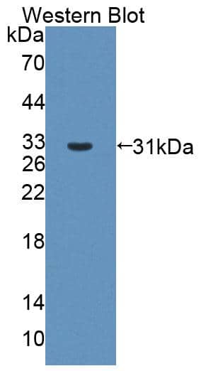
| SDS-PAGE | 120-200 kDa, reducing conditions |
|
| Publications |
|
| Storage | Use a manual defrost freezer and avoid repeated freeze-thaw cycles.
|
| Buffer | Lyophilized from a 0.2 μm filtered solution in PBS. |
| Purity | >95%, by SDS-PAGE under reducing conditions and visualized by silver stain |
| Reconstitution Instructions | Reconstitute at 300 μg/mL in PBS. |


![Immunoprecipitation CD44 Antibody (2C5) [Unconjugated] - s Pan Specific](https://images.novusbio.com/images/antibody/bba10_human-cd44s-pan-specific-mab-clone-2c5-immunoprecipitation-2852025105258..png)
![Knockout Validated CD44 Antibody (2C5) [Unconjugated] - s Pan Specific](https://images.novusbio.com/images/antibody/bba10_human-cd44s-pan-specific-mab-clone-2c5-knockout-validated-285202510529..png)
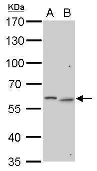
![Western Blot CD44 Antibody (2C5) [Unconjugated] - s Pan Specific](https://images.novusbio.com/images/antibody/CD44_BBA10_Western_Blot_21459.jpg)


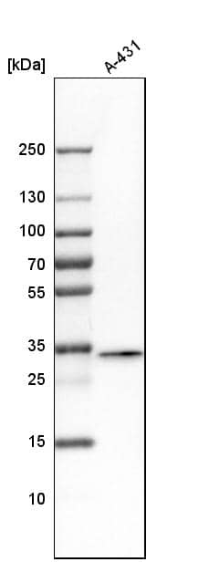
![Western Blot Decorin Antibody [Unconjugated]](https://images.novusbio.com/images/antibody/Decorin_AF1060_Western_Blot_19006.jpg)
The concentration calculator allows you to quickly calculate the volume, mass or concentration of your vial. Simply enter your mass, volume, or concentration values for your reagent and the calculator will determine the rest.


![Immunohistochemistry Brevican Antibody [Unconjugated]](https://images.novusbio.com/images/antibody/Brevican_AF4009_Immunohistochemistry_19694.jpg)
![Western Blot Brevican Antibody [Unconjugated]](https://images.novusbio.com/images/af4009_human-rat-brevican-affinity-purified-polyclonal-ab-western-blot-121220259302820.jpg)
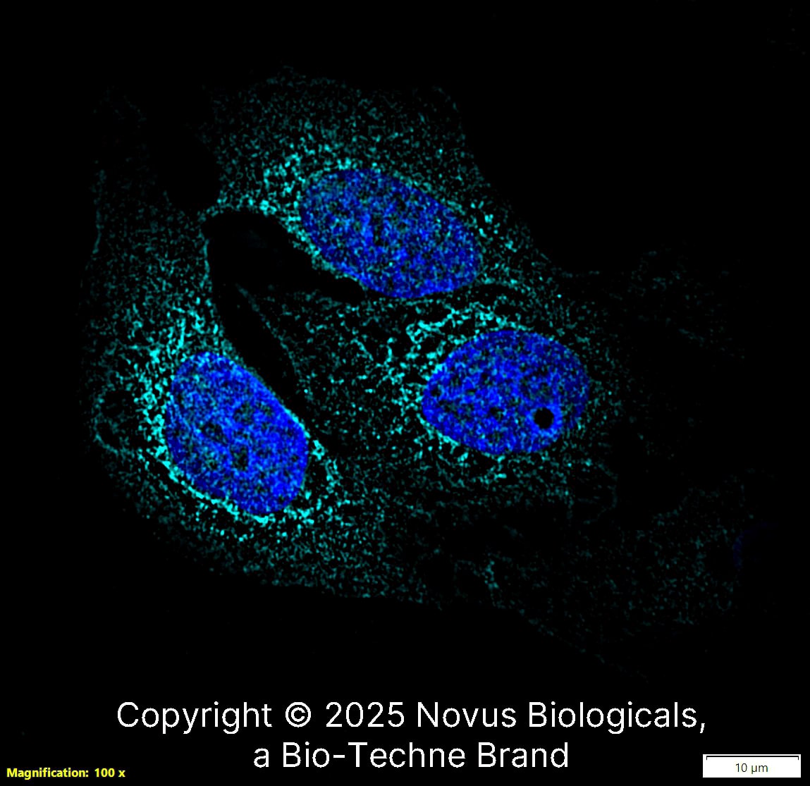
![Immunocytochemistry Brevican Antibody [Unconjugated]](https://images.novusbio.com/images/antibody/Brevican_AF4009_Immunocytochemistry__Immunofluorescence_19520.jpg)

![Western Blot Tenascin R Antibody (619) [Unconjugated]](https://images.novusbio.com/images/mab1624_mouse-rat-tenascin-r-mab-clone-619-western-blot-35202385637..jpg)
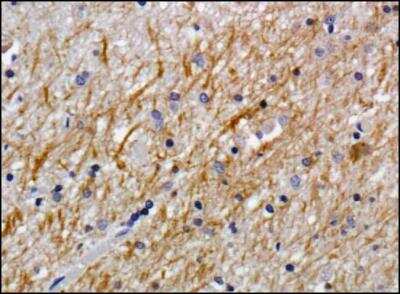
![Immunohistochemistry Tenascin R Antibody (619) [Unconjugated]](https://images.novusbio.com/images/antibody/Tenascin_R_MAB1624_Immunohistochemistry_10138.jpg)


![Immunohistochemistry Contactin-2/TAG1 Antibody [Unconjugated]](https://images.novusbio.com/images/antibody/Contactin-2_AF4439_Immunohistochemistry_10516.jpg)
![Simple Western Contactin-2/TAG1 Antibody [Unconjugated]](https://images.novusbio.com/images/antibody/Contactin2_AF4439_Simple_Western_17121.jpg)
![Western Blot Contactin-2/TAG1 Antibody [Unconjugated]](https://images.novusbio.com/images/antibody/Contactin-2_AF4439_Western_Blot_9589.jpg)
![N/A MMP-2 [HRP]](https://images.novusbio.com/images/elisa/DATA_MMP2_MMP200_ELISA_1003.jpg)
![N/A MMP-2 [HRP]](https://images.novusbio.com/images/elisa/MMP2_MMP200_ELISA_474.jpg)


