| Reactivity | MuSpecies Glossary |
| Applications | Bioactivity |
| Format | Carrier-Free |
| Details of Functionality | Measured by its ability to modulate collagen fibrillogenesis. 20 µg/mL of rmDermatopontin can significantly enhance the rate of collagen fibrillogenesis. |
| Source | Mouse myeloma cell line, NS0-derived mouse Dermatopontin protein Gln19-Val201 with a C-terminal 6-His tag |
| Accession # | |
| N-terminal Sequence | No results obtained: Gln19 predicted |
| Protein/Peptide Type | Recombinant Proteins |
| Gene | Dpt |
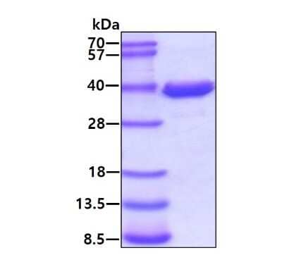
| Purity | >95%, by SDS-PAGE visualized with Silver Staining and quantitative densitometry by Coomassie® Blue Staining |
| Endotoxin Note | <0.10 EU per 1 μg of the protein by the LAL method. |
| Dilutions |
|
|
| Theoretical MW | 22.8 kDa. Disclaimer note: The observed molecular weight of the protein may vary from the listed predicted molecular weight due to post translational modifications, post translation cleavages, relative charges, and other experimental factors. |
|
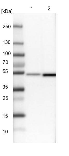
| SDS-PAGE | 19-25 kDa, reducing conditions |
|
| Publications |
|
| Storage | Use a manual defrost freezer and avoid repeated freeze-thaw cycles.
|
| Buffer | Lyophilized from a 0.2 μm filtered solution in PBS. |
| Purity | >95%, by SDS-PAGE visualized with Silver Staining and quantitative densitometry by Coomassie® Blue Staining |
| Reconstitution Instructions | Reconstitute at 500 μg/mL in PBS. |

![Intracellular Staining by Flow Cytometry AKT [p Ser473] Antibody [Unconjugated] - Pan Specific](https://images.novusbio.com/images/antibody/Akt3_AF887_Flow_Cytometry_8283.jpg)
![Western Blot AKT [p Ser473] Antibody [Unconjugated] - Pan Specific](https://images.novusbio.com/images/af887_phospho-akt-s473-pan-specific-affinity-purified-pab-41202410485440.jpg)
![Western Blot AKT [p Ser473] Antibody [Unconjugated] - Pan Specific](https://images.novusbio.com/images/af887_phospho-akt-s473-pan-specific-affinity-purified-pab-8120255552843.jpg)


The concentration calculator allows you to quickly calculate the volume, mass or concentration of your vial. Simply enter your mass, volume, or concentration values for your reagent and the calculator will determine the rest.


![Western Blot Androgen R/NR3C4 [p Ser213, p Ser210] Antibody (156C135.2) - BSA Free](https://images.novusbio.com/images/Androgen-R-NR3C4-p-Ser213-p-Ser210-Antibody-156C135-2-Western-Blot-NB100-56603-img0004.jpg)
![SDS-Page TNF-alpha [Unconjugated]](https://images.novusbio.com/images/protein/TNF-alpha_210-TA_256.jpg)
![Bioactivity TNF-alpha [Unconjugated]](https://images.novusbio.com/images/protein/TNFalpha_210TA_1658.jpg)
![SEC-MALS TNF-alpha [Unconjugated]](https://images.novusbio.com/images/210-ta_recombinant-human-tnf-alpha-protein-sec-mals-35202312244..jpg)


![Bioactivity CTLA-4 [Unconjugated]](https://images.novusbio.com/images/protein/CTLA4_7268CT_2293.jpg)
![Bioactivity IGF-I/IGF-1 [Unconjugated]](https://images.novusbio.com/images/protein/IGF-I_291-G1_41.jpg)
![Mass Spectrometry IGF-I/IGF-1 [Unconjugated]](https://images.novusbio.com/images/protein/IGF-I_291-G1_42.jpg)
![SEC-MALS IGF-I/IGF-1 [Unconjugated]](https://images.novusbio.com/images/291-g1_recombinant-human-igf-i-igf-1-protein-cf-sec-mals-224202691859.jpg)
![N/A Kallikrein 3/PSA [HRP]](https://images.novusbio.com/images/elisa/DATA_Kallikrein_3_DKK300_ELISA_730.jpg)
![N/A Kallikrein 3/PSA [HRP]](https://images.novusbio.com/images/elisa/DATA_Kallikrein_3_DKK300_ELISA_729.jpg)
![N/A Kallikrein 3/PSA [HRP]](https://images.novusbio.com/images/elisa/Kallikrein_3_DKK300_ELISA_124.jpg)
![TRAIL/TNFSF10 [Unconjugated]](/sites/all/modules/enterprise-tech/et_datasheets/images/novus_guarantee.png "TRAIL/TNFSF10 [Unconjugated]")
![SDS-Page Sonic Hedgehog/Shh [Unconjugated]](https://images.novusbio.com/images/protein/Sonic_Hedgehog_464-SH_341.jpg)
![Cell Culture Sonic Hedgehog/Shh [Unconjugated]](https://images.novusbio.com/images/464-sh_recombinant-mouse-sonic-hedgehog-shh-c25ii-n-terminus-cell-culture-105202415719..jpg)
![SEC-MALS Sonic Hedgehog/Shh [Unconjugated]](https://images.novusbio.com/images/464-sh_recombinant-mouse-sonic-hedgehog-shh-c25ii-n-terminus-sec-mals-642023224324.jpg)