| Reactivity | HuSpecies Glossary |
| Applications | Binding Activity |
| Format | Carrier-Free |
| Details of Functionality | Measured by its binding ability in a functional ELISA. Immobilized recombinant human SHH at 4 µg/mL can bind Recombinant Human BOC with an apparent Kd <10 nM. |
| Source | Mouse myeloma cell line, NS0-derived human BOC protein Asp31-Asp852 & Leu417-Asp852, both with a C-terminal 6-His tag |
| Accession # | |
| N-terminal Sequence | Asp31 & Leu417 |
| Protein/Peptide Type | Recombinant Proteins |
| Gene | BOC |
| Purity | >85%, by SDS-PAGE under reducing conditions and visualized by silver stain |
| Endotoxin Note | <0.10 EU per 1 μg of the protein by the LAL method. |
| Dilutions |
|
|
| Theoretical MW | 91.1 kDa. Disclaimer note: The observed molecular weight of the protein may vary from the listed predicted molecular weight due to post translational modifications, post translation cleavages, relative charges, and other experimental factors. |
|
| SDS-PAGE | 135 kDa, reducing conditions |
|
| Publications |
|
| Storage | Use a manual defrost freezer and avoid repeated freeze-thaw cycles.
|
| Buffer | Lyophilized from a 0.2 μm filtered solution in PBS. |
| Purity | >85%, by SDS-PAGE under reducing conditions and visualized by silver stain |
| Reconstitution Instructions | Reconstitute at 100 μg/mL in sterile PBS. |
BOC (Brother of CDO [CAM-related/down-regulated by oncogenes]) is a member of the Immunoglobulin (Ig) superfamily, Ig/Fibronectin (FN) type III repeat family of cell surface proteins (1). Human BOC is a type I transmembrane (TM) protein. It is synthesized as a 1114 amino acid (aa) precursor that contains a 30 aa signal sequence, an 825 aa extracellular domain (ECD), a 21 aa TM segment and a 238 aa cytoplasmic region (1, 2). The ECD contains four Ig-like domains, followed by three FN type III repeats. The third (or juxtramembrane) FN type III repeat (aa 712 - 809) binds SHH (3). The intracellular region is not essential for BOC-containing receptor complex signaling (1). However, it appears both the ECD and intracellular regions of BOC are used to form functional subunit interactions in cis-oriented receptor complexes (1, 4). One 157 aa BOC alternate splice form is reported that shows a 32 aa substitution for aa 126 - 1114. The ECD of human BOC is 92% aa identical to mouse BOC ECD. BOC is found in the embryo associated with muscle precursors, limb mesenchyme, early chondrocytes and neurons (2, 5, 6). It appears to promote muscle differentiation and axon guidance (2, 6). BOC contributes to two multi-subunit receptor complexes. On myocytes, a BOC-associated complex includes CDO, neogenin, netrin, and at least two cadherin homodimers formed by either M- or N-cadherin (2). A second complex on neurons, somewhat ill‑defined, potentially includes BOC, CDO and Gas1. Here, BOC and/or CDO interact with SHH, with subsequent "transfer" or presentation of SHH to PTCH1 (6, 7).


The concentration calculator allows you to quickly calculate the volume, mass or concentration of your vial. Simply enter your mass, volume, or concentration values for your reagent and the calculator will determine the rest.


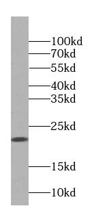
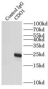
![Western Blot CDO Antibody [Unconjugated]](https://images.novusbio.com/images/antibody/CDO_AF2429_Western_Blot_17538.jpg)
![SDS-Page Sonic Hedgehog/Shh [Unconjugated]](https://images.novusbio.com/images/protein/Sonic_Hedgehog_464-SH_341.jpg)
![Cell Culture Sonic Hedgehog/Shh [Unconjugated]](https://images.novusbio.com/images/464-sh_recombinant-mouse-sonic-hedgehog-shh-c25ii-n-terminus-cell-culture-105202415719..jpg)
![SEC-MALS Sonic Hedgehog/Shh [Unconjugated]](https://images.novusbio.com/images/464-sh_recombinant-mouse-sonic-hedgehog-shh-c25ii-n-terminus-sec-mals-642023224324.jpg)


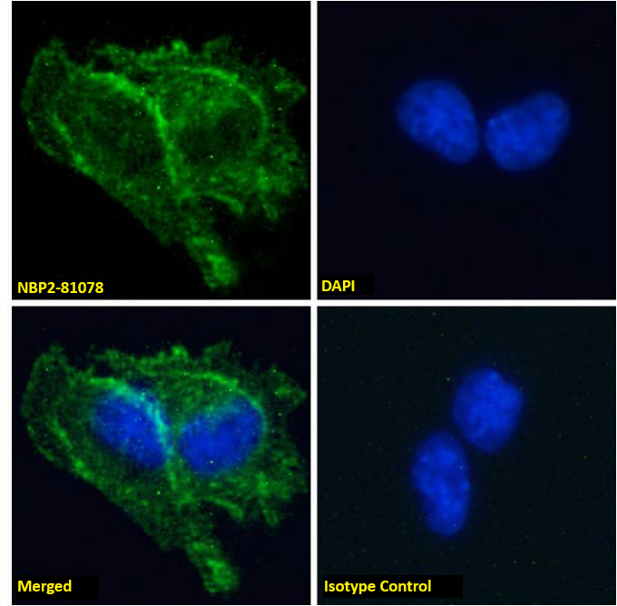
![Immunocytochemistry/ Immunofluorescence Gas1 Antibody [Unconjugated]](https://images.novusbio.com/images/af2644_mouse-gas1-affinity-purified-polyclonal-ab-412024124024.jpg)
![Western Blot Gas1 Antibody [Unconjugated]](https://images.novusbio.com/images/antibody/Gas1_AF2644_Western_Blot_18066.jpg)
![Western Blot Gas1 Antibody [Unconjugated]](https://images.novusbio.com/images/af2644_mouse-gas1-affinity-purified-polyclonal-ab-41202412394947.jpg)


![GDF-5/BMP-14 [Unconjugated]](/sites/all/modules/enterprise-tech/et_datasheets/images/novus_guarantee.png "GDF-5/BMP-14 [Unconjugated]")
![Cell Culture FGF-8 [Unconjugated]](https://images.novusbio.com/images/423-f8_recombinant-human-mouse-fgf-8b-protein-cell-culture-1052024145159..jpg)
![Bioactivity FGF-8 [Unconjugated]](https://images.novusbio.com/images/protein/FGF8_423F8_1438.jpg)
![Cell Culture FGF-8 [Unconjugated]](https://images.novusbio.com/images/423-f8_recombinant-human-mouse-fgf-8b-protein-cell-culture-1052024145958..jpg)


![N/A Erythropoietin/EPO [HRP]](https://images.novusbio.com/images/elisa/Erythropoietin_MEP00_ELISA_421.jpg)
![N/A Erythropoietin/EPO [HRP]](https://images.novusbio.com/images/elisa/DATA_Erythropoietin_MEP00_ELISA_970.jpg)
![Western Blot Ret Antibody [Unconjugated]](https://images.novusbio.com/images/af482_mouse-ret-affinity-purified-polyclonal-ab-8120255541732.jpg)
![Western Blot Ret Antibody [Unconjugated]](https://images.novusbio.com/images/af482_mouse-ret-affinity-purified-polyclonal-ab-8120255541771.jpg)
![Western Blot Ret Antibody [Unconjugated]](https://images.novusbio.com/images/af482_mouse-ret-affinity-purified-polyclonal-ab-8120255534722.jpg)