 at 8 µg/mL for 3 hours at room temperature. Cells were stained using the NorthernLights™ 557-conjugated Anti-Rat IgG Secondary Antibody (red; NL013) and counterstained with DAPI (blue). Specific staining was localized to cell surface. Staining was performed using our protocol for Fluorescent ICC Staining of Non-adherent Cells.")
| Reactivity | MuSpecies Glossary |
| Applications | IHC, ICC/IF |
| Clone | 1039504 |
| Clonality | Monoclonal |
| Host | Rat |
| Conjugate | Unconjugated |
| Immunogen | Mouse myeloma cell line NS0-derived mouse PDGF R alpha Leu25-Glu524 Accession # P26618.3 |
| Specificity | Detects mouse PDGF R alpha in direct ELISAs |
| Source | N/A |
| Isotype | IgG2b |
| Clonality | Monoclonal |
| Host | Rat |
| Purity Statement | Protein A or G purified from hybridoma culture supernatant |
| Innovator's Reward | Test in a species/application not listed above to receive a full credit towards a future purchase. |
| Storage | Use a manual defrost freezer and avoid repeated freeze-thaw cycles.
|
| Buffer | Lyophilized from a 0.2 μm filtered solution in PBS with Trehalose. |
| Reconstitution Instructions | Reconstitute at 0.5 mg/mL in sterile PBS. |
PDGF R alpha (platelet-derived growth factor receptor alpha) is a type I transmembrane glycoprotein in the class III subfamily of receptor tyrosine kinases (RTK) (1-3). PDGF R alpha and PDGF R beta can form homo- or hetero-dimeric receptors when engaged by dimers of the PDGF family of growth factors, which include disulfide-linked homodimers of PDGF-A, B, C or D, or the heterodimer PDGF-AB that is mainly found in human platelets. While multiple in vitro ligand-receptor combinations have been identified, in vivo evidence indicates that PDGF R alpha primarily binds PDGF-AA and PDGF-CC, while PDGF R beta primarily binds PDGF-BB and probably PDGF-DD. Like all class III RTKs, the extracellular domain (ECD) of mouse PDGF R alpha (amino acids 25-525) contains five immunoglobulin-like domains, while the intracellular region contains a split tyrosine kinase domain (aa 593‑954). Within the ECD, mouse PDGF R alpha shares 85%, 93%, 84%, 84%, and 81% amino acid sequence identity with human, rat, equine, canine and bovine PDGF R alpha respectively. PDGF R alpha autophosphorylates upon dimerization, activating signaling cascades in PI 3-kinase Ras-MAP kinase, and PLC-gamma pathways (1, 2). Signaling is down‑regulated by SHP-2 phosphatase activity and by receptor endocytosis and lysosomal degradation. PDGF R alpha is expressed at low levels in most mesenchymal cells, but is strongly expressed in oligodendrocyte, lung, skin and intestinal progenitor cells and induced by inflammation or growth in culture (1-3). During development, mesenchymal cells expressing PDGF R alpha respond to local gradients of epithelially produced PDGF-AA or PDGF-CC during formation of the cranial and cardiac neural crest, retina, gonads, lung alveoli, intestinal villi, skin, hair follicles, skeleton, teeth, palate, and interstitial kidney mesenchyme (1, 4). Deletion of PDGF R alpha in mice severely impairs mesenchymal derivatives in both embryo and extraembryonic tissues, and high or low PDGF R alpha signaling in humans may result in spina bifida or cleft palate‑type malformations. Postnatally, PDGF R alpha is implicated in gliomas and fibrotic disorders of lung, heart and skin (scleroderma) (5- 7).
![Flow Cytometry CD117/c-kit Antibody [Unconjugated]](https://images.novusbio.com/images/af1356_human-mouse-cd117-c-kit-affinity-purified-polyclonal-ab-81202555532.jpg)
![Immunohistochemistry CD117/c-kit Antibody [Unconjugated]](https://images.novusbio.com/images/antibody/af1356_human-mouse-cd117-c-kit-affinity-purified-polyclonal-ab-immunohistochemistry-812202575731.png)
![Immunocytochemistry/ Immunofluorescence CD117/c-kit Antibody [Unconjugated]](https://images.novusbio.com/images/af1356_human-mouse-cd117-c-kit-affinity-purified-polyclonal-ab-41202410481199.jpg)
![Immunocytochemistry EGFR Antibody [Unconjugated]](https://images.novusbio.com/images/antibody/EGF_R_AF231_Immunocytochemistry__Immunofluorescence_21143.jpg)
![Flow Cytometry EGFR Antibody [Unconjugated]](https://images.novusbio.com/images/antibody/EGF_R_AF231_Flow_Cytometry_20401.jpg)
![Western Blot EGFR Antibody [Unconjugated]](https://images.novusbio.com/images/antibody/EGF_R_AF231_Western_Blot_19925.jpg)


![Immunohistochemistry VEGFR2/KDR/Flk-1 Antibody [Unconjugated]](https://images.novusbio.com/images/antibody/VEGF_R2_AF644_Immunohistochemistry_6888.jpg)
![Immunohistochemistry VEGFR2/KDR/Flk-1 Antibody [Unconjugated]](https://images.novusbio.com/images/af644_mouse-vegf-r2-flk-1-affinity-purified-polyclonal-ab-4120241048248.jpg)
![In-situ Hybridization VEGFR2/KDR/Flk-1 Antibody [Unconjugated]](https://images.novusbio.com/images/antibody/af644_mouse-vegf-r2-flk-1-affinity-purified-polyclonal-ab-in-situ-hybridization-235202421442..jpg)


![Intracellular Staining by Flow Cytometry AKT [p Ser473] Antibody [Unconjugated] - Pan Specific](https://images.novusbio.com/images/antibody/Akt3_AF887_Flow_Cytometry_8283.jpg)
![Western Blot AKT [p Ser473] Antibody [Unconjugated] - Pan Specific](https://images.novusbio.com/images/af887_phospho-akt-s473-pan-specific-affinity-purified-pab-41202410485440.jpg)
![Western Blot AKT [p Ser473] Antibody [Unconjugated] - Pan Specific](https://images.novusbio.com/images/af887_phospho-akt-s473-pan-specific-affinity-purified-pab-8120255552843.jpg)


Secondary Antibodies |
Isotype Controls |
The concentration calculator allows you to quickly calculate the volume, mass or concentration of your vial. Simply enter your mass, volume, or concentration values for your reagent and the calculator will determine the rest.
| Uniprot |
|


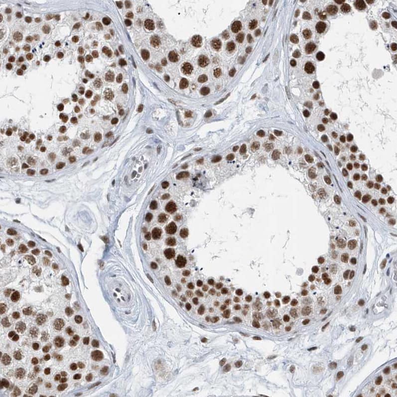
 at 5 µg/mL for 1 hour at room temperature followed by incubation with the Anti-Rat IgG VisUCyte™ HRP Polymer Antibody (VC005). Before incubation with the primary antibody, tissue was subjected to heat-induced epitope retrieval using Antigen Retrieval Reagent-Basic (CTS013). Tissue was stained using DAB (brown) and counterstained with hematoxylin (blue). Specific staining was localized to embryonic kidney. Staining was performed using our protocol for IHC Staining with VisUCyte HRP Polymer Detection Reagents.")
![Immunohistochemistry PDGF R beta Antibody [Unconjugated]](https://images.novusbio.com/images/af1042_mouse-pdgf-r-beta-affinity-purified-polyclonal-ab-immunohistochemistry-121220259315424.jpg)
![Immunohistochemistry PDGF R beta Antibody [Unconjugated]](https://images.novusbio.com/images/af1042_mouse-pdgf-r-beta-affinity-purified-polyclonal-ab-immunohistochemistry-121220258463418.jpg)
![Immunohistochemistry PDGF R beta Antibody [Unconjugated]](https://images.novusbio.com/images/af1042_mouse-pdgf-r-beta-affinity-purified-polyclonal-ab-immunohistochemistry-12122025857822.jpg)
![Cell Culture PDGF-AA [Unconjugated]](https://images.novusbio.com/images/221-aa_recombinant-human-pdgf-aa-protein-cf-cell-culture-1052024153816..jpg)
![Bioactivity PDGF-AA [Unconjugated]](https://images.novusbio.com/images/protein/221-aa_recombinant-human-pdgf-aa-protein-cf-bioactivity-1410202017288.jpg)
![SCF/c-kit Ligand [Unconjugated]](/sites/all/modules/enterprise-tech/et_datasheets/images/novus_guarantee.png "SCF/c-kit Ligand [Unconjugated]")


![Immunohistochemistry Desmin Antibody [Unconjugated]](https://images.novusbio.com/images/antibody/Desmin_AF3844_Immunohistochemistry_10689.jpg)
![Immunohistochemistry Desmin Antibody [Unconjugated]](https://images.novusbio.com/images/antibody/Desmin_AF3844_Immunohistochemistry_6808.jpg)
![Immunocytochemistry/ Immunofluorescence Desmin Antibody [Unconjugated]](https://images.novusbio.com/images/af3844_human-mouse-desmin-affinity-purified-polyclonal-ab-41202410331920.jpg)
![N/A VEGF [HRP]](https://images.novusbio.com/images/elisa/VEGF_DVE00_ELISA_208.jpg)
![N/A VEGF [HRP]](https://images.novusbio.com/images/elisa/DATA_VEGF_DVE00_ELISA_871.jpg)
![N/A VEGF [HRP]](https://images.novusbio.com/images/elisa/DATA_VEGF_DVE00_ELISA_872.jpg)
![Neutralization PDGF-C Antibody [Unconjugated]](https://images.novusbio.com/images/antibody/PDGF-C_AF1447_Block_Neutralize_8780.jpg)
![Immunocytochemistry/ Immunofluorescence PDGF-C Antibody [Unconjugated]](https://images.novusbio.com/images/af1447_mouse-pdgf-c-affinity-purified-polyclonal-ab-44202415425060.jpg)