 1433-OP) was serially diluted and captured by Mouse Anti-Human Osteopontin/OPN Monoclonal Antibody (Catalog # MAB14332) coated on a Clear Polystyrene Microplate (Catalog # DY990). Goat Anti-Human Osteopontin/OPN Antigen Affinity-purified Polyclonal Antibody (Catalog # AF1433) was biotinylated and incubated with the protein captured on the plate. Detection of the standard curve was achieved by incubating Streptavidin-HRP (Catalog # DY998)" title="Recombinant Human Osteopontin/OPN (Catalog # 1433-OP) was serially diluted and captured by Mouse Anti-Human Osteopontin/OPN Monoclonal Antibody (Catalog # MAB14332) coated on a Clear Polystyrene Microplate (Catalog # DY990). Goat Anti-Human Osteopontin/OPN Antigen Affinity-purified Polyclonal Antibody (Catalog # AF1433) was biotinylated and incubated with the protein captured on the plate. Detection of the standard curve was achieved by incubating Streptavidin-HRP (Catalog # DY998)" />
| Reactivity | HuSpecies Glossary |
| Applications | ELISA |
| Clone | 223112 |
| Clonality | Monoclonal |
| Host | Mouse |
| Conjugate | Unconjugated |
| Concentration | LYOPH |
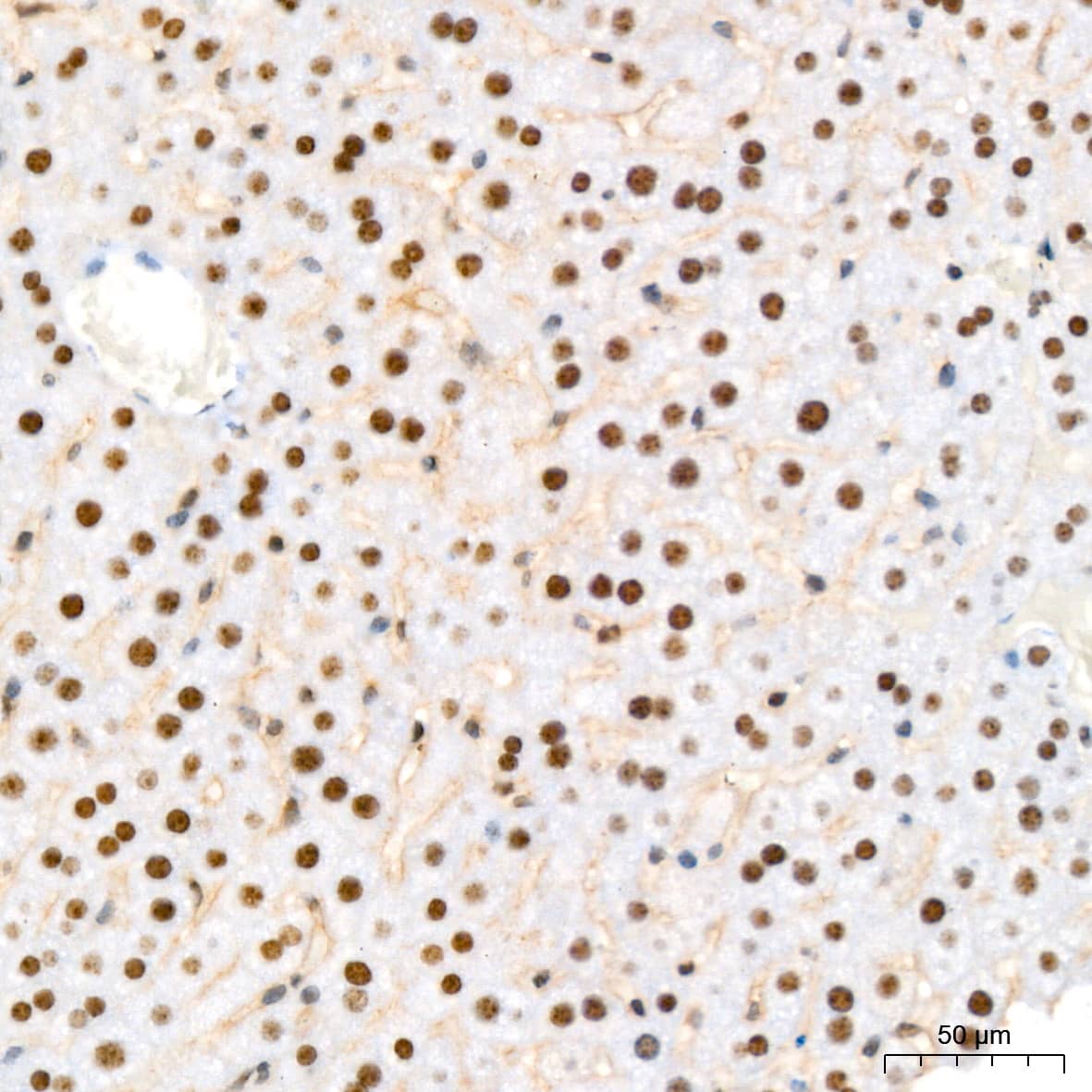
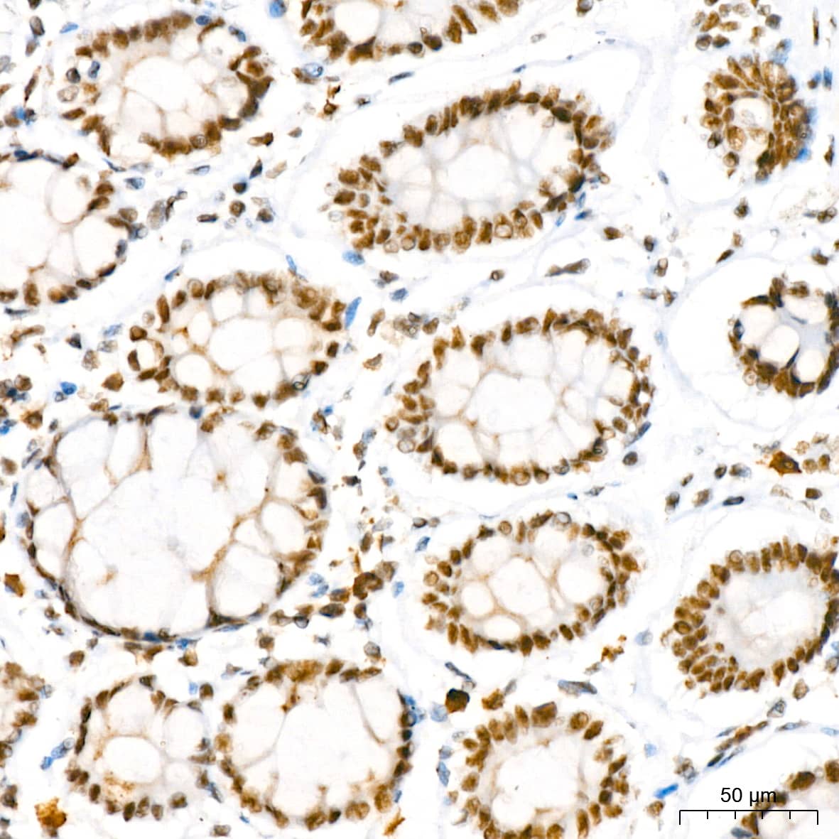
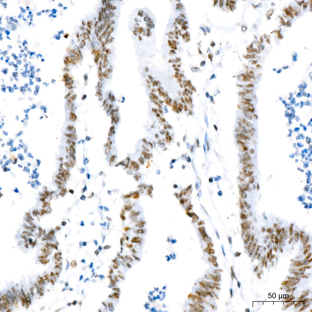
| Immunogen | Mouse myeloma cell line NS0-derived recombinant human Osteopontin Ile17-Asn300 Accession # NP_000573.1 |
| Specificity | Detects human Osteopontin in ELISAs. In ELISAs, this antibody does not cross-react with recombinant mouse Osteopontin. |
| Source | N/A |
| Isotype | IgG2a |
| Clonality | Monoclonal |
| Host | Mouse |
| Gene | SPP1 |
| Purity Statement | Protein A or G purified from hybridoma culture supernatant |
| Innovator's Reward | Test in a species/application not listed above to receive a full credit towards a future purchase. |
| Dilutions |
|
|
| Application Notes | ELISA Capture: Human Osteopontin/OPN Antibody (Catalog # MAB14332) ELISA Detection: Human Osteopontin/OPN Biotinylated Antibody (Catalog # BAF1433) Standard: Recombinant Human Osteopontin/OPN (Catalog # 1433-OP) |
|
| Publications |
|
| Storage | Use a manual defrost freezer and avoid repeated freeze-thaw cycles.
|
| Buffer | Lyophilized from a 0.2 μm filtered solution in PBS with Trehalose. *Small pack size (SP) is supplied either lyophilized or as a 0.2 µm filtered solution in PBS. |
| Preservative | No Preservative |
| Concentration | LYOPH |
| Reconstitution Instructions | Reconstitute at 0.5 mg/mL in sterile PBS. |
Osteopontin (OPN, previously also referred to as transformation-associated secreted phosphoprotein, bone sialoprotein I, 2ar, 2B7, early T lymphocyte activation 1 protein, minopotin, calcium oxalate crystal growth inhibitor protein), is a secreted, highly acidic, calcium-binding, RGD-containing, phosphorylated glycoprotein originally isolated from bone matrix (1). Subsequently, OPN has been found in kidney, placenta, blood vessels and various tumor tissues. Many cell types (including macrophages, osteoclasts, activated T-cells, fibroblasts, epithelial cells, vascular smooth muscle cells, and natural killer cells) can express OPN in response to activation by cytokines, growth factors or inflammatory mediators. Elevated expression of OPN has also been associated with numerous pathobiological conditions such as atherosclerotic plaques, renal tubulointerstitial fibrosis, granuloma formations in tuberculosis and silicosis, neointimal formation associated with balloon catheterization, metastasizing tumors, and cerebral ischemia. Human OPN cDNA encodes a 314 amino acid (aa) residue precursor protein with a 16 aa residue predicted signal peptide that is cleaved to yield a 298 aa residue mature protein with an integrin binding sequence (RGD), and N- and O-glycosylation sites. By alternative splicing, at least three human OPN isoforms exist. OPN has been shown to bind to different cell types through RGD-mediated interaction with the integrins alpha v beta 1, alpha v beta 3, alpha v beta 5, and non-RGD-mediated interaction with CD44 and the integrins alpha 8 beta 1 or alpha 9 beta 1. OPN exists both as a component of extracellular matrix and as a soluble molecule. Functionally, OPN is chemotactic for macrophages, smooth muscle cells, endothelial cells and glial cells. OPN has also been shown to inhibit nitric oxide production and cytotoxicity by activated macrophages. Human, mouse, rat, pig and bovine OPN share from approximately 40% - 80% amino acid sequence identity. Osteopontin is a substrate for proteolytic cleavage by thrombin, enterokinase, MMP-3 and MMP-7. The functions of OPN in a variety of cell types were shown to be modified as a result of proteolytic cleavage (2, 3).


![Immunoprecipitation CD44 Antibody (2C5) [Unconjugated] - s Pan Specific](https://images.novusbio.com/images/antibody/bba10_human-cd44s-pan-specific-mab-clone-2c5-immunoprecipitation-2852025105258..png)
![Knockout Validated CD44 Antibody (2C5) [Unconjugated] - s Pan Specific](https://images.novusbio.com/images/antibody/bba10_human-cd44s-pan-specific-mab-clone-2c5-knockout-validated-285202510529..png)
![Western Blot CD44 Antibody (2C5) [Unconjugated] - s Pan Specific](https://images.novusbio.com/images/antibody/CD44_BBA10_Western_Blot_21459.jpg)


")
Secondary Antibodies |
Isotype Controls |
|
Successful Transplantation of Friedreich Ataxia Induced Pluripotent Stem Cell (iPSC)-Derived Sensory Neurons in Dorsal Root Ganglia of Adult Rodents Jamshed Arslan, Pharm D, PhD The dorsal root ganglia (DRG) are a collection of cell bodies of sensory nerves carrying sensory information – including nociception, mechanoreception and proprioception – from periphera... Read full blog post. |
The concentration calculator allows you to quickly calculate the volume, mass or concentration of your vial. Simply enter your mass, volume, or concentration values for your reagent and the calculator will determine the rest.
| Gene Symbol | SPP1 |
| Uniprot |


![Bioactivity IBSP/Sialoprotein II [Unconjugated]](https://images.novusbio.com/images/4014-sp_recombinant-human-ibsp-sialoprotein-ii-protein-cf-bioactivity-4420249391..jpg)
![Immunocytochemistry SPARC Antibody [Unconjugated]](https://images.novusbio.com/images/antibody/SPARC_AF942_Immunocytochemistry__Immunofluorescence_23321.jpg)
![Intracellular Staining by Flow Cytometry SPARC Antibody [Unconjugated]](https://images.novusbio.com/images/antibody/af942_mouse-sparc-affinity-purified-polyclonal-ab-intracellular-staining-by-flow-cytometry-22122020141853.jpg)
![Immunohistochemistry SPARC Antibody [Unconjugated]](https://images.novusbio.com/images/antibody/SPARC_AF942_Immunohistochemistry_10419.jpg)
![Western Blot RUNX2/CBFA1 Antibody (232902) [Unconjugated]](https://images.novusbio.com/images/mab2006_human-runx2-cbfa1-mab-clone-232902-western-blot-1212202597422.jpg)
![Western Blot RUNX2/CBFA1 Antibody (232902) [Unconjugated]](https://images.novusbio.com/images/mab2006_human-runx2-cbfa1-mab-clone-232902-western-blot-121220259172010.jpg)
![Immunocytochemistry RUNX2/CBFA1 Antibody (232902) [Unconjugated]](https://images.novusbio.com/images/antibody/RUNX2_MAB2006_Immunocytochemistry_13309.jpg)
![N/A SLPI [HRP]](https://images.novusbio.com/images/elisa/SLPI_DPI00_ELISA_160.jpg)
![N/A SLPI [HRP]](https://images.novusbio.com/images/elisa/DATA_SLPI_DPI00_ELISA_792.jpg)
![N/A CCL27/CTACK [HRP]](https://images.novusbio.com/images/elisa/DATA_CCL27_DCC270_ELISA_615.jpg)
![N/A CCL27/CTACK [HRP]](https://images.novusbio.com/images/elisa/CCL27_DCC270_ELISA_54.jpg)


![N/A Osteoprotegerin/TNFRSF11B [Biotin]](https://images.novusbio.com/images/elisa/DATA_Osteoprotegerin_DY805_ELISA_2238.jpg)
![Bioactivity BMP-2 [Unconjugated]](https://images.novusbio.com/images/protein/355-bm_recombinant-human-mouse-rat-bmp-2-protein-bioactivity-181120209742.jpg)
![SDS-Page TNF-alpha [Unconjugated]](https://images.novusbio.com/images/protein/TNF-alpha_210-TA_256.jpg)
![Bioactivity TNF-alpha [Unconjugated]](https://images.novusbio.com/images/protein/TNFalpha_210TA_1658.jpg)
![SEC-MALS TNF-alpha [Unconjugated]](https://images.novusbio.com/images/210-ta_recombinant-human-tnf-alpha-protein-sec-mals-35202312244..jpg)
![N/A VEGF [HRP]](https://images.novusbio.com/images/elisa/VEGF_DVE00_ELISA_208.jpg)
![N/A VEGF [HRP]](https://images.novusbio.com/images/elisa/DATA_VEGF_DVE00_ELISA_871.jpg)
![N/A VEGF [HRP]](https://images.novusbio.com/images/elisa/DATA_VEGF_DVE00_ELISA_872.jpg)
![N/A IL-6 [HRP]](https://images.novusbio.com/images/elisa/DATA_IL6_M6000_ELISA_936.jpg)
![N/A IL-6 [HRP]](https://images.novusbio.com/images/elisa/IL-6_M6000_ELISA_415.jpg)
![N/A IL-6 [HRP]](https://images.novusbio.com/images/m6000b_mouse-il-6-quantikine-elisa-kit-1752025024034.jpg)

 followed by 30 min incubation with Goat anti Mouse HRP conjugated secondary antibodies (Catalog # HAF007) at 1:20 dilution + DAB chromogen (brown). The tissue was counterstained with Hematoxylin (blue). Control was done by omitting primary antibody.")