| Reactivity | Mu, RtSpecies Glossary |
| Applications | WB, ELISA |
| Clonality | Polyclonal |
| Host | Chicken |
| Conjugate | Unconjugated |
| Format | BSA Free |
| Concentration | 1 mg/ml |
| Immunogen | Synthetic peptides from mouse green opsin and zebra finch red opsin. |
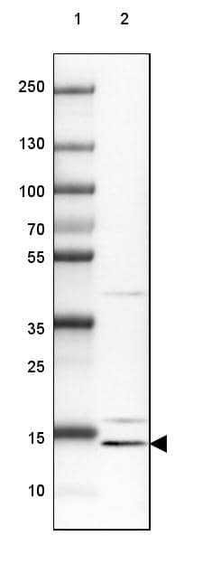
| Specificity | Opsin, red and green. The antibody reacts with a protein of ~40 kDa. |
| Clonality | Polyclonal |
| Host | Chicken |
| Gene | OPN1MW |
| Purity | Immunogen affinity purified |
| Innovator's Reward | Test in a species/application not listed above to receive a full credit towards a future purchase. |
| Dilutions |
|
|
| Application Notes | ImmunoBlotting: on mouse brain extract. To minimize background staining it is suggested that you include a higher content of detergent (Tween-20) in your dilution and washing solutions. Immunohistochemistry: not tested. It is recommended that you try the antibody at 1:50-1:1,000. |
|
| Reviewed Applications |
|
| Storage | Aliquot and store at -20C or -80C. Avoid freeze-thaw cycles. |
| Buffer | PBS |
| Preservative | 0.02% Sodium Azide |
| Concentration | 1 mg/ml |
| Purity | Immunogen affinity purified |


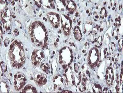
![Immunohistochemistry ACE/CD143 Antibody [Unconjugated]](https://images.novusbio.com/images/antibody/ACE_AF1513_Immunohistochemistry_6703.jpg)
![Western Blot ACE/CD143 Antibody [Unconjugated]](https://images.novusbio.com/images/antibody/ACE_AF1513_Flow_Cytometry_20028.jpg)
![Simple Western ACE/CD143 Antibody [Unconjugated]](https://images.novusbio.com/images/antibody/ACE_AF1513_Simple_Western_20109.jpg)

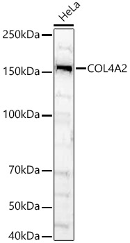


![Western Blot RFC1 Antibody [Unconjugated]](https://images.novusbio.com/images/antibody/RFC1_AF6457_Western_Blot_9874.jpg)


 Antibody - BSA Free")
| Images | Ratings | Applications | Species | Date | Details | ||||||||
|---|---|---|---|---|---|---|---|---|---|---|---|---|---|

Enlarge |
reviewed by:
Verified Customer |
IHC-Fr | Mouse | 12/27/2016 |
Summary
Comments
|
||||||||

Enlarge |
reviewed by:
Verified Customer |
Immunohistochemistry-Whole Mount | Mouse | 12/27/2016 |
Summary
Comments
|
Secondary Antibodies |
Isotype Controls |
Research Areas for Opsin 1 (Medium Wave) Antibody (NBP2-29858)Find related products by research area.
|
The concentration calculator allows you to quickly calculate the volume, mass or concentration of your vial. Simply enter your mass, volume, or concentration values for your reagent and the calculator will determine the rest.
5 | |
4 | |
3 | |
2 | |
1 |
| Verified Customer 12/27/2016 |
||
| Application: | IHC-Fr | |
| Species: | Mouse |
| Verified Customer 12/27/2016 |
||
| Application: | Immunohistochemistry-Whole Mount | |
| Species: | Mouse |


![SDS-Page Sonic Hedgehog/Shh [Unconjugated]](https://images.novusbio.com/images/protein/Sonic_Hedgehog_464-SH_341.jpg)
![Cell Culture Sonic Hedgehog/Shh [Unconjugated]](https://images.novusbio.com/images/464-sh_recombinant-mouse-sonic-hedgehog-shh-c25ii-n-terminus-cell-culture-105202415719..jpg)
![SEC-MALS Sonic Hedgehog/Shh [Unconjugated]](https://images.novusbio.com/images/464-sh_recombinant-mouse-sonic-hedgehog-shh-c25ii-n-terminus-sec-mals-642023224324.jpg)


![SDS-Page TNF-alpha [Unconjugated]](https://images.novusbio.com/images/protein/TNF-alpha_210-TA_256.jpg)
![Bioactivity TNF-alpha [Unconjugated]](https://images.novusbio.com/images/protein/TNFalpha_210TA_1658.jpg)
![SEC-MALS TNF-alpha [Unconjugated]](https://images.novusbio.com/images/210-ta_recombinant-human-tnf-alpha-protein-sec-mals-35202312244..jpg)