![Immunohistochemistry-Frozen: MMR/CD206/Mannose Receptor Antibody (15-2) [NB600-1415] - Frozen sections of human tonsil. NB600-1415 was used ina dilution of 1/25 and stains endothelia of lymph vesselsstrongly.](http://images.novusbio.com/fullsize/MMR-CD206-Mannose-Receptor-Antibody-15-2-Immunohistochemistry-Frozen-NB600-1415-img0003.jpg "Immunohistochemistry-Frozen: MMR/CD206/Mannose Receptor Antibody (15-2) [NB600-1415] - Frozen sections of human tonsil. NB600-1415 was used ina dilution of 1/25 and stains endothelia of lymph vesselsstrongly.")
| Reactivity | HuSpecies Glossary |
| Applications | WB, Flow, Func, IHC |
| Clone | 15-2 |
| Clonality | Monoclonal |
| Host | Mouse |
| Conjugate | Unconjugated |
| Concentration | 0.1 mg/ml |
| Additional Information | Clone 15-2 was used by HLDA to establish CD designation. |
| Immunogen | Purified human mannose receptor from human placental tissue |
| Isotype | IgG1 |
| Clonality | Monoclonal |
| Host | Mouse |
| Gene | MRC1 |
| Purity | Protein G purified |
| Innovator's Reward | Test in a species/application not listed above to receive a full credit towards a future purchase. |
| Dilutions |
|
|
| Application Notes | For IHC-F: Tissue sections were fixed in acetone/chloroform. Sections were preincubated with normal rabbit serum. For FC: Antibody 15-2 stains the extracellular domain of MR. WB: A non-reduced sample treatment and 6% SDS-Page was used. The band size is 175 kDa. |
|
| Publications |
|
| Storage | Store at 4C. |
| Buffer | PBS containing 0.1% BSA |
| Preservative | 0.02% Sodium Azide |
| Concentration | 0.1 mg/ml |
| Purity | Protein G purified |


")
The concentration calculator allows you to quickly calculate the volume, mass or concentration of your vial. Simply enter your mass, volume, or concentration values for your reagent and the calculator will determine the rest.


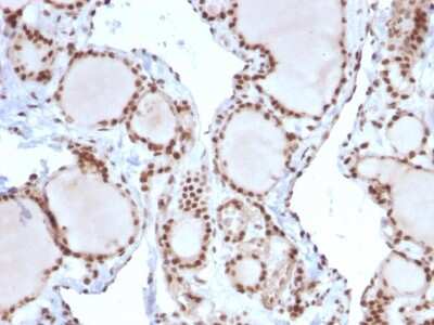
![Immunohistochemistry-Frozen: MMR/CD206/Mannose Receptor Antibody (15-2) [NB600-1415] - staining Mannose Receptor in human mammary gland tissue section by Immunohistochemistry (Frozen sections). Tissue samples were fixed with 70% Ethanol and blocking for 10 minutes at RT was performed. The sample was incubated with primary antibody (1/10) for 1 hour. A HRP-conjugated mouse polyclonal to mouse IgG was used undiluted as secondary antibody.](http://images.novusbio.com/fullsize/MMR-CD206-Mannose-Receptor-Antibody-15-2-Immunohistochemistry-Frozen-NB600-1415-img0002.jpg "Immunohistochemistry-Frozen: MMR/CD206/Mannose Receptor Antibody (15-2) [NB600-1415] - staining Mannose Receptor in human mammary gland tissue section by Immunohistochemistry (Frozen sections). Tissue samples were fixed with 70% Ethanol and blocking for 10 minutes at RT was performed. The sample was incubated with primary antibody (1/10) for 1 hour. A HRP-conjugated mouse polyclonal to mouse IgG was used undiluted as secondary antibody.")


![N/A IL-10 [Biotin]](https://images.novusbio.com/images/elisa/DATA_IL10_DY417_ELISA_2014.jpg)
![SDS-Page TNF-alpha [Unconjugated]](https://images.novusbio.com/images/protein/TNF-alpha_210-TA_256.jpg)
![Bioactivity TNF-alpha [Unconjugated]](https://images.novusbio.com/images/protein/TNFalpha_210TA_1658.jpg)
![SEC-MALS TNF-alpha [Unconjugated]](https://images.novusbio.com/images/210-ta_recombinant-human-tnf-alpha-protein-sec-mals-35202312244..jpg)
![Bioactivity IL-4 [Unconjugated]](https://images.novusbio.com/images/protein/6507-ilcf_recombinant-human-il-4-cho-expressed-protein-cf-bioactivity-272020133214.jpg)
![Bioactivity CTLA-4 [Unconjugated]](https://images.novusbio.com/images/protein/CTLA4_7268CT_2293.jpg)

 followed by 30 min incubation with Goat anti Mouse HRP conjugated secondary antibodies (Catalog # HAF007) at 1:20 dilution + DAB chromogen (brown). The tissue was counterstained with Hematoxylin (blue). Control was done by omitting primary antibody.")
![SDS-Page: Mouse IgG1 Isotype Control (MG1) [NBP1-97005] - Lane 1: , Non-reduced. M: Opal Pre-stained Ladder. Lane 2: , Reduced. Load: 1.0 ug per lane. Predicted/Observed: 120 kDa Non-reduced, 55 and 25 Reduced.](https://images.novusbio.com/images/Mouse-IgG1-Isotype-Control-MG1-SDS-Page-NBP1-97005-img0002.jpg "SDS-Page: Mouse IgG1 Isotype Control (MG1) [NBP1-97005] - Lane 1: , Non-reduced. M: Opal Pre-stained Ladder. Lane 2: , Reduced. Load: 1.0 ug per lane. Predicted/Observed: 120 kDa Non-reduced, 55 and 25 Reduced.")