HAF017). A specific band was detected for p300 at approximately 300 kDa (as indicated). This experiment was conducted using Immunoblot Buffer Group 1." title="Western blot shows nuclear extracts of A431 human epithelial carcinoma cell line epidermoid carcinoma and HeLa human cervical epithelial carcinoma cell line cervical carcinoma cell lines. PVDF membrane was probed with 1 µg/mL of Goat Anti-Human p300 Antigen Affinity-purified Polyclonal Antibody (Catalog # AF3789) followed by HRP-conjugated Anti-Goat IgG Secondary Antibody (Catalog # HAF017). A specific band was detected for p300 at approximately 300 kDa (as indicated). This experiment was conducted using Immunoblot Buffer Group 1." />
| Reactivity | HuSpecies Glossary |
| Applications | WB, Simple Western, IHC, ChIP, ICC/IF |
| Clonality | Polyclonal |
| Host | Goat |
| Conjugate | Unconjugated |
| Concentration | LYOPH |
| Immunogen | E. coli-derived recombinant human p300 His2283-His2414 Accession # Q09472 |
| Specificity | Detects human p300 in Western blots. |
| Source | N/A |
| Isotype | IgG |
| Clonality | Polyclonal |
| Host | Goat |
| Gene | EP300 |
| Purity Statement | Antigen Affinity-purified |
| Innovator's Reward | Test in a species/application not listed above to receive a full credit towards a future purchase. |
| Dilutions |
|
|
| Publications |
|
| Storage | Use a manual defrost freezer and avoid repeated freeze-thaw cycles.
|
| Buffer | Lyophilized from a 0.2 μm filtered solution in PBS with Trehalose. *Small pack size (SP) is supplied either lyophilized or as a 0.2 µm filtered solution in PBS. |
| Preservative | No Preservative |
| Concentration | LYOPH |
| Reconstitution Instructions | Reconstitute at 0.2 mg/mL in sterile PBS. |
The E1A-binding protein p300 is a histone acetyltransferase that also acts to acetylate other proteins, such as p53. p300 acts as a transcriptional coactivator that can serve as an adaptor molecule to bridge to transcriptional regulators. In addition, p300 binds to PCNA and may participate in chromatin remodeling.






Secondary Antibodies |
Isotype Controls |
The concentration calculator allows you to quickly calculate the volume, mass or concentration of your vial. Simply enter your mass, volume, or concentration values for your reagent and the calculator will determine the rest.


 AB-108-C) for 15 minutes in an ultrasonic bath, followed by Biotinylated Anti-Goat IgG Secondary Antibody (Catalog #
  NL001) and counterstained with DAPI (blue). Specific staining was localized to nuclei. View our protocol for
  HAF109). This experiment was conducted under reducing conditions and using the 66-440 kDa separation system." title="Simple Western lane view shows lysates of HeLa human cervical epithelial carcinoma cell line, loaded at 0.2 mg/mL. A specific band was detected for p300 at approximately 308 kDa (as indicated) using 50 µg/mL of Goat Anti-Human p300 Antigen Affinity-purified Polyclonal Antibody (Catalog # AF3789) followed by 1:50 dilution of HRP-conjugated Anti-Goat IgG Secondary Antibody (Catalog #
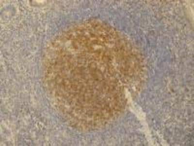
  VC004). Tissue was stained using DAB (brown) and counterstained with hematoxylin (blue). Specific staining was localized to nuclei. View our protocol for

![Immunocytochemistry eIF4E Antibody (299910) [Unconjugated]](https://images.novusbio.com/images/antibody/eIF4E_MAB3228_Immunocytochemistry_6605.jpg)
![Western Blot eIF4E Antibody (299910) [Unconjugated]](https://images.novusbio.com/images/mab3228_human-mouse-rat-eif4e-mab-clone-299910-western-blot-12122025824121.jpg)
![Western Blot eIF4E Antibody (299910) [Unconjugated]](https://images.novusbio.com/images/mab3228_human-mouse-rat-eif4e-mab-clone-299910-western-blot-12122025745315.jpg)


![Immunocytochemistry/ Immunofluorescence c-jun [p Ser63] Antibody (SY0297)](https://images.novusbio.com/images/c-jun-p-Ser63-Antibody-SY0297-Immunocytochemistry-Immunofluorescence-NBP2-67471-img0006.jpg)
![Western Blot c-jun [p Ser63] Antibody (SY0297)](https://images.novusbio.com/images/c-jun-p-Ser63-Antibody-SY0297-Western-Blot-NBP2-67471-img0008.jpg)
![Western Blot c-jun [p Ser63] Antibody (SY0297)](https://images.novusbio.com/images/c-jun-p-Ser63-Antibody-SY0297-Western-Blot-NBP2-67471-img0009.jpg)