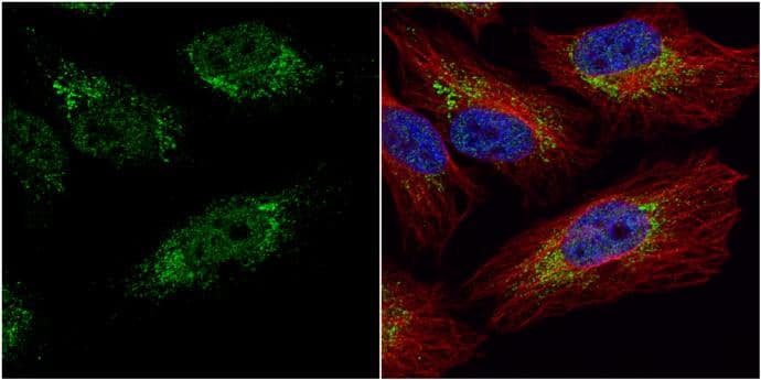
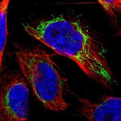
![Immunocytochemistry/Immunofluorescence: IDH3A Antibody [NBP1-32396] - HeLa cells were fixed in ice-cold MeOH for 5 min. Green: IDH3A stained by IDH3A antibody diluted at 1:500. Blue: Hoechst 33342 staining. Scale bar= 10 um.](http://images.novusbio.com/fullsize/IDH3A-Antibody-Immunocytochemistry-Immunofluorescence-NBP1-32396-img0007.jpg "Immunocytochemistry/Immunofluorescence: IDH3A Antibody [NBP1-32396] - HeLa cells were fixed in ice-cold MeOH for 5 min. Green: IDH3A stained by IDH3A antibody diluted at 1:500. Blue: Hoechst 33342 staining. Scale bar= 10 um.")
| Reactivity | Hu, Mu, Rt, Ze, Bv, Ch, RM, XpSpecies Glossary |
| Applications | WB, ICC/IF, IHC |
| Clonality | Polyclonal |
| Host | Rabbit |
| Conjugate | Unconjugated |
| Format | Azide Free |
| Immunogen | Recombinant protein encompassing a sequence within the center region of human IDH3A. The exact sequence is proprietary. |
| Localization | Mitochondrion |
| Predicted Species | Rhesus Macaque (99%), Bovine (97%), Chicken (94%), Xenopus (91%). Backed by our 100% Guarantee. |
| Isotype | IgG |
| Clonality | Polyclonal |
| Host | Rabbit |
| Gene | IDH3A |
| Purity | Antigen Affinity-purified |
| Innovator's Reward | Test in a species/application not listed above to receive a full credit towards a future purchase. |
| Dilutions |
|
|
| Theoretical MW | 40 kDa. Disclaimer note: The observed molecular weight of the protein may vary from the listed predicted molecular weight due to post translational modifications, post translation cleavages, relative charges, and other experimental factors. |
|
| Publications |
|
| Storage | Aliquot and store at -20C or -80C. Avoid freeze-thaw cycles. |
| Buffer | PBS, 1% BSA, 20% Glycerol |
| Preservative | 0.025% Proclin 300 |
| Purity | Antigen Affinity-purified |


| Publications using NBP1-32396 | Applications | Species |
|---|---|---|
| Tai Y, Engels D, Locatelli G et al. Targeting the TCA cycle can ameliorate widespread axonal energy deficiency in neuroinflammatory lesions bioRxiv 2023-04-04 (IHC, Mouse) Details: Dilutions: 1:400 |
IHC | Mouse |
| Tai YH, Engels D, Locatelli G et al. Targeting the TCA cycle can ameliorate widespread axonal energy deficiency in neuroinflammatory lesions Nature Metabolism 2023-07-10 [PMID: 37430025] (In vivo assay) | In vivo assay |
Secondary Antibodies |
Isotype Controls |
Research Areas for IDH3A Antibody (NBP1-32396)Find related products by research area.
|
The concentration calculator allows you to quickly calculate the volume, mass or concentration of your vial. Simply enter your mass, volume, or concentration values for your reagent and the calculator will determine the rest.
| Gene Symbol | IDH3A |


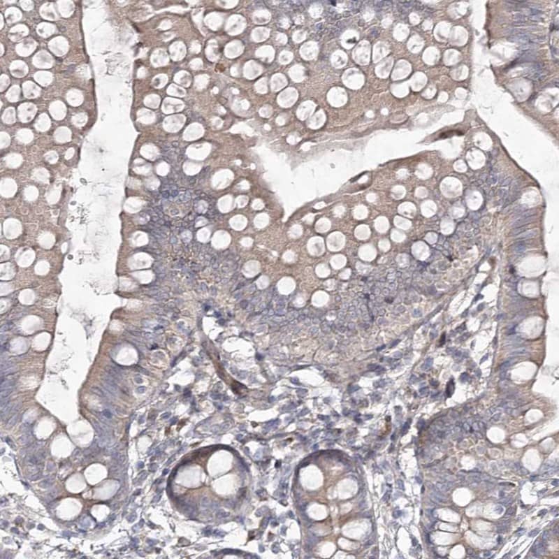
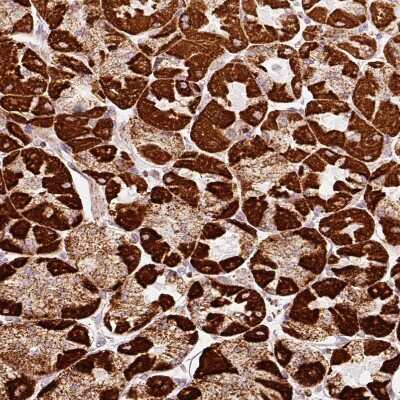
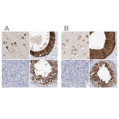
![Immunohistochemistry-Paraffin: IDH3A Antibody [NBP1-32396] - Human lung SCC, using IDH3A antibody at 1:100 dilution. Antigen Retrieval: Trilogy™ (EDTA based, pH 8.0) buffer, 15min.](http://images.novusbio.com/fullsize/IDH3A-Antibody-Immunohistochemistry-Paraffin-NBP1-32396-img0003.jpg "Immunohistochemistry-Paraffin: IDH3A Antibody [NBP1-32396] - Human lung SCC, using IDH3A antibody at 1:100 dilution. Antigen Retrieval: Trilogy™ (EDTA based, pH 8.0) buffer, 15min.")
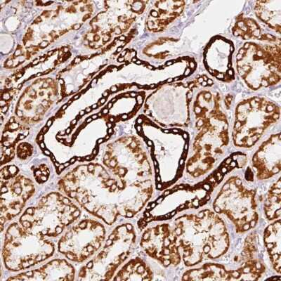
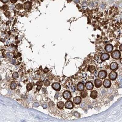
![Immunohistochemistry-Paraffin: IDH3A Antibody [NBP1-32396] - Analysis of paraffin-embedded zebrafish tissue, using IDH3A antibody at 1:300 dilution.](http://images.novusbio.com/fullsize/antibody/nbp1-32396_rabbit-polyclonal-idh3a-antibody-immunohistochemistry-paraffin-271120239124..jpg "Immunohistochemistry-Paraffin: IDH3A Antibody [NBP1-32396] - Analysis of paraffin-embedded zebrafish tissue, using IDH3A antibody at 1:300 dilution.")
 was separated by 10% SDS-PAGE, and the membrane was blotted with IDH3A antibody (NBP1-32396) diluted at 1:10000. The HRP-conjugated anti-rabbit IgG antibody was used to detect the primary antibody.")
 were separated by 10% SDS-PAGE, and the membrane was blotted with IDH3A antibody (NBP1-32396) diluted at 1:500. The HRP-conjugated anti-rabbit IgG antibody was used to detect the primary antibody.")
 was separated by 10% SDS-PAGE, and the membrane was blotted with IDH3A antibody (NBP1-32396) diluted at 1:10000. The HRP-conjugated anti-rabbit IgG antibody was used to detect the primary antibody.")
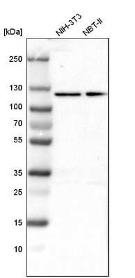
 were separated by 10% SDS-PAGE, and the membrane was blotted with IDH3A antibody (NBP1-32396) diluted at 1:500. The HRP-conjugated anti-rabbit IgG antibody was used to detect the primary antibody. Corresponding RNA expression data for the same cell lines are based on Human Protein Atlas program.")
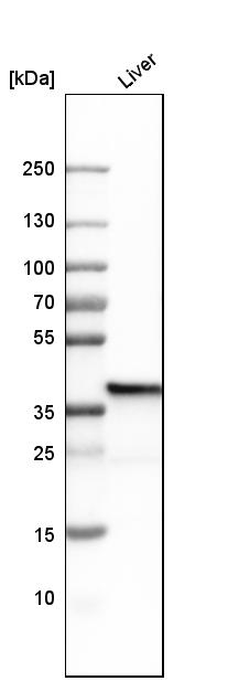
 was separated by 10% SDS-PAGE, and the membrane was blotted with IDH3A antibody (NBP1-32396) diluted at 1:1000. The HRP-conjugated anti-rabbit IgG antibody was used to detect the primary antibody.")
 was separated by 10% SDS-PAGE, and the membrane was blotted with IDH3A antibody (NBP1-32396) diluted at 1:10000. The HRP-conjugated anti-rabbit IgG antibody was used to detect the primary antibody.")
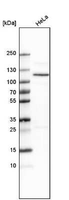
 were separated by 10% SDS-PAGE, and the membrane was blotted with IDH3A antibody (NBP1-32396) diluted at 1:500. The HRP-conjugated anti-rabbit IgG antibody was used to detect the primary antibody. Corresponding RNA expression data for the same cell lines are based on Human Protein Atlas program.")
 were separated by 10% SDS-PAGE, and the membrane was blotted with IDH3A antibody (NBP1-32396) diluted at 1:500. The HRP-conjugated anti-rabbit IgG antibody was used to detect the primary antibody.")


![Western Blot HTRA2/Omi Antibody [Unconjugated]](https://images.novusbio.com/images/af1458_human-mouse-rat-htra2-omi-affinity-purified-polyclonal-ab-8220249504633.jpg)
![Simple Western HTRA2/Omi Antibody [Unconjugated]](https://images.novusbio.com/images/antibody/16328.jpg)
![Western Blot HTRA2/Omi Antibody [Unconjugated]](https://images.novusbio.com/images/af1458_human-mouse-rat-htra2-omi-affinity-purified-polyclonal-ab-822024951145.jpg)
![Immunohistochemistry Isocitrate Dehydrogenase 1/IDH1 Antibody (843219) [Unconjugated]](https://images.novusbio.com/images/antibody/Isocitrate_Dehydrogenase_1_MAB7049_Immunohistochemistry_19973.jpg)
![Knockout Validated Isocitrate Dehydrogenase 1/IDH1 Antibody (843219) [Unconjugated]](https://images.novusbio.com/images/antibody/Isocitrate_Dehydrogenase_1_MAB7049_Knockout_Validated_22540.jpg)
![Western Blot Isocitrate Dehydrogenase 1/IDH1 Antibody (843219) [Unconjugated]](https://images.novusbio.com/images/antibody/Isocitrate_Dehydrogenase_1_MAB7049_Western_Blot_13853.jpg)


![Western Blot: Goat anti-Rabbit IgG (H+L) Secondary Antibody [HRP] [NB7160] - Western blot showing vemurafenib treatment in BRAFV600E CRC cells inhibits fission mediator DRP1 with no significant effect on fusion proteins (Mfn1 & 2) using MFN-1 antibody (NBP1-51841) and corresponding secondary antibody, goat anti-rabbit IgG-HRP (NB7160). Image collected and cropped by CiteAb from the following publication (https://pubmed.ncbi.nlm.nih.gov/33738242).](https://images.novusbio.com/images/Goat-anti-Rabbit-IgG-H+L-Secondary-Antibody-HRP-Western-Blot-NB7160-img0001.jpg "Western Blot: Goat anti-Rabbit IgG (H+L) Secondary Antibody [HRP] [NB7160] - Western blot showing vemurafenib treatment in BRAFV600E CRC cells inhibits fission mediator DRP1 with no significant effect on fusion proteins (Mfn1 & 2) using MFN-1 antibody (NBP1-51841) and corresponding secondary antibody, goat anti-rabbit IgG-HRP (NB7160). Image collected and cropped by CiteAb from the following publication (https://pubmed.ncbi.nlm.nih.gov/33738242).")
 followed by 30 min incubation with Goat anti Rabbit HRP conjugated secondary antibodies (Catalog # HAF008) at 1:20 dilution + DAB chromogen (brown). The tissue was counterstained with Hematoxylin (blue). Control was done by omitting primary antibody.")
![Flow Cytometry: Rabbit IgG Isotype Control [NBP2-24891] - An intracellular stain was performed on Raji cells with Adiponectin antibody NB100-65810 (blue) and a matched isotype control NBP2-24893 (orange). Cells were fixed with 4% PFA and then permeablized with 0.1% saponin. Cells were incubated in an antibody dilution of 1 ug/mL for 30 minutes at room temperature, followed by Dylight488-conjugated anti-rabbit secondary antibody. Image using the Azide Free form of this antibody.](https://images.novusbio.com/images/Rabbit--Mouse-IgG-Isotype-Control-Flow-Cytometry-NBP2-24891-img0006.jpg "Flow Cytometry: Rabbit IgG Isotype Control [NBP2-24891] - An intracellular stain was performed on Raji cells with Adiponectin antibody NB100-65810 (blue) and a matched isotype control NBP2-24893 (orange). Cells were fixed with 4% PFA and then permeablized with 0.1% saponin. Cells were incubated in an antibody dilution of 1 ug/mL for 30 minutes at room temperature, followed by Dylight488-conjugated anti-rabbit secondary antibody. Image using the Azide Free form of this antibody.")