HAF016). A specific band was detected for Gastrokine 1 at approximately 18-20 kDa (as indicated). This experiment was conducted under reducing conditions and using Immunoblot Buffer Group 1." title="Western blot shows lysates of mouse stomach tissue and rat stomach tissue. PVDF membrane was probed with 0.1 µg/mL of Sheep Anti-Human/Mouse Gastrokine 1 Antigen Affinity-purified Polyclonal Antibody (Catalog # AF7287) followed by HRP-conjugated Anti-Sheep IgG Secondary Antibody (Catalog # HAF016). A specific band was detected for Gastrokine 1 at approximately 18-20 kDa (as indicated). This experiment was conducted under reducing conditions and using Immunoblot Buffer Group 1." />
| Reactivity | Hu, MuSpecies Glossary |
| Applications | WB, IHC |
| Clonality | Polyclonal |
| Host | Sheep |
| Conjugate | Unconjugated |
| Concentration | LYOPH |
| Immunogen | E. coli-derived recombinant mouse Gastrokine 1 Tyr38-Tyr201 Accession # Q9CR36 |
| Specificity | Detects mouse Gastrokine 1 in direct ELISAs. In direct ELISAs, approximately 10% cross-reactivity with recombinant human Gastrokine 1 is observed. |
| Source | N/A |
| Isotype | IgG |
| Clonality | Polyclonal |
| Host | Sheep |
| Gene | GKN1 |
| Purity Statement | Antigen Affinity-purified |
| Innovator's Reward | Test in a species/application not listed above to receive a full credit towards a future purchase. |
| Dilutions |
|
|
| Publications |
|
| Storage | Use a manual defrost freezer and avoid repeated freeze-thaw cycles.
|
| Buffer | Lyophilized from a 0.2 μm filtered solution in PBS with Trehalose. *Small pack size (SP) is supplied either lyophilized or as a 0.2 µm filtered solution in PBS. |
| Preservative | No Preservative |
| Concentration | LYOPH |
| Reconstitution Instructions | Sterile PBS to a final concentration of 0.2 mg/mL. |
Gastrokine-1 (GKN1; also CA11 and AMP-18) is an 18-20 kDa member of the Gastrokine protein family. It has limited expression, being restricted to mucous secreting pyloric antrum epithelial cells. Gastrokine-1 appears to promote epithelial cell proliferation and migration, and induce the formation of tight junctions between epithelial cells. By contrast, gastrokine-1 induces Fas expression in tumor cells, resulting in apoptosis. Mature mouse gastrokine-1 is 166 amino acids (aa) in length (aa 36-201). Based on the SwissProt sequence, it possesses one BRICHOS domain (aa 71-165) that contains a mitogenic sequence (aa 113-131). There is one potential alternative start site at Met18. Over aa 38-201, mouse gastrokine-1 shares 65% and 92% aa sequence identity with human and rat gastrokine-1, respectively.


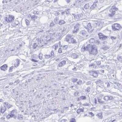


![Western Blot Carbonic Anhydrase VIII/CA8 Antibody [Unconjugated]](https://images.novusbio.com/images/antibody/Carbonic_Anhydrase_VIII_AF2187_Western_Blot_20462.jpg)
![Simple Western Carbonic Anhydrase VIII/CA8 Antibody [Unconjugated]](https://images.novusbio.com/images/antibody/Carbonic_Anhydrase_VIII_AF2187_Simple_Western_20463.jpg)






Secondary Antibodies |
Isotype Controls |
The concentration calculator allows you to quickly calculate the volume, mass or concentration of your vial. Simply enter your mass, volume, or concentration values for your reagent and the calculator will determine the rest.


 NL010) and counterstained with DAPI (blue). Specific staining was localized to islets. View our protocol for
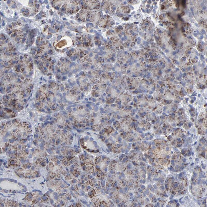
  CTS013). Tissue was stained using the Anti-Sheep HRP-DAB Cell & Tissue Staining Kit (brown; Catalog #




![N/A TFPI [HRP]](https://images.novusbio.com/images/elisa/TFPI_DTFP10_ELISA_190.jpg)
![N/A TFPI [HRP]](https://images.novusbio.com/images/elisa/DATA_TFPI_DTFP10_ELISA_841.jpg)