 HAF016). A specific band was detected for Cytokeratin 19 at approximately 40 kDa (as indicated). This experiment was conducted under reducing conditions and using Immunoblot Buffer Group 1." title="Western blot shows lysates of HT-29 human colon adenocarcinoma cell line, DU145 human prostate carcinoma cell line, HCT-116 human colorectal carcinoma cell line, and MCF-7 human breast cancer cell line. PVDF membrane was probed with 0.1 µg/mL of Sheep Anti-Human Cytokeratin 19 Antigen Affinity-purified Polyclonal Antibody (Catalog # AF3506) followed by HRP-conjugated Anti-Sheep IgG Secondary Antibody (Catalog # HAF016). A specific band was detected for Cytokeratin 19 at approximately 40 kDa (as indicated). This experiment was conducted under reducing conditions and using Immunoblot Buffer Group 1." />
| Reactivity | HuSpecies Glossary |
| Applications | WB, Simple Western, IHC, Flow, ICC/IF |
| Clonality | Polyclonal |
| Host | Sheep |
| Conjugate | Unconjugated |
| Concentration | LYOPH |
| Immunogen | E. coli-derived recombinant human Cytokeratin 19 Thr2-Leu400 Accession # P08727 |
| Specificity | Detects human Cytokeratin 19 in direct ELISAs and Western blots. In direct ELISAs, less than 15% cross-reactivity with recombinant human Cytokeratin 14 is observed. |
| Source | N/A |
| Isotype | IgG |
| Clonality | Polyclonal |
| Host | Sheep |
| Gene | KRT19 |
| Purity Statement | Antigen Affinity-purified |
| Innovator's Reward | Test in a species/application not listed above to receive a full credit towards a future purchase. |
| Dilutions |
|
|
| Publications |
|
| Storage | Use a manual defrost freezer and avoid repeated freeze-thaw cycles.
|
| Buffer | Lyophilized from a 0.2 μm filtered solution in PBS with Trehalose. *Small pack size (SP) is supplied either lyophilized or as a 0.2 µm filtered solution in PBS. |
| Preservative | No Preservative |
| Concentration | LYOPH |
| Reconstitution Instructions | Sterile PBS to a final concentration of 0.2 mg/mL. |
Cytokeratin 19 (Keratin, type I cytoskeletal 19; also KRT-19, CK19 and Keratin-19) is a 40-45 kDa, acidic Class I keratin member of the intermediate filament family of proteins. Individual keratins are always expressed in tandem with a second keratin, and these are found in all epithelial cells. The class I KRT-19 heterodimerizes/polymerizes with 50-52 kDa class II KRT-8 (plus KRT-5 and -7) to form 8-10 nm filaments in epidermal stem cells, secretory gland (sweat; mammary; bile duct) simple epithelium, and neuroendocrine epidermal Merkel cells. It may represent a viable marker for skin stem cells. In skin, Cytokeratin 19 forms filaments in the fetal epithelium, and then progressively decreases with age, being virtually absent by age 17. Human Cytokeratin 19 is 400 amino acids (aa) in length. It contains an N-terminal "head" region (aa 1-79) and a subsequent "rod" region (aa 80-387), but is absent a typical C-terminal tail region. Cytokeratin 19 possesses at least 5 utilized phosphorylation sites plus one acetylated Lys residue. Based on other keratins, and the presence of an Asp at position 238, there may be caspase cleavage-generated isoforms. Full length human Cytokeratin 19 (aa 2-400) shares 82% aa sequence identity with mouse Cytokeratin 19.



![Western Blot alpha-Fetoprotein/AFP Antibody (189502) [Unconjugated]](https://images.novusbio.com/images/antibody/alphaFetoprotein_MAB1368_Western_Blot_20276.jpg)
![Immunocytochemistry/ Immunofluorescence alpha-Fetoprotein/AFP Antibody (189502) [Unconjugated]](https://images.novusbio.com/images/mab1368_human-mouse-alpha-fetoprotein-mab-clone-189502-41202410325519.jpg)
![Knockout Validated alpha-Fetoprotein/AFP Antibody (189502) [Unconjugated]](https://images.novusbio.com/images/antibody/alphaFetoprotein_MAB1368_Knockout_Validated_21503.jpg)




![Immunohistochemistry Albumin Antibody (188835) [Unconjugated] - Serum](https://images.novusbio.com/images/antibody/Albumin_MAB1455_Immunohistochemistry_23317.jpg)
![Immunocytochemistry Albumin Antibody (188835) [Unconjugated] - Serum](https://images.novusbio.com/images/antibody/Albumin_MAB1455_Immunocytochemistry__Immunofluorescence_17777.jpg)
![Intracellular Staining by Flow Cytometry Albumin Antibody (188835) [Unconjugated] - Serum](https://images.novusbio.com/images/antibody/Albumin_MAB1455_Flow_Cytometry_15110.jpg)


Secondary Antibodies |
Isotype Controls |
The concentration calculator allows you to quickly calculate the volume, mass or concentration of your vial. Simply enter your mass, volume, or concentration values for your reagent and the calculator will determine the rest.


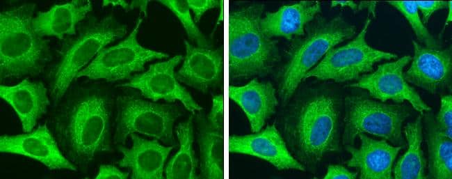
 NL010) and counterstained with DAPI (blue). Specific staining was localized to cytoplasm. View our protocol for
  CTS019) and counterstained with hematoxylin (blue). Specific staining was localized to cytoplasm in convoluted tubules. View our protocol for
  5-001-A, open histogram) followed by Anti-Sheep IgG PE-conjugated secondary antibody (Catalog #
  HAF016). This experiment was conducted under reducing conditions and using the 12-230 kDa separation system." title="Simple Western lane view shows lysates of HT-29 human colon adenocarcinoma cell line, loaded at 0.2 mg/mL. A specific band was detected for Cytokeratin 19 at approximately 50 kDa (as indicated) using 1 µg/mL of Sheep Anti-Human Cytokeratin 19 Antigen Affinity-purified Polyclonal Antibody (Catalog # AF3506) followed by 1:50 dilution of HRP-conjugated Anti-Sheep IgG Secondary Antibody (Catalog #


![Western Blot CEACAM7 Antibody [Unconjugated]](https://images.novusbio.com/images/antibody/CEACAM-7_AF4478_Western_Blot_5475.jpg)
![Immunohistochemistry CEACAM7 Antibody [Unconjugated]](https://images.novusbio.com/images/antibody/CEACAM7_AF4478_Immunohistochemistry_20616.jpg)


![Bioactivity ErbB2/Her2 [Unconjugated]](https://images.novusbio.com/images/protein/1129-er_recombinant-human-erbb2-her2-fc-chimera-protein-cf-bioactivity-7122020142841.jpg)
![SDS-Page ErbB2/Her2 [Unconjugated]](https://images.novusbio.com/images/protein/ErbB2_1129-ER_70.jpg)
![Simple Western CKAP4/p63 Antibody [Unconjugated]](https://images.novusbio.com/images/antibody/af7355_human-ckap4-p63-affinity-purified-polyclonal-ab-simple-western-157202493849.jpg)
![Western Blot CKAP4/p63 Antibody [Unconjugated]](https://images.novusbio.com/images/antibody/CKAP4_AF7355_Western_Blot_11997.jpg)
![Immunocytochemistry CKAP4/p63 Antibody [Unconjugated]](https://images.novusbio.com/images/antibody/CKAP4_AF7355_Immunocytochemistry__Immunofluorescence_17000.jpg)


