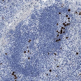
 VC001). Before incubation with the primary antibody, tissue was subjected to heat-induced epitope retrieval using Antigen Retrieval Reagent-Basic (CTS013). Tissue was stained using DAB (brown) and counterstained with hematoxylin (blue). Specific staining was localized to cytoplasm. View our protocol for IHC Staining with VisUCyte HRP Polymer Detection Reagents." title="CXCL13/BLC/BCA-1 was detected in immersion fixed paraffin-embedded sections of human tonsil using Mouse Anti-Human CXCL13/BLC/BCA-1 Monoclonal Antibody (Catalog # MAB8012) at 1.7 µg/mL for 1 hour at room temperature followed by incubation with the Anti-Mouse IgG VisUCyte™ HRP Polymer Antibody VC001). Before incubation with the primary antibody, tissue was subjected to heat-induced epitope retrieval using Antigen Retrieval Reagent-Basic (CTS013). Tissue was stained using DAB (brown) and counterstained with hematoxylin (blue). Specific staining was localized to cytoplasm. View our protocol for IHC Staining with VisUCyte HRP Polymer Detection Reagents." />
| Reactivity | HuSpecies Glossary |
| Applications | IHC, B/N, Flow |
| Clone | 53602 |
| Clonality | Monoclonal |
| Host | Mouse |
| Conjugate | Unconjugated |
| Immunogen | E. coli-derived recombinant human CXCL13/BLC/BCA-1 Val23-Arg94 Accession # O43927 |
| Specificity | Detects human CXCL13/BLC/BCA-1 in direct ELISAs. |
| Source | N/A |
| Isotype | IgG1 |
| Clonality | Monoclonal |
| Host | Mouse |
| Purity Statement | Protein A or G purified from hybridoma culture supernatant |
| Endotoxin Note | <0.10 EU per 1 μg of the antibody by the LAL method. |
| Innovator's Reward | Test in a species/application not listed above to receive a full credit towards a future purchase. |
| Dilutions |
|
|
| Publications |
|
| Storage | Use a manual defrost freezer and avoid repeated freeze-thaw cycles.
|
| Buffer | Lyophilized from a 0.2 μm filtered solution in PBS with Trehalose. *Small pack size (SP) is supplied either lyophilized or as a 0.2 µm filtered solution in PBS. |
| Reconstitution Instructions | Reconstitute at 0.5 mg/mL in sterile PBS. |
CXCL13, also known as B-lymphocyte chemoattractant (BLC), is a CXC chemokine that is constitutively expressed in secondary lymphoid organs. BCA-1 cDNA encodes a protein of 109 amino acid residues with a leader sequence of 22 residues. Mature human BCA-1 shares 64% amino acid sequence similarity with the mouse protein and 23-34% amino acid sequence identity with other known CXC chemokines. Recombinant or chemically synthesized BCA-1 is a potent chemoattractant for B lymphocytes but not T lymphocytes, monocytes or neutrophils. BLR1, a G protein-coupled receptor originally isolated from Burkitt’s lymphoma cells, has now been shown to be the specific receptor for BCA-1. Among cells of the hematopoietic lineages, the expression of BLR1, now designated CXCR5, is restricted to B lymphocytes and a subpopulation of T helper memory cells. Mice lacking BLR1 have been shown to lack inguinal lymph nodes. These mice were also found to have impaired development of Peyer’s patches and defective formation of primary follicles and germinal centers in the spleen as a result of the inability of B lymphocytes to migrate into B cell areas.


![Immunohistochemistry CCR7 Antibody (150503) [Unconjugated]](https://images.novusbio.com/images/antibody/CCR7_MAB197_Immunohistochemistry_22800.jpg)
![Flow Cytometry CCR7 Antibody (150503) [Unconjugated]](https://images.novusbio.com/images/mab197_human-ccr7-mab-clone-150503-41202410485427.jpg)
![Neutralization CCR7 Antibody (150503) [Unconjugated]](https://images.novusbio.com/images/antibody/CCR7_MAB197_Block_Neutralize_9048.jpg)


![CXCL13/BLC/BCA-1 Antibody (53602) [Unconjugated]](/sites/all/modules/enterprise-tech/et_datasheets/images/novus_guarantee.png "CXCL13/BLC/BCA-1 Antibody (53602) [Unconjugated]")
Secondary Antibodies |
Isotype Controls |
The concentration calculator allows you to quickly calculate the volume, mass or concentration of your vial. Simply enter your mass, volume, or concentration values for your reagent and the calculator will determine the rest.
| Uniprot |
|


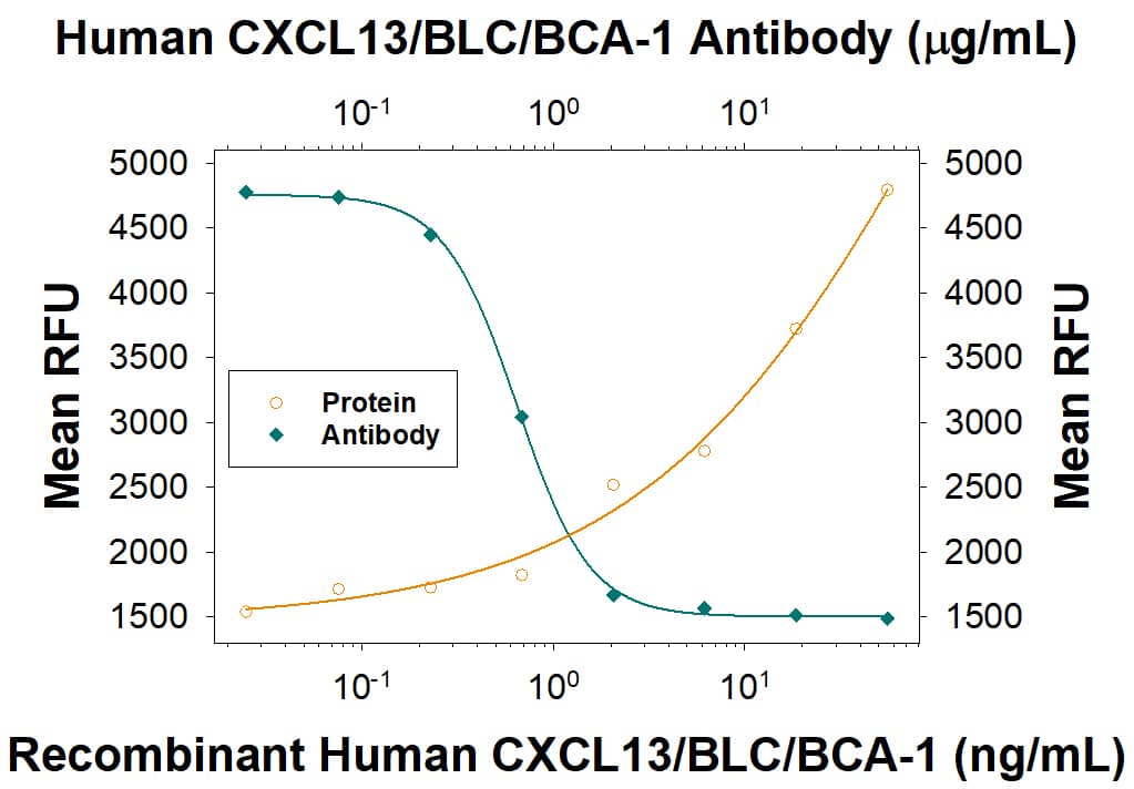 801-CX) chemoattracts the BaF3 mouse pro-B cell line transfected with human CXCR5 in a dose-dependent manner (orange line). The amount of cells that migrated through to the lower chemotaxis chamber was measured by Resazurin (
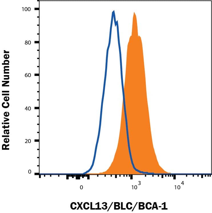 204-IL) and 50 ng/mL Recombinant Human GM-CSF Protein (
 204-IL) and 50 ng/mL Recombinant Human GM-CSF Protein (
 MAB002, open histogram), followed by Allophycocyanin-conjugated Anti-Mouse IgG Secondary Antibody (Catalog #
 ![Immunohistochemistry CXCR5 Antibody (51505) [Unconjugated]](https://images.novusbio.com/images/antibody/CXCR5_MAB190_Flow_Cytometry_18756.jpg)
![Flow Cytometry CXCR5 Antibody (51505) [Unconjugated]](https://images.novusbio.com/images/antibody/CXCR5_MAB190_Flow_Cytometry_22006.jpg)
![Immunocytochemistry CXCR5 Antibody (51505) [Unconjugated]](https://images.novusbio.com/images/antibody/CXCR5_MAB190_Immunocytochemistry_9842.jpg)
![Neutralization CCL21/6Ckine Antibody [Unconjugated]](https://images.novusbio.com/images/antibody/CCL21_AF457_Block_Neutralize_8883.jpg)
![Immunohistochemistry CCL21/6Ckine Antibody [Unconjugated]](https://images.novusbio.com/images/af457_mouse-ccl21-6ckine-affinity-purified-polyclonal-ab-8120255531640.jpg)
![Immunohistochemistry CCL21/6Ckine Antibody [Unconjugated]](https://images.novusbio.com/images/antibody/CCL21_AF457_Immunohistochemistry_10649.jpg)
![Bioactivity CXCL12/SDF-1 alpha [Unconjugated]](https://images.novusbio.com/images/protein/CXCL12SDF1_alpha_350NS_1178.jpg)
![SDS-Page CXCL12/SDF-1 alpha [Unconjugated]](https://images.novusbio.com/images/protein/CXCL12_SDF-1_alpha_350-NS_308.jpg)
![SDS-Page TNF-alpha [Unconjugated]](https://images.novusbio.com/images/protein/TNF-alpha_210-TA_256.jpg)
![Bioactivity TNF-alpha [Unconjugated]](https://images.novusbio.com/images/protein/TNFalpha_210TA_1658.jpg)
![SEC-MALS TNF-alpha [Unconjugated]](https://images.novusbio.com/images/210-ta_recombinant-human-tnf-alpha-protein-sec-mals-35202312244..jpg)
![Bioactivity Lymphotoxin-alpha/TNF-beta [Unconjugated]](https://images.novusbio.com/images/protein/Lymphotoxin-alpha_211-TBB_CF_697.jpg)
![SDS-Page Lymphotoxin-alpha/TNF-beta [Unconjugated]](https://images.novusbio.com/images/protein/Lymphotoxin-alpha_211-TBB_CF_695.jpg)
![N/A CXCL10/IP-10/CRG-2 [HRP]](https://images.novusbio.com/images/elisa/CXCL10_DIP100_ELISA_121.jpg)
![N/A CXCL10/IP-10/CRG-2 [HRP]](https://images.novusbio.com/images/elisa/DATA_CXCL10_DIP100_ELISA_723.jpg)
![N/A CXCL10/IP-10/CRG-2 [HRP]](https://images.novusbio.com/images/elisa/DATA_CXCL10_DIP100_ELISA_724.jpg)
![Bioactivity CTLA-4 [Unconjugated]](https://images.novusbio.com/images/protein/CTLA4_7268CT_2293.jpg)
![Western Blot CXCR4 Antibody (44716) [Unconjugated]](https://images.novusbio.com/images/mab172_human-cxcr4-fusin-mab-clone-44716-western-blot-12122025854115.jpg)
![Western Blot CXCR4 Antibody (44716) [Unconjugated]](https://images.novusbio.com/images/mab172_human-cxcr4-fusin-mab-clone-44716-western-blot-121220258554119.jpg)
![Western Blot CXCR4 Antibody (44716) [Unconjugated]](https://images.novusbio.com/images/mab172_human-cxcr4-fusin-mab-clone-44716-western-blot-121220259205019.jpg)
![N/A IL-10 [Biotin]](https://images.novusbio.com/images/elisa/DATA_IL10_DY417_ELISA_2014.jpg)
![N/A CCL5/RANTES [HRP]](https://images.novusbio.com/images/elisa/CCL5_DRN00_ELISA_393.jpg)
![N/A CCL5/RANTES [HRP]](https://images.novusbio.com/images/elisa/DATA_CCL5_DRN00_ELISA_811.jpg)
![N/A CCL5/RANTES [HRP]](https://images.novusbio.com/images/elisa/DATA_CCL5_DRN00_ELISA_810.jpg)
![N/A CCL2/JE/MCP-1 [HRP]](https://images.novusbio.com/images/elisa/CCL2_DCP00_ELISA_67.jpg)
![N/A CCL2/JE/MCP-1 [HRP]](https://images.novusbio.com/images/elisa/DATA_CCL2_DCP00_ELISA_633.jpg)
![N/A CCL2/JE/MCP-1 [HRP]](https://images.novusbio.com/images/elisa/DATA_CCL2_DCP00_ELISA_634.jpg)
![N/A IL-6 [HRP]](https://images.novusbio.com/images/elisa/DATA_IL6_M6000_ELISA_936.jpg)
![N/A IL-6 [HRP]](https://images.novusbio.com/images/elisa/IL-6_M6000_ELISA_415.jpg)
![N/A IL-6 [HRP]](https://images.novusbio.com/images/m6000b_mouse-il-6-quantikine-elisa-kit-1752025024034.jpg)
![Bioactivity GM-CSF [Unconjugated]](https://images.novusbio.com/images/protein/7954-gmcf_recombinant-human-gm-csf-cho-expressed-protein-cf-bioactivity-272020121514.jpg)


