HAF017). A specific band was detected for CD31/PECAM-1 at approximately 130 kDa (as indicated). This experiment was conducted under reducing conditions and using
Immunoblot Buffer Group 1." class="big_lightbox">
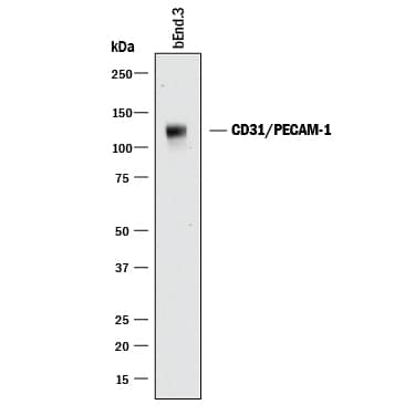
HAF017). A specific band was detected for CD31/PECAM-1 at approximately 130 kDa (as indicated). This experiment was conducted under reducing conditions and using
Immunoblot Buffer Group 1." alt="Western blot shows lysates of bEnd.3 mouse endothelioma cell line. PVDF membrane was probed with 0.5 µg/mL of Goat Anti-Human/Mouse/Rat CD31/PECAM-1 Antigen Affinity-purified Polyclonal Antibody (Catalog # AF3628) followed by HRP-conjugated Anti-Goat IgG Secondary Antibody (
HAF017). A specific band was detected for CD31/PECAM-1 at approximately 130 kDa (as indicated). This experiment was conducted under reducing conditions and using
Immunoblot Buffer Group 1." class="big_thumb" />

AB-108-C, open histogram), followed by Allophycocyanin-conjugated Anti-Goat IgG Secondary Antibody (
F0108)." class="big_lightbox">
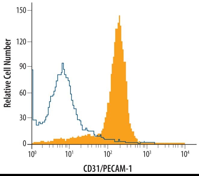
AB-108-C, open histogram), followed by Allophycocyanin-conjugated Anti-Goat IgG Secondary Antibody (
F0108)." class="big_thumb" />
AB-108-C, open histogram), followed by Allophycocyanin-conjugated Anti-Goat IgG Secondary Antibody (
F0108)." class="big_lightbox">
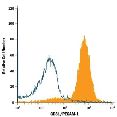
AB-108-C, open histogram), followed by Allophycocyanin-conjugated Anti-Goat IgG Secondary Antibody (
F0108)." class="big_thumb" />
VC001). Before incubation with the primary antibody, tissue was subjected to heat-induced epitope retrieval using Antigen Retrieval Reagent-Basic (
CTS013). Tissue was stained using DAB (brown) and counterstained with hematoxylin (blue). Specific staining was localized to endothelial cells in vasculature." class="big_lightbox">
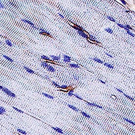
VC001). Before incubation with the primary antibody, tissue was subjected to heat-induced epitope retrieval using Antigen Retrieval Reagent-Basic (
CTS013). Tissue was stained using DAB (brown) and counterstained with hematoxylin (blue). Specific staining was localized to endothelial cells in vasculature." class="big_thumb" />
VC004). Before incubation with the primary antibody, tissue was subjected to heat-induced epitope retrieval using VisUCyte Antigen Retrieval Reagent-Basic (Catalog #
VCTS021). Tissue was stained using DAB (brown) and counterstained with hematoxylin (blue). Specific staining was localized to endothelial cells in chorionic villi. View our protocol for
IHC Staining with VisUCyte HRP Polymer Detection Reagents." class="big_lightbox">

VC004). Before incubation with the primary antibody, tissue was subjected to heat-induced epitope retrieval using VisUCyte Antigen Retrieval Reagent-Basic (Catalog #
VCTS021). Tissue was stained using DAB (brown) and counterstained with hematoxylin (blue). Specific staining was localized to endothelial cells in chorionic villi. View our protocol for
IHC Staining with VisUCyte HRP Polymer Detection Reagents." class="big_thumb" />
AF3628) followed by HRP-conjugated Anti-Goat IgG Secondary Antibody (Catalog #
HAF017). A specific band was detected for CD31/PECAM‑1 at approximately 130 kDa (as indicated). This experiment was conducted under reducing conditions and using
Western Blot Buffer Group 1." class="big_lightbox">

AF3628) followed by HRP-conjugated Anti-Goat IgG Secondary Antibody (Catalog #
HAF017). A specific band was detected for CD31/PECAM‑1 at approximately 130 kDa (as indicated). This experiment was conducted under reducing conditions and using
Western Blot Buffer Group 1." class="big_thumb" />
 HAF017). A specific band was detected for CD31/PECAM-1 at approximately 130 kDa (as indicated). This experiment was conducted under reducing conditions and using Immunoblot Buffer Group 1." alt="Western blot shows lysates of bEnd.3 mouse endothelioma cell line. PVDF membrane was probed with 0.5 µg/mL of Goat Anti-Human/Mouse/Rat CD31/PECAM-1 Antigen Affinity-purified Polyclonal Antibody (Catalog # AF3628) followed by HRP-conjugated Anti-Goat IgG Secondary Antibody (HAF017). A specific band was detected for CD31/PECAM-1 at approximately 130 kDa (as indicated). This experiment was conducted under reducing conditions and using Immunoblot Buffer Group 1." class="big_thumb" />
 HAF016). A specific band was detected for CD31/PECAM-1 at approximately 130 kDa (as indicated). This experiment was conducted under reducing conditions and using Immunoblot Buffer Group 1." alt="Western blot shows lysates of HepG2 human hepatocellular carcinoma cell line. PVDF membrane was probed with 1 µg/mL of Sheep Anti-Human CD31/PECAM-1 Antigen Affinity-purified Polyclonal Antibody (Catalog # AF806) followed by HRP-conjugated Anti-Sheep IgG Secondary Antibody (HAF016). A specific band was detected for CD31/PECAM-1 at approximately 130 kDa (as indicated). This experiment was conducted under reducing conditions and using Immunoblot Buffer Group 1." class="big_thumb" />
 HAF018). A specific band was detected for CD31/PECAM-1 at approximately 130 kDa (as indicated). This experiment was conducted under reducing conditions and using Immunoblot Buffer Group 1." alt="Western blot shows lysates of HUVEC human umbilical vein endothelial cells. PVDF membrane was probed with 1 µg/mL of Mouse Anti-Human CD31/PECAM-1 Monoclonal Antibody (Catalog # BBA7) followed by HRP-conjugated Anti-Mouse IgG Secondary Antibody (HAF018). A specific band was detected for CD31/PECAM-1 at approximately 130 kDa (as indicated). This experiment was conducted under reducing conditions and using Immunoblot Buffer Group 1." class="big_thumb" />


 AB-108-C, open histogram), followed by Allophycocyanin-conjugated Anti-Goat IgG Secondary Antibody (
 AB-108-C, open histogram), followed by Allophycocyanin-conjugated Anti-Goat IgG Secondary Antibody (
 NL003) and counterstained with DAPI (blue). Specific staining was localized to cell membrane. View our protocol for
  NL001) and counterstained with DAPI (blue). Specific staining was localized to developing endothelium. View our protocol for
  VC004). Tissue was stained using DAB (brown) and counterstained with hematoxylin (blue). Specific staining was localized to developing guts. View our protocol for
  VC001). Before incubation with the primary antibody, tissue was subjected to heat-induced epitope retrieval using Antigen Retrieval Reagent-Basic (
 VC004). Before incubation with the primary antibody, tissue was subjected to heat-induced epitope retrieval using VisUCyte Antigen Retrieval Reagent-Basic (Catalog #
  AB-108-C, open histogram), followed by Phycoerythrin-conjugated Anti-Goat IgG Secondary Antibody (Catalog #
  AF3628) followed by HRP-conjugated Anti-Goat IgG Secondary Antibody (Catalog #


 AB-108-C, open histogram) followed by Phycoerythrin-conjugated Anti-Goat IgG Secondary Antibody (Catalog #


![Immunohistochemistry: Rabbit Polyclonal CD31/PECAM-1 Antibody [NB100-2284] - Staining of CD31 in human lung tissue. Nuclei in blue and CD31 (vessel) in green. Mouse anti-CD31 (1:100, overnight incubation at +4°C), followed by donkey anti-mouse 488 (1:400, 3h at RT). Image from a verified customer review.](http://images.novusbio.com/fullsize/antibody/nb100-2284_rabbit-polyclonal-cd31-pecam-1-antibody-immunohistochemistry-49202494658..jpg)
![Immunohistochemistry: CD31/PECAM-1 Antibody [NB100-2284] - Antibodies selected for characterization of stromal cells & vasculature in FFPE specimens are species-specific. (A, B) Labeling of a patient colon tumor (A) & Colo205 human cell line xenograft (B) with anti-human mitochondrial antibody (single arrow- tumor cells; double arrows- stroma). (C, D) Labeling of a patient pancreatic tumor (C) & Colo205 human cell line xenograft (D) with anti-human CD34. (E, F) Labeling of a patient lung tumor (E) & Colo205 xenograft (F) with anti-human CD31. (G, H) Labeling of a patient lung tumor (G) & Colo205 xenograft (H) with anti-mouse CD34. (bars = 50 μ). Image collected & cropped by CiteAb from the following publication (//translational-medicine.biomedcentral.com/articles/10.1186/1479-5876-11-110), licensed under a CC-BY license. Not internally tested by Novus Biologicals.](http://images.novusbio.com/fullsize/nb100-2284_rabbit-cd31-pecam-1-pab-310202415371939.jpg)
![Immunohistochemistry: CD31/PECAM-1 Antibody [NB100-2284] - At the time of second passage, vessels in successfully growing xenografts of four different tumor types surveyed were of murine origin: A-C Colon; D-F Lung; G-I Pancreatic; J-L Renal Cell Carcinoma. For each tumor type, a representative section of an original patient specimen labeled with anti-huCD31 is shown (A, D, G, J). For each tumor, sections of the first passage xenografted tumor when it was resected are also shown; while no huCD31(+) vessels were identified in the xenografts (B, E, H, K), msCD34(+) vessels were abundant (C, F, I, L). (bars = 50 μ). Image collected & cropped by CiteAb from the following publication (//translational-medicine.biomedcentral.com/articles/10.1186/1479-5876-11-110), licensed under a CC-BY license. Not internally tested by Novus Biologicals.](http://images.novusbio.com/fullsize/nb100-2284_rabbit-cd31-pecam-1-pab-3102024153970.jpg)
![Immunohistochemistry: CD31/PECAM-1 Antibody [NB100-2284] - Vascularization of an engrafted patient colon tumor. A patient colon tumor was implanted in a cohort of mice & vessel development was analyzed over 8 weeks. Vessels in the original patient specimen labeled strongly for huCD31 (A) & not for msCD34 (D). Representative sections showing loss of huCD31(+) vessels (B- 4 weeks, C- 7 weeks) & presence of msCD34(+) vessels (E- 4 weeks, F- 7 weeks) are shown. The graph (G) summarizes this process; huCD31(+) vessels were rapidly lost, & by one week, mouse vessels were the predominate vessels present in the colon tumors (no data for msCD34 at 2 weeks; bars = 100 μ). Image collected & cropped by CiteAb from the following publication (//translational-medicine.biomedcentral.com/articles/10.1186/1479-5876-11-110), licensed under a CC-BY license. Not internally tested by Novus Biologicals.](http://images.novusbio.com/fullsize/nb100-2284_rabbit-cd31-pecam-1-pab-31020241535625.jpg)
![Western Blot: CD31/PECAM-1 Antibody [NB100-2284] - KCl pulse decreased membrane, but increased nuclear, detection of NHE1 in b.End3 endothelial cells.(A) Representative immunofluorescence images of bEnd.3 cells at two time-points after KCl or aCSF pulse. (B) Representative immunoblots of NHE1, alpha -tubulin, PECAM, & lamin B in cytosol, membrane, & nuclear fractions of bEnd.3 cells harvested at 5 min after KCl or aCSF pulse. (C = cytosol, M = membrane, N = nuclear) Values represent the mean ratio of NHE1 detection ± SEM (n = 11–12). (C) Representative immunoblots indicating NHE1 & alpha -tubulin as a loading control in whole cell lysate of bEnd.3 cells harvested at 5 min after KCl or aCSF pulse. Values represent the % of aCSF-treated relative expression ± SEM (n = 6). (D) Representative immunoblots of NHE1 & alpha -tubulin as a loading control in whole lysate of microvessels harvested at 90 min after cortical injection of KCl or aCSF. Values represent the % of naive relative expression ± SEM (n = 6). # denotes significantly different vs naïve (p<0.01), as assessed by one-way ANOVA (E) Intracellular pH during aCSF or KCl pulse. All data represent mean ± SEM (n = 45). (F) Extracellular pH during aCSF or KCl pulse. All data represent mean ± SEM (n = 6) in triplicate. Image collected & cropped by CiteAb from the following publication (//pubmed.ncbi.nlm.nih.gov/32469979), licensed under a CC-BY license. Not internally tested by Novus Biologicals.](http://images.novusbio.com/fullsize/nb100-2284_rabbit-cd31-pecam-1-pab-310202415541933.jpg)
![Immunocytochemistry/ Immunofluorescence: CD31/PECAM-1 Antibody [NB100-2284] - Impact of T‐AuNPs on endothelial cells in vivo. Immunofluorescent staining for endothelial cells using an anti‐CD 31 antibody revealed an intact endothelial cell monolayer in the uninjured control carotid artery (N = 3). As expected post balloon angioplasty, there is no endothelial cell monolayer of the injured left carotid arteries in the injury alone, injury + T‐AuNP, & injury + S‐AuNP treatment conditions. Green indicates autofluorescence of the elastic lamina. Magenta indicates endothelial cells. Images obtained using 25× magnification with exposure time of 400 msec. TUNEL staining for apoptosis revealed no evidence of endothelial cell apoptosis in the uninjured control carotid arteries, with a prominent endothelial cell monolayer (white arrowhead). Mild apoptosis was noted in the media of balloon‐injured left carotid arteries, as expected. Black arrows indicate apoptotic cells. Representative images obtained using 40× magnification. Image collected & cropped by CiteAb from the following publication (//pubmed.ncbi.nlm.nih.gov/28242820), licensed under a CC-BY license. Not internally tested by Novus Biologicals.](http://images.novusbio.com/fullsize/nb100-2284_rabbit-cd31-pecam-1-pab-3102024165737.jpg)
![Immunohistochemistry: CD31/PECAM-1 Antibody [NB100-2284] - The growth of a patient mesothelioma xenograft was supported by development of a murine vascular network. A patient mesothelioma was implanted in a cohort of mice & monitored for tumor growth. Once tumors began to actively grow, representative tumors were resected at weekly intervals & analyzed for vessel content. Staining for huCD31 was prominent in the original patient specimen (A), much reduced at 4 weeks (B) & negligible at 9 weeks (C). In contrast, patient specimens were not stained for msCD34 (D), whereas at 4 weeks large numbers of vessels stained for msCD34 (E) & by 9 weeks, msCD34 labeled vessels were predominant (F). The graph (G) summarizes the loss of detectable human vessels & acquisition of murine vessels over a 9 week period. (bars = 100 μ). Image collected & cropped by CiteAb from the following publication (//translational-medicine.biomedcentral.com/articles/10.1186/1479-5876-11-110), licensed under a CC-BY license. Not internally tested by Novus Biologicals.](http://images.novusbio.com/fullsize/nb100-2284_rabbit-cd31-pecam-1-pab-31020241612684.jpg)
![Immunocytochemistry/ Immunofluorescence: CD31/PECAM-1 Antibody [NB100-2284] - TGF‐ beta 1 released by MSCs contributes to induced myofibroblast differentiation & granulation tissue formationA–C2.5 × 105 of TGF‐ beta 1 siRNA or control siRNA‐transfected AT‐MSCs were intradermally injected around each of CD18−/− murine wound. PBS mock injection served as negative control. Wound tissue was harvested at day 2, 5, & 7 post‐wounding for quantification of human TGF‐ beta 1 mRNA (A) at day 2 by qPCR, total TGF‐ beta 1 (B), & active TGF‐ beta 1 (C) protein at day 5 by ELISA. Data are expressed as mean ± SEM, n = 3 wounds per group, **P < 0.01, by one‐way ANOVA with Tukey's test.D–GExpression of alpha ‐SMA (D & E) & CD31 (F & G) at days 5 & 7 by immunostaining on tissue sections. The dashed lines indicate the border of the wound bed & epidermis or eschar. e, epidermis; es, eschar; wb, wound bed. Scale bars: 100 μm. Quantification data are expressed as mean ± SEM, n = 3 wounds per group, *P < 0.05, ***P < 0.001, by one‐way ANOVA with Tukey's test.Source data are available online for this figure. Image collected & cropped by CiteAb from the following publication (//pubmed.ncbi.nlm.nih.gov/32080965), licensed under a CC-BY license. Not internally tested by Novus Biologicals.](http://images.novusbio.com/fullsize/nb100-2284_rabbit-cd31-pecam-1-pab-310202415532085.jpg)
![Immunohistochemistry-Paraffin: Rabbit Polyclonal CD31/PECAM-1 Antibody [NB100-2284] - CD31 positive vessels in a canine splenic hemangiosarcoma.Tissue slides were loaded into automated research stainer, dewaxed and pretreated with EDTA-based epitope retrieval ER2 solution for 20 mins at 100°C. After a peroxide incubation for 5 min, the rabbit antibody against CD31 (NB100-2284) (1:100) was incubated for 60 mins at room temperature followed by Bond Polymer (anti-rabbit HRP) incubation for 8 min at room temperature. Mixed DAB reagent (Polymer Refine Detection Kit) was incubated for 10 mins, and Hematoxylin (Refine Detection Kit) counterstaining for 10 mins. After staining, sample slides were washed in water, dehydrated using ethanol gradient (70%, 90%, 100%), washed three times, and mounted in mounting medium. Image from a verified customer review.](http://images.novusbio.com/fullsize/antibody/nb100-2284_rabbit-polyclonal-cd31-pecam-1-antibody-immunohistochemistry-paraffin-203202593412..jpg)
 NL010) and counterstained with DAPI (blue). View our protocol for
  HAF016) in Milk-free Antibody Diluent (Catalog #
  NL010) and counterstained with DAPI (blue). Specific staining was localized to cell membrane. Staining was performed using our protocol for Fluorescent ICC Staining of Non-adherent Cells." class="big_thumb" />
 VC004). Before incubation with the primary antibody, tissue was subjected to heat-induced epitope retrieval using VisUCyte Antigen Retrieval Reagent-Basic (Catalog #
  VC004). Before incubation with the primary antibody, tissue was subjected to heat-induced epitope retrieval using VisUCyte Antigen Retrieval Reagent-Basic (Catalog #


![Immunocytochemistry/ Immunofluorescence: CD31/PECAM-1 Antibody (JC/70A) - BSA Free [NB600-562] - Immunohistochemial comparison of the expression of NMDAR1 on vessels & EndoMT markers in the urinary bladder.Biopsy specimens of frozen sections from the urinary bladders of (A, C, E) normal control subjects & (B, D, F) KC patients. (A, B) Double-immunofluorescence staining with antibody to endothelial marker CD31 (green) & NMDAR1 (red); DAPI was used as a nuclear stain (blue); co-expression of CD31 & NMDAR1 was evident in two groups (yellow, denoted by white arrows). (C–F) Double-immunofluorescence staining with antibody to endothelial marker CD31 (green) & two mesenchymal markers: FSP1 (red, in panels C & D) & alpha -SMA (red, in panels E & F); DAPI was used as a nuclear stain (blue); co-expression of CD31 & FSP1 or alpha -SMA was evident in three groups (yellow, denoted by white arrows) (white bars: 50 μm). Image collected & cropped by CiteAb from the following publication (//pubmed.ncbi.nlm.nih.gov/27529746), licensed under a CC-BY license. Not internally tested by Novus Biologicals.](http://images.novusbio.com/fullsize/nb600-562_mouse-monoclonal-cd31-pecam-1-antibody-jc-70a-310202416171426.jpg)
![Immunocytochemistry/ Immunofluorescence: CD31/PECAM-1 Antibody (JC/70A) - BSA Free [NB600-562] - Immunohistochemial comparison of the expression of NMDAR1 on vessels & EndoMT markers in the urinary bladder.Biopsy specimens of frozen sections from the urinary bladders of (A, C, E) normal control subjects & (B, D, F) KC patients. (A, B) Double-immunofluorescence staining with antibody to endothelial marker CD31 (green) & NMDAR1 (red); DAPI was used as a nuclear stain (blue); co-expression of CD31 & NMDAR1 was evident in two groups (yellow, denoted by white arrows). (C–F) Double-immunofluorescence staining with antibody to endothelial marker CD31 (green) & two mesenchymal markers: FSP1 (red, in panels C & D) & alpha -SMA (red, in panels E & F); DAPI was used as a nuclear stain (blue); co-expression of CD31 & FSP1 or alpha -SMA was evident in three groups (yellow, denoted by white arrows) (white bars: 50 μm). Image collected & cropped by CiteAb from the following publication (//pubmed.ncbi.nlm.nih.gov/27529746), licensed under a CC-BY license. Not internally tested by Novus Biologicals.](http://images.novusbio.com/fullsize/nb600-562_mouse-monoclonal-cd31-pecam-1-antibody-jc-70a-31020241618941.jpg)
![Immunocytochemistry/ Immunofluorescence: CD31/PECAM-1 Antibody (JC/70A) - BSA Free [NB600-562] - Immunohistochemial comparison of the expression of NMDAR1 on vessels & EndoMT markers in the urinary bladder.Biopsy specimens of frozen sections from the urinary bladders of (A, C, E) normal control subjects & (B, D, F) KC patients. (A, B) Double-immunofluorescence staining with antibody to endothelial marker CD31 (green) & NMDAR1 (red); DAPI was used as a nuclear stain (blue); co-expression of CD31 & NMDAR1 was evident in two groups (yellow, denoted by white arrows). (C–F) Double-immunofluorescence staining with antibody to endothelial marker CD31 (green) & two mesenchymal markers: FSP1 (red, in panels C & D) & alpha -SMA (red, in panels E & F); DAPI was used as a nuclear stain (blue); co-expression of CD31 & FSP1 or alpha -SMA was evident in three groups (yellow, denoted by white arrows) (white bars: 50 μm). Image collected & cropped by CiteAb from the following publication (//pubmed.ncbi.nlm.nih.gov/27529746), licensed under a CC-BY license. Not internally tested by Novus Biologicals.](http://images.novusbio.com/fullsize/nb600-562_mouse-monoclonal-cd31-pecam-1-antibody-jc-70a-310202416173525.jpg)
![Immunocytochemistry/ Immunofluorescence: CD31/PECAM-1 Antibody (JC/70A) - BSA Free [NB600-562] - Immunohistochemial comparison of the expression of NMDAR1 on vessels & EndoMT markers in the urinary bladder.Biopsy specimens of frozen sections from the urinary bladders of (A, C, E) normal control subjects & (B, D, F) KC patients. (A, B) Double-immunofluorescence staining with antibody to endothelial marker CD31 (green) & NMDAR1 (red); DAPI was used as a nuclear stain (blue); co-expression of CD31 & NMDAR1 was evident in two groups (yellow, denoted by white arrows). (C–F) Double-immunofluorescence staining with antibody to endothelial marker CD31 (green) & two mesenchymal markers: FSP1 (red, in panels C & D) & alpha -SMA (red, in panels E & F); DAPI was used as a nuclear stain (blue); co-expression of CD31 & FSP1 or alpha -SMA was evident in three groups (yellow, denoted by white arrows) (white bars: 50 μm). Image collected & cropped by CiteAb from the following publication (//pubmed.ncbi.nlm.nih.gov/27529746), licensed under a CC-BY license. Not internally tested by Novus Biologicals.](http://images.novusbio.com/fullsize/nb600-562_mouse-monoclonal-cd31-pecam-1-antibody-jc-70a-31020241616378.jpg)
![Immunocytochemistry/ Immunofluorescence: CD31/PECAM-1 Antibody (JC/70A) - BSA Free [NB600-562] - Immunohistochemial comparison of the expression of NMDAR1 on vessels & EndoMT markers in the urinary bladder.Biopsy specimens of frozen sections from the urinary bladders of (A, C, E) normal control subjects & (B, D, F) KC patients. (A, B) Double-immunofluorescence staining with antibody to endothelial marker CD31 (green) & NMDAR1 (red); DAPI was used as a nuclear stain (blue); co-expression of CD31 & NMDAR1 was evident in two groups (yellow, denoted by white arrows). (C–F) Double-immunofluorescence staining with antibody to endothelial marker CD31 (green) & two mesenchymal markers: FSP1 (red, in panels C & D) & alpha -SMA (red, in panels E & F); DAPI was used as a nuclear stain (blue); co-expression of CD31 & FSP1 or alpha -SMA was evident in three groups (yellow, denoted by white arrows) (white bars: 50 μm). Image collected & cropped by CiteAb from the following publication (//pubmed.ncbi.nlm.nih.gov/27529746), licensed under a CC-BY license. Not internally tested by Novus Biologicals.](http://images.novusbio.com/fullsize/nb600-562_mouse-monoclonal-cd31-pecam-1-antibody-jc-70a-3102024161609.jpg)
![Immunocytochemistry/ Immunofluorescence: CD31/PECAM-1 Antibody (JC/70A) - BSA Free [NB600-562] - Immunohistochemial comparison of the expression of NMDAR1 on vessels & EndoMT markers in the urinary bladder.Biopsy specimens of frozen sections from the urinary bladders of (A, C, E) normal control subjects & (B, D, F) KC patients. (A, B) Double-immunofluorescence staining with antibody to endothelial marker CD31 (green) & NMDAR1 (red); DAPI was used as a nuclear stain (blue); co-expression of CD31 & NMDAR1 was evident in two groups (yellow, denoted by white arrows). (C–F) Double-immunofluorescence staining with antibody to endothelial marker CD31 (green) & two mesenchymal markers: FSP1 (red, in panels C & D) & alpha -SMA (red, in panels E & F); DAPI was used as a nuclear stain (blue); co-expression of CD31 & FSP1 or alpha -SMA was evident in three groups (yellow, denoted by white arrows) (white bars: 50 μm). Image collected & cropped by CiteAb from the following publication (//pubmed.ncbi.nlm.nih.gov/27529746), licensed under a CC-BY license. Not internally tested by Novus Biologicals.](http://images.novusbio.com/fullsize/nb600-562_mouse-monoclonal-cd31-pecam-1-antibody-jc-70a-310202416163753.jpg)
![Immunocytochemistry/ Immunofluorescence: CD31/PECAM-1 Antibody (JC/70A) - BSA Free [NB600-562] - Immunohistochemial comparison of the expression of NMDAR1 on vessels & EndoMT markers in the urinary bladder.Biopsy specimens of frozen sections from the urinary bladders of (A, C, E) normal control subjects & (B, D, F) KC patients. (A, B) Double-immunofluorescence staining with antibody to endothelial marker CD31 (green) & NMDAR1 (red); DAPI was used as a nuclear stain (blue); co-expression of CD31 & NMDAR1 was evident in two groups (yellow, denoted by white arrows). (C–F) Double-immunofluorescence staining with antibody to endothelial marker CD31 (green) & two mesenchymal markers: FSP1 (red, in panels C & D) & alpha -SMA (red, in panels E & F); DAPI was used as a nuclear stain (blue); co-expression of CD31 & FSP1 or alpha -SMA was evident in three groups (yellow, denoted by white arrows) (white bars: 50 μm). Image collected & cropped by CiteAb from the following publication (//pubmed.ncbi.nlm.nih.gov/27529746), licensed under a CC-BY license. Not internally tested by Novus Biologicals.](http://images.novusbio.com/fullsize/nb600-562_mouse-monoclonal-cd31-pecam-1-antibody-jc-70a-310202416171483.jpg)

 NL007) and counterstained with DAPI (blue). View our protocol for
  NL007) and counterstained with DAPI (blue). Specific staining was localized to cell membrane. Staining was performed using our protocol for Fluorescent ICC Staining of Non-adherent Cells." class="big_thumb" />
 VC001). Before incubation with the primary antibody, tissue was subjected to heat-induced epitope retrieval using VisUCyte Antigen Retrieval Reagent-Basic (Catalog #

![Immunohistochemistry-Frozen: CD31/PECAM-1 Antibody (TLD-3A12) [NB100-64796] - Analysis using the FITC conjugate of CD31/PECAM-1 Antibody (TLD-3A12) (NB100-64796). Staining of rat brain cryosection with Mouse.](http://images.novusbio.com/fullsize/CD31-PECAM-1-Antibody-TLD-3A12-Immunohistochemistry-Frozen-NB100-64796-img0012.jpg "Immunohistochemistry-Frozen: CD31/PECAM-1 Antibody (TLD-3A12) [NB100-64796] - Analysis using the FITC conjugate of CD31/PECAM-1 Antibody (TLD-3A12) (NB100-64796). Staining of rat brain cryosection with Mouse.")
![Flow Cytometry: CD31/PECAM-1 Antibody (TLD-3A12) [NB100-64796] - Analysis using the Alexa Fluor (R) 488 conjugate of CD31/PECAM-1 Antibody (TLD-3A12) (NB100-64796). Staining of PBMC's (Monocyte gate at 1 x 10^6 cells/ml) with CD31 antibody (orange) stained at a dilution of 1:500. Shown with mIgG (AF488) isotype control (blue).](http://images.novusbio.com/fullsize/CD31-PECAM-1-Antibody-TLD-3A12-Flow-Cytometry-NB100-64796-img0007.jpg)
![Immunohistochemistry-Paraffin: CD31/PECAM-1 Antibody (TLD-3A12) [NB100-64796] - Analysis of CD31 in 4th day cutaneous wound section of diabetic rat using CD31/PECAM-1 Antibody (TLD-3A12). Image from verified customer review.](http://images.novusbio.com/fullsize/CD31-PECAM-1-Antibody-TLD-3A12-Immunohistochemistry-Paraffin-NB100-64796-img0005.jpg)
![Immunohistochemistry-Frozen: CD31/PECAM-1 Antibody (TLD-3A12) [NB100-64796] - Analysis using the FITC conjugate of CD31/PECAM-1 Antibody (TLD-3A12) (NB100-64796). Staining of rat spleen cryosection.](http://images.novusbio.com/fullsize/CD31-PECAM-1-Antibody-TLD-3A12-Immunohistochemistry-Frozen-NB100-64796-img0013.jpg)


![Immunocytochemistry/Immunofluorescence: CD31/PECAM-1 Antibody (MEC13.3) [NB600-1475] - Mouse MS1 cells were fixed in 4% paraformaldehyde for 10 minutes and permeabilized in 0.05% Triton X-100 in PBS for 5 minutes. The cells were incubated with CD31/PECAM-1 Antibody [MEC13.3] (NB600-1475) at 1ug/ml overnight at 4C and detected with an anti-rat DyLight 488 (Green) at a 1:1000 dilution for 60 minutes. Nuclei were counterstained with DAPI (Blue). Cells were imaged using a 100X objective and digitally deconvolved.](http://images.novusbio.com/fullsize/CD31-PECAM-1-Antibody-MEC13-3-Immunocytochemistry-Immunofluorescence-NB600-1475-img0019.jpg "Immunocytochemistry/Immunofluorescence: CD31/PECAM-1 Antibody (MEC13.3) [NB600-1475] - Mouse MS1 cells were fixed in 4% paraformaldehyde for 10 minutes and permeabilized in 0.05% Triton X-100 in PBS for 5 minutes. The cells were incubated with CD31/PECAM-1 Antibody [MEC13.3] (NB600-1475) at 1ug/ml overnight at 4C and detected with an anti-rat DyLight 488 (Green) at a 1:1000 dilution for 60 minutes. Nuclei were counterstained with DAPI (Blue). Cells were imaged using a 100X objective and digitally deconvolved.")
![Immunohistochemistry-Frozen: CD31/PECAM-1 Antibody (MEC13.3) [NB600-1475] - 4T1 tumor sections were stained for CD31 (green) and CD105 (red).](http://images.novusbio.com/fullsize/CD31-PECAM-1-Antibody-MEC13-3-Immunohistochemistry-Frozen-NB600-1475-img0006.jpg)
![Flow (Cell Surface): CD31/PECAM-1 Antibody (MEC13.3) [NB600-1475] - A surface stain was performed on WEHI-3 Cells with CD31/PECAM-1 Antibody (MEC13.3) (NB600-1475, blue) and a matched isotype control (orange). Cells were incubated in an antibody dilution of 2.5 ug/mL for 20 minutes at room temperature, followed by rat F(ab)2 IgG (H+L) APC-conjugated secondary antibody (F0113, R&D Systems).](http://images.novusbio.com/fullsize/CD31-PECAM-1-Antibody-MEC13-3-Flow-Cell-Surface-NB600-1475-img0009.jpg)
![Flow Cytometry: CD31/PECAM-1 Antibody (MEC13.3) [NB600-1475] - C57BL/6 mouse splenocytes stained with purified CD31/PECAM-1 Antibody (MEC13.3), followed by anti-rat IgG FITC](http://images.novusbio.com/fullsize/CD31-PECAM-1-Antibody-MEC13-3-Flow-Cytometry-NB600-1475-img0010.jpg)
![Immunocytochemistry/Immunofluorescence: CD31/PECAM-1 Antibody (MEC13.3) [NB600-1475] - Mouse MS1 cells were fixed in 4% paraformaldehyde for 10 minutes and permeabilized in 0.05% Triton X-100 in PBS for 5 minutes. The cells were incubated with CD31/PECAM-1 Antibody [MEC13.3] conjugated to DyLight 488 (NB600-1475G) at 5 ug/ml for 1 hour at room temperature. Nuclei were counterstained with DAPI (Blue). Cells were imaged using a 100X objective and digitally deconvolved.](http://images.novusbio.com/fullsize/CD31-PECAM-1-Antibody-MEC13-3-Immunocytochemistry-Immunofluorescence-NB600-1475-img0017.jpg)
![Immunocytochemistry/Immunofluorescence: CD31/PECAM-1 Antibody (MEC13.3) [NB600-1475] - MS1 cells were fixed for 10 minutes using 10% formalin and then permeabilized for 5 minutes using 1X TBS + 0.5% Triton-X100. The cells were incubated with at 5.0 ug/ml overnight at 4C and detected with an anti-mouse Dylight 488 (Green) at a 1:500 dilution. Actin was detected with Phalloidin 568 (Red) at a 1:200 dilution. Nuclei were counterstained with DAPI (Blue). Cells were imaged using a 40X objective.](http://images.novusbio.com/fullsize/CD31-PECAM-1-Antibody-MEC13-3-Immunocytochemistry-Immunofluorescence-NB600-1475-img0007.jpg)
![Immunocytochemistry/Immunofluorescence: CD31/PECAM-1 Antibody (MEC13.3) [NB600-1475] - was tested in Wehi-3 cells with Dylight 488 (green). Nuclei and beta-tubulin were counterstained with DAPI (blue) and Dylight 550 (red).](http://images.novusbio.com/fullsize/CD31-PECAM-1-Antibody-MEC13-3-Immunocytochemistry-Immunofluorescence-NB600-1475-img0011.jpg)

![Flow Cytometry: CD31/PECAM-1 Antibody (MEC13.3) [NB600-1475] - A surface stain was performed on MS1 cells with CD31/PECAM-1 [MEC13.3] Antibody NB600-1475R (blue) and a matched isotype control (orange). Cells were incubated in an antibody dilution of 2.5 ug/mL for 30 minutes at room temperature. Both antibodies were conjugated to DyLight 550.](http://images.novusbio.com/fullsize/CD31-PECAM-1-Antibody-MEC13-3-Flow-Cytometry-NB600-1475-img0022.jpg)
![Immunocytochemistry/Immunofluorescence: CD31/PECAM-1 Antibody (MEC13.3) [NB600-1475] - Mouse MS1 cells were fixed in 4% paraformaldehyde for 10 minutes and permeabilized in 0.05% Triton X-100 in PBS for 5 minutes. The cells were incubated with CD31/PECAM-1 Antibody [MEC13.3] conjugated to Alexa Fluor 647 (NB600-1475AF647) at 5 ug/ml for 1 hour at room temperature. Nuclei were counterstained with DAPI (Blue). Cells were imaged using a 100X objective and digitally deconvolved.](http://images.novusbio.com/fullsize/CD31-PECAM-1-Antibody-MEC13-3-Immunocytochemistry-Immunofluorescence-NB600-1475-img0016.jpg)
![Immunocytochemistry/Immunofluorescence: CD31/PECAM-1 Antibody (MEC13.3) [NB600-1475] - Mouse MS1 cells were fixed in 4% paraformaldehyde for 10 minutes and permeabilized in 0.05% Triton X-100 in PBS for 5 minutes. The cells were incubated with CD31/PECAM-1 Antibody [MEC13.3] conjugated to DyLight 550 (NB600-1475R) at 5 ug/ml for 1 hour at room temperature. Nuclei were counterstained with DAPI (Blue). Cells were imaged using a 100X objective and digitally deconvolved.](http://images.novusbio.com/fullsize/CD31-PECAM-1-Antibody-MEC13-3-Immunocytochemistry-Immunofluorescence-NB600-1475-img0018.jpg)
![Immunohistochemistry-Frozen: CD31/PECAM-1 Antibody (MEC13.3) [NB600-1475] - Staining of mouse CD31 in non-small cell lung cancer tumor sections from a human NSCLC tumor-bearing mouse. This image was submitted via customer Review.](http://images.novusbio.com/fullsize/CD31-PECAM-1-Antibody-MEC13-3-Immunohistochemistry-Frozen-NB600-1475-img0012.jpg)
![Flow (Cell Surface): CD31/PECAM-1 Antibody (MEC13.3) [NB600-1475] - A surface stain was performed on WEHI-3 Cells with CD31 (MEC13.3) antibody NB600-1475 (blue) and a matched isotype control (orange). Cells were incubated in an antibody dilution of 2.5 ug/mL for 20 minutes at room temperature, followed by rat F(ab)2 IgG (H+L) APC-conjugated secondary antibody (F0113, R&D Systems).](http://images.novusbio.com/fullsize/CD31-PECAM-1-Antibody-MEC13-3-Flow-Cell-Surface-NB600-1475-img0013.jpg)
![Flow Cytometry: CD31/PECAM-1 Antibody (MEC13.3) [NB600-1475] - A surface stain was performed on MS-1 cells with CD31/PECAM-1 Antibody (MEC13.3) (NB600-1475AF647, blue) and a matched isotype control (orange). Cells were incubated in an antibody dilution of 5 ug/mL for 20 minutes at room temperature. Both antibodies were conjugated to Alexa Fluor 647.](http://images.novusbio.com/fullsize/CD31-PECAM-1-Antibody-MEC13-3-Flow-Cytometry-NB600-1475-img0014.jpg)
![Flow Cytometry: CD31/PECAM-1 Antibody (MEC13.3) [NB600-1475] - A surface stain was performed on MS1 cells with CD31/PECAM-1 [MEC13.3] Antibody NB600-1475DL594 (blue) and a matched isotype control (orange). Cells were incubated in an antibody dilution of 2.5 ug/mL for 30 minutes at room temperature. Both antibodies were conjugated to DyLight 594.](http://images.novusbio.com/fullsize/CD31-PECAM-1-Antibody-MEC13-3-Flow-Cytometry-NB600-1475-img0020.jpg)
![Flow Cytometry: CD31/PECAM-1 Antibody (MEC13.3) [NB600-1475] - A surface stain was performed on MS1 cells with CD31/PECAM-1 [MEC13.3] Antibody NB600-1475G (blue) and a matched isotype control (orange). Cells were incubated in an antibody dilution of 5 ug/mL for 30 minutes at room temperature. Both antibodies were conjugated to DyLight 488.](http://images.novusbio.com/fullsize/CD31-PECAM-1-Antibody-MEC13-3-Flow-Cytometry-NB600-1475-img0021.jpg)
![Immunocytochemistry/ Immunofluorescence: CD31/PECAM-1 Antibody (MEC13.3) - BSA Free [NB600-1475] - (A) Representative Western blots depicting phosphorylated JAK3 (pJAK3), total JAK3, & corresponding beta -actin levels in the ipsilateral (CXI) versus the contralateral (CXC) cortex at 24 h in sham & mice subjected to permanent middle cerebral artery occlusion (pMCAO). (B,C) Quantified pJAK3 & JAK3 values normalized to beta -actin in the cortex of sham & mice subjected to stroke. Both pJAK3 & JAK3 are significantly increased in the ipsilateral cortex of stroked animals (*P < 0.05 & **P < 0.01 with respect to sham ipsilateral/contralateral & stroke contralateral; sham, n = 5; stroke, n = 13). Data were analyzed using a two-way ANOVA (ipsilateral/contralateral or sham/stroke) with Bonferroni post-test. (D) Diagram showing peri-infarct brain region from which images were taken (n = 3). Scale bar is 10 µm, & magnification is 60×. (E) JAK3 colocalized with microglia/macrophages labeled with Iba-1. (F) JAK3 colocalized with neurons labeled with NeuN. (G) JAK3 colocalized with endothelial cells labeled with CD31. (H) JAK3 is not colocalized with astrocytes labeled with GFAP. Image collected & cropped by CiteAb from the following publication (//pubmed.ncbi.nlm.nih.gov/28790974), licensed under a CC-BY license. Not internally tested by Novus Biologicals.](http://images.novusbio.com/fullsize/nb600-1475_rat-monoclonal-cd31-pecam-1-antibody-mec13-3-310202416171428.jpg)

![Immunocytochemistry/Immunofluorescence: CD31/PECAM-1 Antibody (MEC 7.46) [NB100-1642] - Mouse MS1 cells were fixed in 4% paraformaldehyde for 10 minutes and permeabilized in 0.05% Triton X-100 in PBS for 5 minutes. The cells were incubated with CD31/PECAM-1 Antibody [MEC 7.46] (NB100-1642) at 1ug/ml overnight at 4C and detected with an anti-rat DyLight 488 (Green) at a 1:1000 dilution for 60 minutes. Nuclei were counterstained with DAPI (Blue). Cells were imaged using a 100X objective and digitally deconvolved.](http://images.novusbio.com/fullsize/CD31-PECAM-1-Antibody-MEC-7-46-Immunocytochemistry-Immunofluorescence-NB100-1642-img0014.jpg "Immunocytochemistry/Immunofluorescence: CD31/PECAM-1 Antibody (MEC 7.46) [NB100-1642] - Mouse MS1 cells were fixed in 4% paraformaldehyde for 10 minutes and permeabilized in 0.05% Triton X-100 in PBS for 5 minutes. The cells were incubated with CD31/PECAM-1 Antibody [MEC 7.46] (NB100-1642) at 1ug/ml overnight at 4C and detected with an anti-rat DyLight 488 (Green) at a 1:1000 dilution for 60 minutes. Nuclei were counterstained with DAPI (Blue). Cells were imaged using a 100X objective and digitally deconvolved.")
![Flow (Cell Surface): CD31/PECAM-1 Antibody (MEC 7.46) [NB100-1642] - A surface stain was performed on MS-1 Cells with CD31/PECAM-1 Antibody (MEC 7.46) NB100-1642(blue) and a matched isotype control (orange). Cells were incubated in an antibody dilution of 2.5 ug/mL for 20 minutes at room temperature, followed by rat F(ab)2 IgG (H+L) APC-conjugated secondary antibody (F0113, R&D Systems).](http://images.novusbio.com/fullsize/CD31-PECAM-1-Antibody-MEC-7-46-Flow-Cell-Surface-NB100-1642-img0005.jpg)

![Immunocytochemistry/Immunofluorescence: CD31/PECAM-1 Antibody (MEC 7.46) [NB100-1642] - Mouse MS1 cells were fixed in 4% paraformaldehyde for 10 minutes and permeabilized in 0.05% Triton X-100 in PBS for 5 minutes. The cells were incubated with CD31/PECAM-1 Antibody [MEC 7.46] conjugated to Alexa Fluor 488 (NB100-1642AF488) at 5 ug/ml for 1 hour at room temperature. Nuclei were counterstained with DAPI (Blue). Cells were imaged using a 100X objective and digitally deconvolved.](http://images.novusbio.com/fullsize/CD31-PECAM-1-Antibody-MEC-7-46-Immunocytochemistry-Immunofluorescence-NB100-1642-img0013.jpg)
![Immunocytochemistry/Immunofluorescence: CD31/PECAM-1 Antibody (MEC 7.46) [NB100-1642] - MS1 cells were fixed for 10 minutes using 10% formalin and then permeabilized for 5 minutes using 1X TBS + 0.5% Triton X-100. The cells were incubated with at 5.0 ug/ml overnight at 4C and detected with an anti-mouse DyLight 488 (Green) at a 1:500 dilution. Actin was detected with Phalloidin 568 (Red) at a 1:200 dilution. Nuclei were counterstained with DAPI (Blue). Cells were imaged using a 40X objective.](http://images.novusbio.com/fullsize/CD31-PECAM-1-Antibody-MEC-7-46-Immunocytochemistry-Immunofluorescence-NB100-1642-img0002.jpg)
![Immunocytochemistry/Immunofluorescence: CD31/PECAM-1 Antibody (MEC 7.46) [NB100-1642] - Mouse MS1 cells were fixed in 4% paraformaldehyde for 10 minutes and permeabilized in 0.05% Triton X-100 in PBS for 5 minutes. The cells were incubated with CD31/PECAM-1 Antibody [MEC 7.46] conjugated to DyLight 550 (NB100-1642R) at 5 ug/ml for 1 hour at room temperature. Nuclei were counterstained with DAPI (Blue). Cells were imaged using a 100X objective and digitally deconvolved.](http://images.novusbio.com/fullsize/CD31-PECAM-1-Antibody-MEC-7-46-Immunocytochemistry-Immunofluorescence-NB100-1642-img0016.jpg)
![<b>Genetic Strategies Validation. </b>Immunohistochemistry-Frozen: CD31/PECAM-1 Antibody (MEC 7.46) [NB100-1642] - Frozen mouse lung tissues of WT B6 and IL1R--/- (knockout) mice were blocked with 1% BSA in PBS and were double stained with Alex647-conjugated CD31/PECAM-1 antibody (MEC 7.46) and PE-conjugated F4/80 for 2 hours at the room temperature. Image using the Alexa Fluor 647 format of this antibody.](http://images.novusbio.com/fullsize/CD31-PECAM-1-Antibody-MEC-7-46-Immunocytochemistry-Immunofluorescence-NB100-1642-img0012.jpg)
![Flow Cytometry: CD31/PECAM-1 Antibody (MEC 7.46) [NB100-1642] - A surface stain was performed on MS1 cells with CD31/PECAM-1 [MEC 7.46] Antibody NB100-1642AF647 (blue) and a matched isotype control (orange). Cells were incubated in an antibody dilution of 2.5 ug/mL for 30 minutes at room temperature. Both antibodies were conjugated to Alexa Fluor 647.](http://images.novusbio.com/fullsize/CD31-PECAM-1-Antibody-MEC-7-46-Flow-Cytometry-NB100-1642-img0015.jpg)

![Flow Cytometry: CD31/PECAM-1 Antibody (MEC 7.46) [NB100-1642] - CD31/PECAM-1 Antibody (MEC 7.46) was tested at 1:250 in WEHI-3 cells with DyLight 488 (green) alongside a matched isotype control (black).](http://images.novusbio.com/fullsize/CD31-PECAM-1-Antibody-MEC-7-46-Flow-Cytometry-NB100-1642-img0006.jpg)


![Immunocytochemistry/Immunofluorescence: CD31/PECAM-1 Antibody [NBP1-71663] - Staining of mouse glioma tissue using CD31/PECAM-1 Antibody (NBP1-71663) at a dilution of 1:2500. Image provided by Dr. Eric Woolf of Dignity Health.](http://images.novusbio.com/fullsize/CD31-PECAM-1-Antibody-Immunocytochemistry-Immunofluorescence-NBP1-71663-img0009.jpg "Immunocytochemistry/Immunofluorescence: CD31/PECAM-1 Antibody [NBP1-71663] - Staining of mouse glioma tissue using CD31/PECAM-1 Antibody (NBP1-71663) at a dilution of 1:2500. Image provided by Dr. Eric Woolf of Dignity Health.")
![Simple Western: CD31/PECAM-1 Antibody [NBP1-71663] - Image shows a specific band for CD31/PECAM 1 in 0.5 mg/mL of HUVEC lysate. This experiment was performed under reducing conditions using the 12-230 kDa separation system.](http://images.novusbio.com/fullsize/CD31-PECAM-1-Antibody-Simple-Western-NBP1-71663-img0010.jpg)


![Immunocytochemistry/Immunofluorescence: CD31/PECAM-1 Antibody [NBP1-71663] - HUVEC cells were fixed for 10 minutes using 10% formalin and then permeabilized for 5 minutes using 1X TBS + 0.5% Triton X-100. The cells were incubated with CD31/PECAM-1 Antibody at 5 ug/mL overnight at 4C and detected with an anti-rabbit Dylight 488 (Green) at a 1:500 dilution. Alpha tubulin (DM1A) NB100-690 was used as a co-stain at a 1:1000 dilution and detected with an anti-mouse Dylight 550 (Red) at a 1:500 dilution. Nuclei were counterstained with DAPI (Blue). Cells were imaged using a 40X objective.](http://images.novusbio.com/fullsize/CD31-PECAM-1-Antibody-Immunocytochemistry-Immunofluorescence-NBP1-71663-img0011.jpg)
![Flow Cytometry: CD31/PECAM-1 Antibody [NBP1-71663] - CD31/PECAM-1 Antibody was tested in WeHi cells with Dylight 488 (green) alongside a matched isotype control (black).](http://images.novusbio.com/fullsize/CD31-PECAM-1-Antibody-Flow-Cytometry-NBP1-71663-img0005.jpg)
![Western Blot: CD31/PECAM-1 Antibody [NBP1-71663] - Detection of CD31 in HUVEC cell lysates.](http://images.novusbio.com/fullsize/CD31-PECAM-1-Antibody-Western-Blot-NBP1-71663-img0008.jpg)
![Western Blot: CD31/PECAM-1 Antibody (C31.3 + C31.7 + C31.10) [NBP2-44342] - Western Blot Analysis of human THP-1 cell lysate using CD31/PECAM-1 Antibody (C31.3 + C31.7 + C31.10)](http://images.novusbio.com/fullsize/CD31-PECAM-1-Antibody-C31-3-+-C31-7-+-C31-10-Western-Blot-NBP2-44342-img0003.jpg "Western Blot: CD31/PECAM-1 Antibody (C31.3 + C31.7 + C31.10) [NBP2-44342] - Western Blot Analysis of human THP-1 cell lysate using CD31/PECAM-1 Antibody (C31.3 + C31.7 + C31.10)")
![Immunohistochemistry-Paraffin: CD31/PECAM-1 Antibody (C31.3 + C31.7 + C31.10) [NBP2-44342] - Formalin-fixed, paraffin-embedded Angiosarcoma stained with CD31/PECAM-1 Antibody (C31.3 + C31.7 + C31.10).](http://images.novusbio.com/fullsize/CD31-PECAM-1-Antibody-C31-3-+-C31-7-+-C31-10-Immunohistochemistry-Paraffin-NBP2-44342-img0002.jpg)
![Immunohistochemistry-Paraffin: CD31/PECAM-1 Antibody (C31.3 + C31.7 + C31.10) [NBP2-44342] - Colon Carcinoma stained with CD31/PECAM-1 Antibody (C31.3 + C31.7 + C31.10).](http://images.novusbio.com/fullsize/CD31-PECAM-1-Antibody-C31-3-+-C31-7-+-C31-10-Immunohistochemistry-Paraffin-NBP2-44342-img0001.jpg)
![Immunohistochemistry-Paraffin: CD31/PECAM-1 Antibody (C31.3 + C31.7 + C31.10) [NBP2-44342] - Cholangiocellular carcinoma (liver cancer tissue) stained with ATTO550 conjugated CD31 antibody. Image from verified customer review.](http://images.novusbio.com/fullsize/nbp2-44342_mouse-monoclonal-cd31-pecam-1-antibody-c31-3-c31-7-c31-10-17720241044653.jpg)
![Immunohistochemistry-Frozen: CD31/PECAM-1 Antibody (C31.3) [NBP2-15202] - Analysis using Azide/BSA FREE version of CD31/PECAM-1 Antibody (C31.3). Formalin-paraffin human Angiosarcoma.](http://images.novusbio.com/fullsize/CD31-PECAM-1-Antibody-C31-3-Immunohistochemistry-Frozen-NBP2-15202-img0013.jpg "Immunohistochemistry-Frozen: CD31/PECAM-1 Antibody (C31.3) [NBP2-15202] - Analysis using Azide/BSA FREE version of CD31/PECAM-1 Antibody (C31.3). Formalin-paraffin human Angiosarcoma.")
![Immunohistochemistry-Paraffin: CD31/PECAM-1 Antibody (C31.3) [NBP2-15202] - Analysis using the Azide and BSA Free version of CD31/PECAM-1 Antibody (C31.3). Staining of human tonsil.](http://images.novusbio.com/fullsize/CD31-PECAM-1-Antibody-C31-3-Immunohistochemistry-Paraffin-NBP2-15202-img0012.jpg)
![Western Blot: CD31/PECAM-1 Antibody (C31.3) [NBP2-15202] - Western Blot Analysis of human Jurkat cell lysate using CD31/PECAM-1 Antibody (C31.3).](http://images.novusbio.com/fullsize/CD31-PECAM-1-Antibody-C31-3-Western-Blot-NBP2-15202-img0016.jpg)
![Immunocytochemistry/Immunofluorescence: CD31/PECAM-1 Antibody (C31.3) [NBP2-15202] - Immunofluorescence Analysis of PFA-fixed Jurkat cells labeled with CD31/PECAM-1 Antibody (C31.3) followed by Goat anti-Mouse IgG-CF488 (Green). The nuclear counterstain is Nucspot (Red)](http://images.novusbio.com/fullsize/CD31-PECAM-1-Antibody-C31-3-Immunocytochemistry-Immunofluorescence-NBP2-15202-img0015.jpg)
![Flow Cytometry: CD31/PECAM-1 Antibody (C31.3) [NBP2-15202] - Flow Cytometric analysis of human CD31 on Jurkat cells. Black: cells alone; grey: Isotype Control; Green: AF488-labeled CD31/PECAM-1 Antibody (C31.3).](http://images.novusbio.com/fullsize/CD31-PECAM-1-Antibody-C31-3-Flow-Cytometry-NBP2-15202-img0014.jpg)
![Immunohistochemistry-Paraffin: CD31/PECAM-1 Antibody (C31.3) [NBP2-15202] - Staining with CD31/PECAM-1 Antibody (C31.3) [Alexa Fluor® 647] (NBP2-33154AF647) using GE Cell DIVE (20x) on Human Rheumatoid Arthritis synovium (10 ug/ml). Image from verified customer review.](http://images.novusbio.com/fullsize/nbp2-15202_mouse-monoclonal-cd31-pecam-1-antibody-c31-3-17720241044649.jpg)


![Flow Cytometry: CD31/PECAM-1 Antibody (CO.3E-1D4) [NB100-65900] - Staining of sheep peripheral blood platelets with Mouse anti Bovine CD31/PECAM-1 Antibody (CO.3E-1D4).](http://images.novusbio.com/fullsize/CD31-PECAM-1-Antibody-CO-3E-1D4-Flow-Cytometry-NB100-65900-img0005.jpg)
![Immunohistochemistry-Frozen: CD31/PECAM-1 Antibody (CO.3E-1D4) [NB100-65900] - Bovine Lung stained with CD31 (green) and DAPI counterstain. Image provided courtesy of Hala Nijmeh, PhD and the PI, Evgenia V. Gerasimovskaya, PhD. Section of Critical Care Medicine, Department of Pediatrics, University of Colorado Anschutz Medical Center.](http://images.novusbio.com/fullsize/CD31-PECAM-1-Antibody-CO-3E-1D4-Immunohistochemistry-Frozen-NB100-65900-img0006.jpg)


