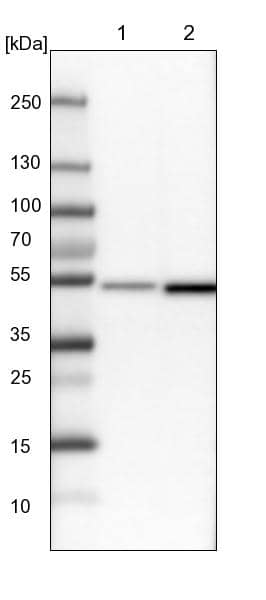
HAF017). A specific band was detected for RNF4 at approximately 34 kDa (as indicated). This experiment was conducted under reducing conditions and using Immunoblot Buffer Group 1." title="Western blot shows lysates of GM00719D human ataxia telangiectasia cell line either mock-treated (-) or exposed (+) to 10 Gy ionizing radiation (IR) and harvested after 1 hour, BaF3 mouse pro-B cell line, and LNCaP human prostate cancer cell line. PVDF membrane was probed with 1 µg/mL of Goat Anti-Human RNF4 Antigen Affinity-purified Polyclonal Antibody (Catalog # AF7964) followed by HRP-conjugated Anti-Goat IgG Secondary Antibody (Catalog # HAF017). A specific band was detected for RNF4 at approximately 34 kDa (as indicated). This experiment was conducted under reducing conditions and using Immunoblot Buffer Group 1." />
| Reactivity | HuSpecies Glossary |
| Applications | WB, IHC, ICC/IF |
| Clonality | Polyclonal |
| Host | Goat |
| Conjugate | Unconjugated |
| Immunogen | E.coli-derived recombinant human RNF4 Met1-Ile190 Accession # P78317 |
| Specificity | Detects human RNF4 in direct ELISAs and Western blots. |
| Source | N/A |
| Isotype | IgG |
| Clonality | Polyclonal |
| Host | Goat |
| Purity Statement | Antigen Affinity-purified |
| Innovator's Reward | Test in a species/application not listed above to receive a full credit towards a future purchase. |
| Dilutions |
|
|
| Publications |
|
| Storage | Use a manual defrost freezer and avoid repeated freeze-thaw cycles.
|
| Buffer | Lyophilized from a 0.2 μm filtered solution in PBS with Trehalose. *Small pack size (SP) is supplied either lyophilized or as a 0.2 µm filtered solution in PBS. |
| Reconstitution Instructions | Reconstitute at 0.2 mg/mL in sterile PBS. |
RNF4 (small nuclear ring finger protein, SNURF) is a RING-finger ubiquitin E3 ligase that ubiquitinates and mediates the proteasomal destruction of targets such as PML, PEA3, CENP1, and PARP1. In addition to the RING domain, RNF4 contains four SUMO-interacting motifs (SIMs) that function to recruit this ligase to poly-sumoylated substrates. RNF4 will autoubiquitinate in vitro, and will also ubiquitinate poly-SUMO chains.




![RNF4 Antibody [Unconjugated]](/sites/all/modules/enterprise-tech/et_datasheets/images/novus_guarantee.png "RNF4 Antibody [Unconjugated]")
Secondary Antibodies |
Isotype Controls |
The concentration calculator allows you to quickly calculate the volume, mass or concentration of your vial. Simply enter your mass, volume, or concentration values for your reagent and the calculator will determine the rest.
| Uniprot |
|


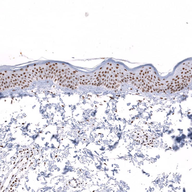
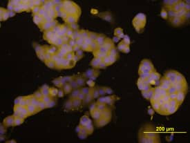
 NL001) and counterstained with DAPI (blue). Specific staining was localized to nuclei and cytoplasm. View our protocol for
  CTS008) and counterstained with hematoxylin (blue). Specific staining was localized to nuclei and cytoplasm. View our protocol for


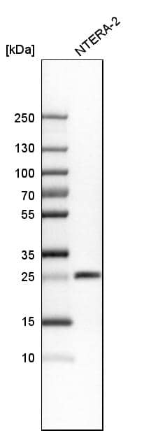
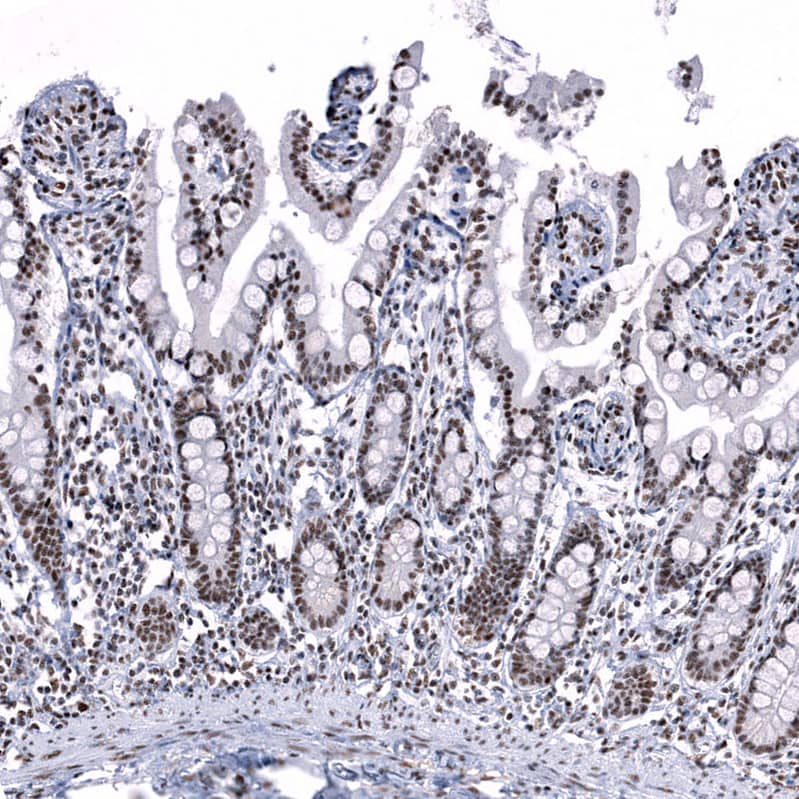
![Western Blot Androgen R/NR3C4 [p Ser213, p Ser210] Antibody (156C135.2) - BSA Free](https://images.novusbio.com/images/Androgen-R-NR3C4-p-Ser213-p-Ser210-Antibody-156C135-2-Western-Blot-NB100-56603-img0004.jpg)


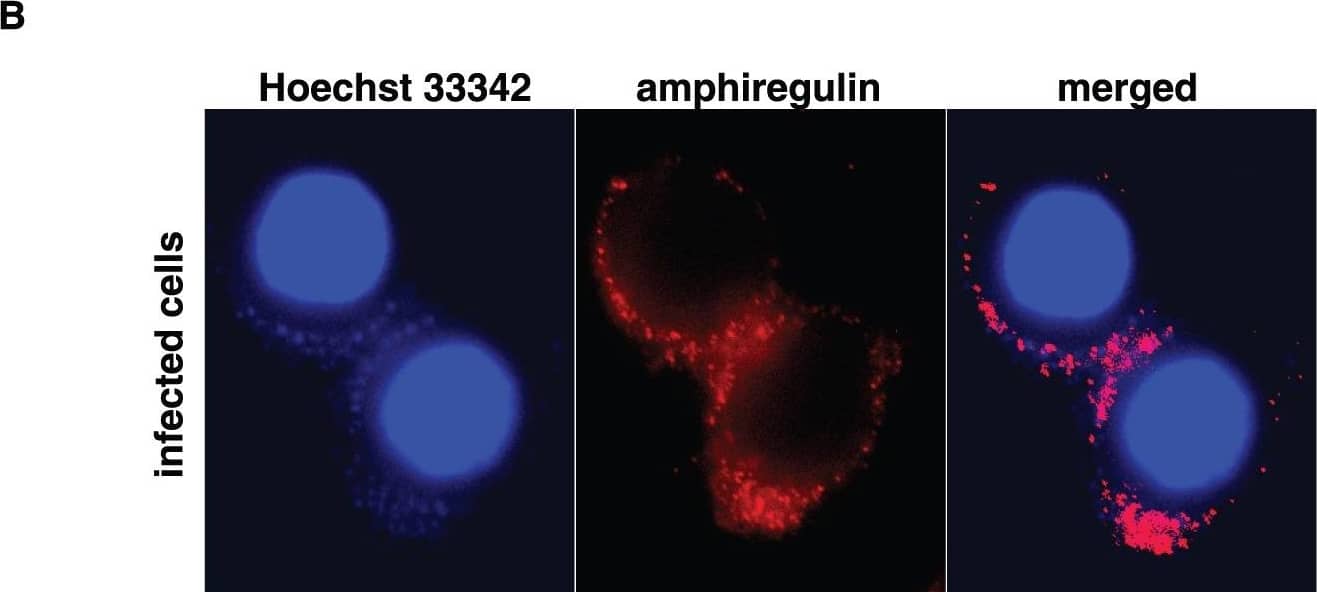
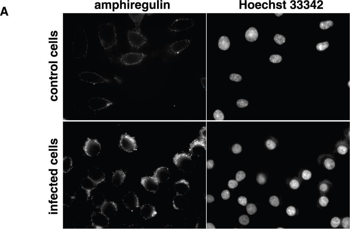


![SDS-Page IGF-II/IGF2 [Unconjugated]](https://images.novusbio.com/images/protein/IGF-II_292-G2_668.jpg)
![Bioactivity IGF-II/IGF2 [Unconjugated]](https://images.novusbio.com/images/protein/IGF-II_292-G2_669.jpg)