| Reactivity | MuSpecies Glossary |
| Applications | Binding Activity |
| Format | Carrier-Free |
| Details of Functionality | Measured by its binding ability in a functional ELISA. When Recombinant Mouse TL1A/TNFSF15 (Catalog # 1896-TL/CF) is immobilized at 1 μg/mL (100 μL/well), the concentration of Recombinant Mouse DR3/TNFRSF25 Fc Chimera that produces 50% of the optimal binding response is approximately 0.75-4 μg/mL |
||||||
| Source | Mouse myeloma cell line, NS0-derived mouse DR3/TNFRSF25 protein
|
||||||
| Accession # | |||||||
| N-terminal Sequence | No results obtained: Gln31 predicted |
||||||
| Structure / Form | Disulfide-linked homodimer |
||||||
| Protein/Peptide Type | Recombinant Proteins |
||||||
| Gene | Tnfrsf25 |
||||||
| Purity | >90%, by SDS-PAGE under reducing conditions and visualized by silver stain |
||||||
| Endotoxin Note | <0.1 EU per 1 μg of the protein by the LAL method. |
| Dilutions |
|
| Theoretical MW | 44.6 kDa (monomer). Disclaimer note: The observed molecular weight of the protein may vary from the listed predicted molecular weight due to post translational modifications, post translation cleavages, relative charges, and other experimental factors. |
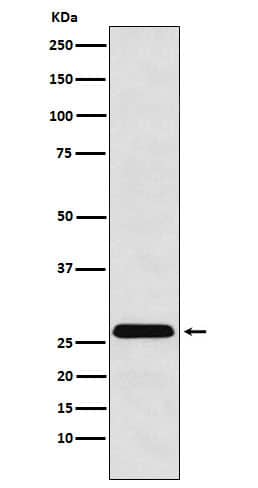
| SDS-PAGE | 55-65 kDa, reducing conditions |
| Storage | Use a manual defrost freezer and avoid repeated freeze-thaw cycles.
|
| Buffer | Lyophilized from a 0.2 μm filtered solution in PBS. |
| Purity | >90%, by SDS-PAGE under reducing conditions and visualized by silver stain |
| Reconstitution Instructions | Reconstitute at 100 μg/mL in sterile PBS. |
Death receptor 3 (DR3), also known as TNFRSF25, LARD, WSL-1, APO3, TRAMP, and TR3, is a 55 kDa TNF receptor superfamily protein that is predominantly expressed by lymphocytes. TNF receptor superfamily members have varying numbers of extracellular cysteine-rich domains (CRDs) with conserved cysteine spacing (1, 2). DR3 contains four CRDs and one cytoplasmic death domain (3, 4). Alternative splicing of mouse DR3 generates an isoform that lacks the fourth CRD and a secreted isoform that consisits of only the extracellular domain (ECD) (3). Human DR3 exists in at least eleven alternate splice forms (5). Within the ECD, mouse and human DR3 share 59% amino acid (aa) sequence identity. DR3 shares 20%-28% aa sequence identity with the ECD of death domain receptors DR5, DR6, EDAR, Fas, NGF R, and TNF RI. Naïve B and T cells preferentially express truncated soluble isoforms of DR3, whereas stimulated lymphocytes preferentially express transmembrane DR3 (5). TL1A/TNFSF15, a high affinity DR3 ligand which also exists in membrane bound and soluble forms, is expressed by activated endothelial cells and T cells (6, 7). TL1A additionally binds to DcR3/TNFRSF6B, a soluble decoy receptor that interferes with DR3 activation (8). DR3 signaling triggers either apoptosis or NF kappa B-induced anti-apoptotic effects depending on the cellular setting (9). Apoptosis is partially impaired during negative selection of thymocytes in DR3-null mice (10). TL1A interactions with DR3 augment T cell proliferation and proinflammatory cytokine secretion (6, 7, 11, 12). DR3 is upregulated by inflammatory stimulation of CCR9+ T cells, a T cell subset important in mucosal immunity (11). T cell and macrophage DR3 expression is prominent in several inflammatory disorders such as Crohn’s disease, inflammatory bowel disease, and atherosclerosis (7, 11-15). DR3 activation on IFN-gamma treated THP-1 cells induces the production of TNF-alpha , CXCL8, CCL2, MMP-1, -9, and -13 (14, 15).


")


The concentration calculator allows you to quickly calculate the volume, mass or concentration of your vial. Simply enter your mass, volume, or concentration values for your reagent and the calculator will determine the rest.


![SDS-Page TNF-alpha [Unconjugated]](https://images.novusbio.com/images/protein/TNF-alpha_210-TA_256.jpg)
![Bioactivity TNF-alpha [Unconjugated]](https://images.novusbio.com/images/protein/TNFalpha_210TA_1658.jpg)
![SEC-MALS TNF-alpha [Unconjugated]](https://images.novusbio.com/images/210-ta_recombinant-human-tnf-alpha-protein-sec-mals-35202312244..jpg)
![Neutralization TRAILR1/TNFRSF10A Antibody [Unconjugated]](https://images.novusbio.com/images/antibody/TRAIL_R1_AF347_Block_Neutralize_875.jpg)
![Western Blot TRAILR1/TNFRSF10A Antibody [Unconjugated]](https://images.novusbio.com/images/antibody/TRAIL_R1_AF347_Western_Blot_5303.jpg)
![Immunohistochemistry TRAILR1/TNFRSF10A Antibody [Unconjugated]](https://images.novusbio.com/images/antibody/TRAIL_R1_AF347_Immunohistochemistry_17355.jpg)


![Bioactivity CTLA-4 [Unconjugated]](https://images.novusbio.com/images/protein/CTLA4_7268CT_2293.jpg)
![N/A TNF RI/TNFRSF1A [HRP]](https://images.novusbio.com/images/elisa/DATA_TNF_RI_DRT100_ELISA_816.jpg)
![N/A TNF RI/TNFRSF1A [HRP]](https://images.novusbio.com/images/elisa/DATA_TNF_RI_DRT100_ELISA_815.jpg)
![N/A TNF RI/TNFRSF1A [HRP]](https://images.novusbio.com/images/elisa/TNF_RI_DRT100_ELISA_175.jpg)
![Immunohistochemistry Insulin Antibody (182410) [Unconjugated]](https://images.novusbio.com/images/antibody/mab1417_human-bovine-mouse-insulin-mab-clone-182410-immunohistochemistry-308202115145.jpg)
![Immunocytochemistry Insulin Antibody (182410) [Unconjugated]](https://images.novusbio.com/images/antibody/Insulin_MAB1417_Immunocytochemistry_9376.jpg)