| Reactivity | HuSpecies Glossary |
| Applications | Bioactivity |
| Format | Carrier-Free |
| Details of Functionality | Measured by its ability to induce adhesion of ATDC5 mouse chondrogenic cells. The ED50 for this effect is 0.5-2.0 μg/mL. Optimal dilutions should be determined by each laboratory for each application. Also measured by its ability to bind Recombinant Human TGF‑ beta 1 (Catalog # 240-B). |
| Source | Mouse myeloma cell line, NS0-derived human CILP-1 protein Met1-Arg720, with a C-terminal 6-His tag |
| Accession # | |
| N-terminal Sequence | Arg22 |
| Protein/Peptide Type | Recombinant Proteins |
| Gene | CILP |
| Purity | >95%, by SDS-PAGE under reducing conditions and visualized by silver stain |
| Endotoxin Note | <0.01 EU per 1 μg of the protein by the LAL method. |
| Dilutions |
|
|
| Theoretical MW | 78.8 kDa. Disclaimer note: The observed molecular weight of the protein may vary from the listed predicted molecular weight due to post translational modifications, post translation cleavages, relative charges, and other experimental factors. |
|
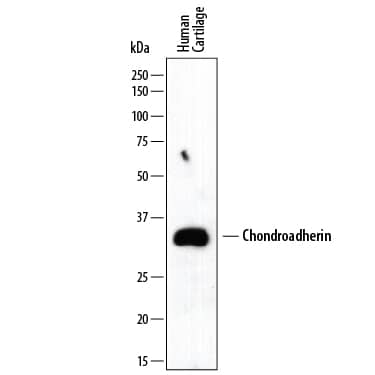
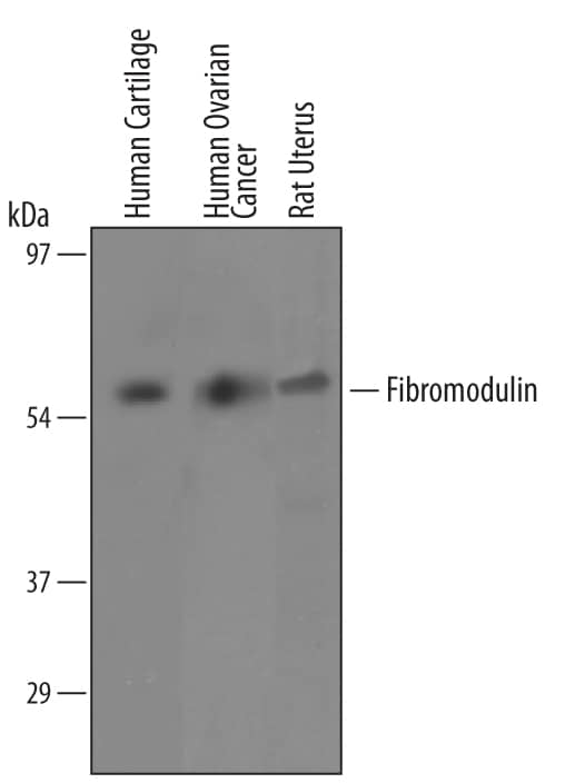
| SDS-PAGE | 85-95 kDa, reducing conditions |
|
| Publications |
|
| Storage | Use a manual defrost freezer and avoid repeated freeze-thaw cycles.
|
| Buffer | Lyophilized from a 0.2 μm filtered solution in PBS. |
| Purity | >95%, by SDS-PAGE under reducing conditions and visualized by silver stain |
| Reconstitution Instructions | Reconstitute at 100 μg/mL in PBS. |


The concentration calculator allows you to quickly calculate the volume, mass or concentration of your vial. Simply enter your mass, volume, or concentration values for your reagent and the calculator will determine the rest.


![N/A COMP/Thrombospondin-5 [HRP]](https://images.novusbio.com/images/elisa/COMP_DCMP0_ELISA_66.jpg)
![N/A COMP/Thrombospondin-5 [HRP]](https://images.novusbio.com/images/elisa/DATA_COMP_DCMP0_ELISA_632.jpg)
![Bioactivity IGF-I/IGF-1 [Unconjugated]](https://images.novusbio.com/images/protein/IGF-I_291-G1_41.jpg)
![Mass Spectrometry IGF-I/IGF-1 [Unconjugated]](https://images.novusbio.com/images/protein/IGF-I_291-G1_42.jpg)
![SEC-MALS IGF-I/IGF-1 [Unconjugated]](https://images.novusbio.com/images/291-g1_recombinant-human-igf-i-igf-1-protein-cf-sec-mals-224202691859.jpg)
![Bioactivity TGF-beta 1 [Unconjugated]](https://images.novusbio.com/images/protein/7754-bhcf_recombinant-human-tgf-beta-1-human-cell-expressed-cf-bioactivity-1811202013946.jpg)


![ENPP-2/Autotaxin [Unconjugated]](/sites/all/modules/enterprise-tech/et_datasheets/images/novus_guarantee.png "ENPP-2/Autotaxin [Unconjugated]")
![Bioactivity BMP-2 [Unconjugated]](https://images.novusbio.com/images/protein/355-bm_recombinant-human-mouse-rat-bmp-2-protein-bioactivity-181120209742.jpg)