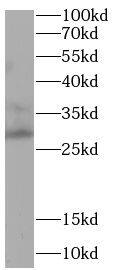
 followed by HRP-conjugated Anti-Goat IgG Secondary Antibody (HAF017). A specific band was detected for MANF at approximately 17 kDa (as indicated). This experiment was conducted under reducing conditions and using Western Blot Buffer Group 1.")
| Reactivity | HuSpecies Glossary |
| Applications | WB, Simple Western, KO |
| Clonality | Polyclonal |
| Host | Goat |
| Conjugate | Unconjugated |
| Concentration | LYOPH |
| Immunogen | Mouse myeloma cell line NS0-derived recombinant human MANF Leu22-Leu179 Accession # P55145 |
| Specificity | Detects human MANF in direct ELISAs and Western blots. |
| Source | N/A |
| Isotype | IgG |
| Clonality | Polyclonal |
| Host | Goat |
| Gene | MANF |
| Purity Statement | Antigen Affinity-purified |
| Innovator's Reward | Test in a species/application not listed above to receive a full credit towards a future purchase. |
| Dilutions |
|
|
| Publications |
|
| Storage | Use a manual defrost freezer and avoid repeated freeze-thaw cycles.
|
| Buffer | Lyophilized from a 0.2 μm filtered solution in PBS with Trehalose. *Small pack size (SP) is supplied either lyophilized or as a 0.2 µm filtered solution in PBS. |
| Preservative | No Preservative |
| Concentration | LYOPH |
| Reconstitution Instructions | Reconstitute at 0.2 mg/mL in sterile PBS. |


![Western Blot Angiopoietin-like Protein 4/ANGPTL4 Antibody [Unconjugated]](https://images.novusbio.com/images/af3485_human-primate-angiopoietin-like-4-affinity-purified-polyl-ab-western-blot-12122025555552.jpg)
![Western Blot Angiopoietin-like Protein 4/ANGPTL4 Antibody [Unconjugated]](https://images.novusbio.com/images/af3485_human-primate-angiopoietin-like-4-affinity-purified-polyl-ab-western-blot-121220258175720.jpg)
![Western Blot Angiopoietin-like Protein 4/ANGPTL4 Antibody [Unconjugated]](https://images.novusbio.com/images/af3485_human-primate-angiopoietin-like-4-affinity-purified-polyl-ab-western-blot-121220257534813.jpg)
![MANF Antibody [Unconjugated]](/sites/all/modules/enterprise-tech/et_datasheets/images/novus_guarantee.png "MANF Antibody [Unconjugated]")
Secondary Antibodies |
Isotype Controls |
The concentration calculator allows you to quickly calculate the volume, mass or concentration of your vial. Simply enter your mass, volume, or concentration values for your reagent and the calculator will determine the rest.


 using 10 µg/mL of Goat Anti-Human MANF Antigen Affinity-purified Polyclonal Antibody (Catalog # AF3748) followed by 1:50 dilution of HRP-conjugated Anti-Goat IgG Secondary Antibody (Catalog # HAF109). This experiment was conducted under reducing conditions and using the 12-230 kDa separation system.")
. PVDF membrane was probed with 1 µg/mL of Goat Anti-Human MANF Antigen Affinity-purified Polyclonal Antibody (Catalog # AF3748) followed by HRP-conjugated Anti-Goat IgG Secondary Antibody (Catalog # HAF017). A specific band was detected for MANF at approximately 17 kDa (as indicated) in the parental HEK293T cell line, but is not detectable in knockout HEK293Tcell line. GAPDH (Catalog # AF5718) is shown as a loading control. This experiment was conducted under reducing conditions and using Immunoblot Buffer Group 1.")

![Western Blot Angiopoietin-like Protein 2/ANGPTL2 Antibody [Unconjugated]](https://images.novusbio.com/images/antibody/Angiopoietinlike_Protein_2_AF1444_Western_Blot_19840.jpg)


![Bioactivity GDNF [Unconjugated]](https://images.novusbio.com/images/protein/GDNF_212GD_2846.jpg)
![SDS-Page GDNF [Unconjugated]](https://images.novusbio.com/images/protein/GDNF_212-GD_62.jpg)
![SEC-MALS GDNF [Unconjugated]](https://images.novusbio.com/images/212-gd_recombinant-human-gdnf-protein-sec-mals-25202393740..jpg)

![Western Blot COUP-TF II/NR2F2 Antibody (H7147) [Unconjugated]](https://images.novusbio.com/images/pp-h7147-00_human-coup-tf-ii-nr2f2-mab-clone-h7147-8120255552858.jpg)
![Knockdown Validated COUP-TF II/NR2F2 Antibody (H7147) [Unconjugated]](https://images.novusbio.com/images/pp-h7147-00_human-coup-tf-ii-nr2f2-mab-clone-h7147-41202412505337.jpg)
![Western Blot COUP-TF II/NR2F2 Antibody (H7147) [Unconjugated]](https://images.novusbio.com/images/pp-h7147-00_human-coup-tf-ii-nr2f2-mab-clone-h7147-8120255541794.jpg)


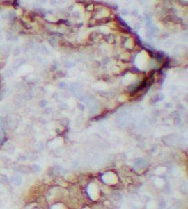
![Immunohistochemistry PITX2 Antibody [Unconjugated]](https://images.novusbio.com/images/antibody/PITX2_AF7388_Immunohistochemistry_12162.jpg)

 or Normal Goat IgG Isotype Control Antibody (Catalog # AB-108-C, open histogram), followed by Phycoerythrin-conjugated Anti-Goat IgG Secondary Antibody (Catalog # F0107).")