 CTS013). Tissue was stained with the Anti-Goat HRP-DAB Cell & Tissue Staining Kit (brown; Catalog # CTS008) and counterstained with hematoxylin (blue). View our protocol for Chromogenic IHC Staining of Paraffin-embedded Tissue Sections." title="GRB2 was detected in immersion fixed paraffin-embedded sections of human breast cancer tissue using 10 µg/mL Human GRB2 Antigen Affinity-purified Polyclonal Antibody (Catalog # AF3846) overnight at 4 °C. Before incubation with the primary antibody tissue was subjected to heat-induced epitope retrieval using Antigen Retrieval Reagent-Basic (Catalog # CTS013). Tissue was stained with the Anti-Goat HRP-DAB Cell & Tissue Staining Kit (brown; Catalog # CTS008) and counterstained with hematoxylin (blue). View our protocol for Chromogenic IHC Staining of Paraffin-embedded Tissue Sections." />
| Reactivity | Hu, Mu, RtSpecies Glossary |
| Applications | WB, IHC |
| Clonality | Polyclonal |
| Host | Goat |
| Conjugate | Unconjugated |
| Concentration | LYOPH |
| Immunogen | E. coli-derived recombinant human GRB2 Met1-Val217 Accession # P62993 |
| Specificity | Detects human, mouse, and rat GRB2. |
| Source | N/A |
| Isotype | IgG |
| Clonality | Polyclonal |
| Host | Goat |
| Gene | GRB2 |
| Purity Statement | Antigen Affinity-purified |
| Innovator's Reward | Test in a species/application not listed above to receive a full credit towards a future purchase. |
| Dilutions |
|
|
| Publications |
|
| Storage | Use a manual defrost freezer and avoid repeated freeze-thaw cycles.
|
| Buffer | Lyophilized from a 0.2 μm filtered solution in PBS with Trehalose. *Small pack size (SP) is supplied either lyophilized or as a 0.2 µm filtered solution in PBS. |
| Preservative | No Preservative |
| Concentration | LYOPH |
| Reconstitution Instructions | Reconstitute at 0.2 mg/mL in sterile PBS. |
GRB-2 (growth factor receptor-bound protein 2), an adaptor protein involved in signal transduction, contains a central SH2 domain flanked by two SH3 domains. The SH2 domain binds to phosphotyrosine residues in RTKs such as PDGF and EGF, non-RTKs such as Bcr/Abl and FAK, and docking proteins such as FRS-2 and Gab1. The SH3 domains associate with proline rich motifs on the guanine nucleotide releasing factor, Sos, stimulating GTP binding to Ras, which in turn activates MAPK and other signaling pathways. GRB-2 also participates in the endocytosis of EGFR through its recruitment of Cbl. Tyrosine phosphorylation of GRB-2 SH3 domains reduces binding to Sos and negatively regulates downstream signaling pathways including Ras, JNK and MAPK.


![Immunocytochemistry EGFR Antibody [Unconjugated]](https://images.novusbio.com/images/antibody/EGF_R_AF231_Immunocytochemistry__Immunofluorescence_21143.jpg)
![Flow Cytometry EGFR Antibody [Unconjugated]](https://images.novusbio.com/images/antibody/EGF_R_AF231_Flow_Cytometry_20401.jpg)
![Western Blot EGFR Antibody [Unconjugated]](https://images.novusbio.com/images/antibody/EGF_R_AF231_Western_Blot_19925.jpg)


![GRB2 Antibody [Unconjugated]](/sites/all/modules/enterprise-tech/et_datasheets/images/novus_guarantee.png "GRB2 Antibody [Unconjugated]")
Secondary Antibodies |
Isotype Controls |
The concentration calculator allows you to quickly calculate the volume, mass or concentration of your vial. Simply enter your mass, volume, or concentration values for your reagent and the calculator will determine the rest.


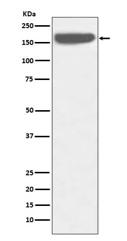
 HAF109). A specific band was detected for GRB2 at approximately 27 kDa (as indicated). This experiment was conducted under reducing conditions and using
 ![Western Blot Src [p Tyr419] Antibody [Unconjugated]](https://images.novusbio.com/images/antibody/Src_AF2685_Western_Blot_5209.jpg)
![Immunohistochemistry Src [p Tyr419] Antibody [Unconjugated]](https://images.novusbio.com/images/af2685_human-phospho-src-y419-affinity-purified-polyclonal-ab-412024124022.jpg)
![Immunohistochemistry Src [p Tyr419] Antibody [Unconjugated]](https://images.novusbio.com/images/antibody/Src_AF2685_Immunohistochemistry_9943.jpg)
![Western Blot ERK2 Antibody [Unconjugated]](https://images.novusbio.com/images/antibody/ERK2_AF1230_Western_Blot_5097.jpg)
![Knockout Validated ERK2 Antibody [Unconjugated]](https://images.novusbio.com/images/antibody/ERK2_AF1230_Knockout_Validated_22864.jpg)
![Immunohistochemistry ERK2 Antibody [Unconjugated]](https://images.novusbio.com/images/antibody/ERK2_AF1230_Immunohistochemistry_20696.jpg)
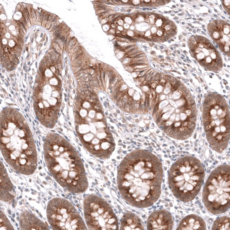
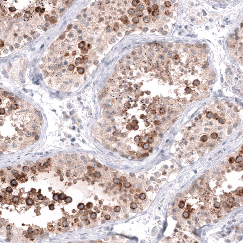
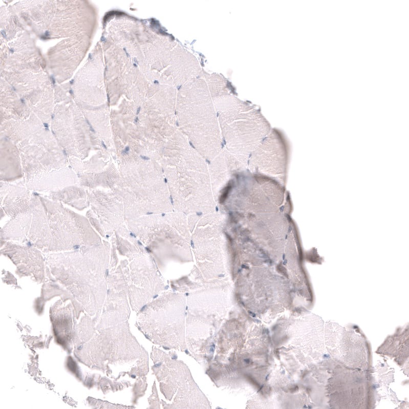


![Bioactivity EGF [Unconjugated]](https://images.novusbio.com/images/protein/EGF_236EG_1570.jpg)
![Cell Culture EGF [Unconjugated]](https://images.novusbio.com/images/236-eg_recombinant-human-egf-protein-cf-25202394526.jpg)
![Cell Culture EGF [Unconjugated]](https://images.novusbio.com/images/236-eg_recombinant-human-egf-protein-cf-bioactivity-25202394053.jpg)
![Immunocytochemistry CD55/DAF Antibody [Unconjugated]](https://images.novusbio.com/images/antibody/CD55_AF2009_Immunocytochemistry__Immunofluorescence_23052.jpg)
![Immunohistochemistry CD55/DAF Antibody [Unconjugated]](https://images.novusbio.com/images/antibody/CD55_AF2009_Immunohistochemistry_6728.jpg)


![Bioactivity CTLA-4 [Unconjugated]](https://images.novusbio.com/images/protein/CTLA4_7268CT_2293.jpg)
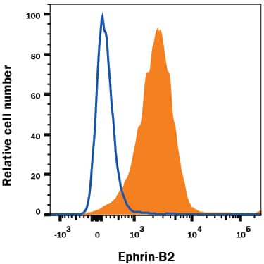
![SDS-Page: Recombinant Human GRB2 Protein [H00002885-P01] - 12.5% SDS-PAGE Stained with Coomassie Blue.](https://images.novusbio.com/images/Recombinant-Human-GRB2-Protein-SDS-Page-H00002885-P01-img0004.jpg "SDS-Page: Recombinant Human GRB2 Protein [H00002885-P01] - 12.5% SDS-PAGE Stained with Coomassie Blue.")

