| Immunogen | Recombinant human Complement 4d protein |
| Localization | Intracytoplasmic vacuoles of endothelial cells and Secreted |
| Marker | Acute Humoral Rejection Marker |
| Specificity | This MAb is specific to Complement 4d (C4d) and it reacts with the secreted as well as cell-bound C4d.C4d is a degradation product of the activated complement factor C4b. Complement 4b is typically activated by binding of Abs to specific target molecules. Following activation and degradation of the C4 molecule, thio-ester groups are exposed, which allow transient, covalent binding of the degradation product Complement 4d to endothelial cell surfaces and extracellular matrix components of vascular basement membranes near the sites of C4 activation. The presence of C4d in peritubular capillaries is a key indicator for acute humoral (i.e. antibody-mediated) rejection of kidney, heart, pancreas and lung allografts. As an established marker of antibody-mediated acute renal allograft rejection and its proclivity for endothelium, this component can be detected in peritubular capillaries in chronic renal allograft rejection as well as hyperacute rejection, acute vascular rejection, acute cellular rejection, and borderline rejection. It has been shown to be a significant predictor of transplant kidney graft survival. Anti-C4d, combined with anti-C3d, can be utilized as a tool for diagnosis of allograft rejection that may warrant a prompt and aggressive anti-rejection treatment. |
| Isotype | IgG1 |
| Clonality | Monoclonal |
| Host | Mouse |
| Gene | C4B |
| Purity | Protein A or G purified |
| Innovator's Reward | Test in a species/application not listed above to receive a full credit towards a future purchase. |
| Dilutions |
|
| Application Notes | Hu-chromosome location: 6p21.3 Molecular weight of antigen: 192kDa (predicted) |
| Theoretical MW | 192 kDa. Disclaimer note: The observed molecular weight of the protein may vary from the listed predicted molecular weight due to post translational modifications, post translation cleavages, relative charges, and other experimental factors. |
| Storage | Store at 4C. |
| Buffer | 10mM PBS and 0.05% BSA |
| Preservative | 0.05% Sodium Azide |
| Concentration | 0.2 mg/ml |
| Purity | Protein A or G purified |


![Simple Western Albumin Antibody (188835) [Unconjugated] - Serum](https://images.novusbio.com/images2/Albumin_MAB1455_Simple_Western_21888.jpg)
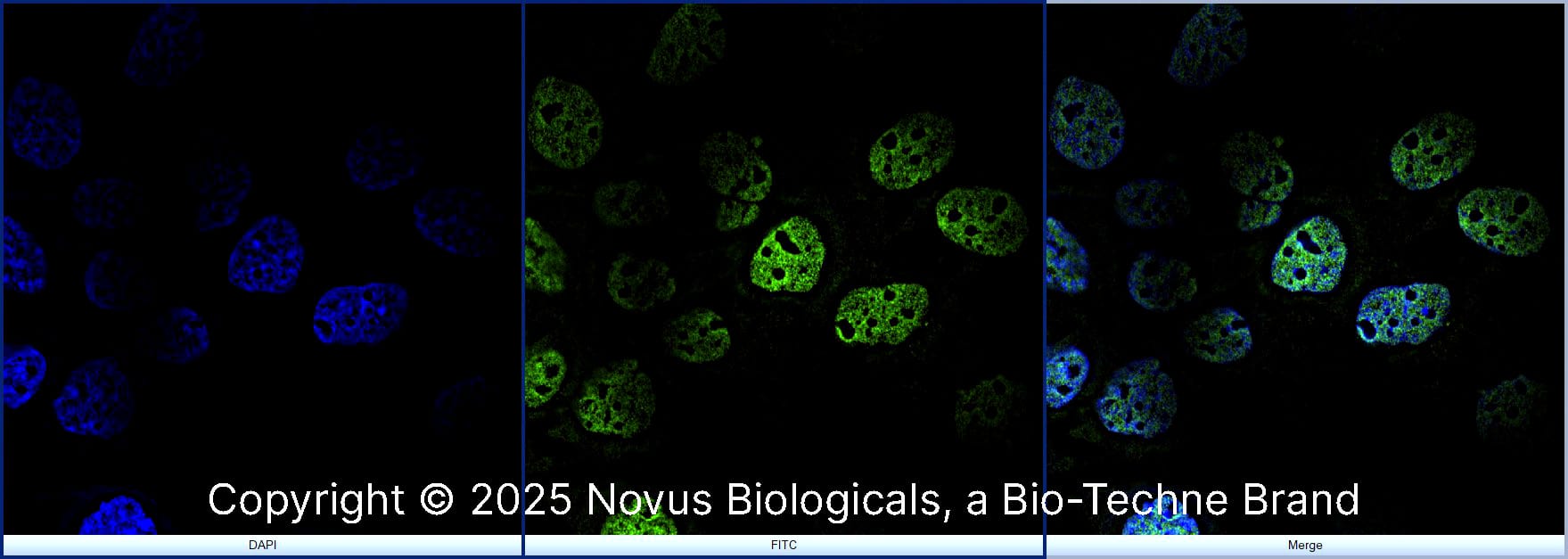
![Immunocytochemistry Albumin Antibody (188835) [Unconjugated] - Serum](https://images.novusbio.com/images2/Albumin_MAB1455_Immunocytochemistry__Immunofluorescence_17777.jpg)
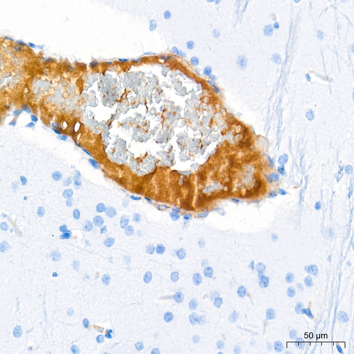
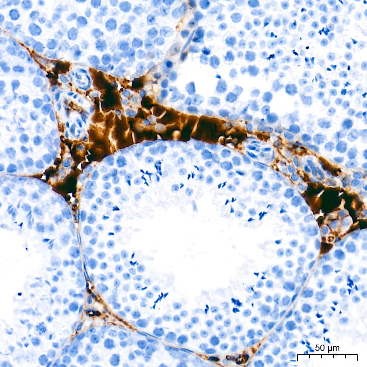
![Immunohistochemistry Albumin Antibody (188835) [Unconjugated] - Serum](https://images.novusbio.com/images2/Albumin_MAB1455_Immunohistochemistry_23317.jpg)


")
Secondary Antibodies |
Isotype Controls |
The concentration calculator allows you to quickly calculate the volume, mass or concentration of your vial. Simply enter your mass, volume, or concentration values for your reagent and the calculator will determine the rest.
| Gene Symbol | C4B |


![Immunocytochemistry Integrin alpha 2/CD49b Antibody (HAS3) [Unconjugated]](https://images.novusbio.com/images2/Integrin_alpha_2_MAB1233_Immunocytochemistry__Immunofluorescence_19196.jpg)
![Flow Cytometry Integrin alpha 2/CD49b Antibody (HAS3) [Unconjugated]](https://images.novusbio.com/images2/Integrin_alpha_2_MAB1233_Flow_Cytometry_19476.jpg)
![Immunohistochemistry CFTR Antibody (24-1) [Unconjugated] - C-terminus](https://images.novusbio.com/images2/CFTR_MAB25031_Immunohistochemistry_6973.jpg)
![Immunohistochemistry CFTR Antibody (24-1) [Unconjugated] - C-terminus](https://images.novusbio.com/images2/CFTR_MAB25031_Immunohistochemistry_9671.jpg)
![Immunoprecipitation CFTR Antibody (24-1) [Unconjugated] - C-terminus](https://images.novusbio.com/images/mab25031_human-cftr-c-terminus-mab-clone-24-1-41202412513815.jpg)


![Immunohistochemistry Complement Factor H Antibody [Unconjugated]](https://images.novusbio.com/images2/Complement_Factor_H_AF4779_Immunohistochemistry_7182.jpg)


![Immunohistochemistry CD55/DAF Antibody [Unconjugated]](https://images.novusbio.com/images2/CD55_AF2009_Immunohistochemistry_6728.jpg)
![Immunocytochemistry CD55/DAF Antibody [Unconjugated]](https://images.novusbio.com/images2/CD55_AF2009_Immunocytochemistry__Immunofluorescence_23052.jpg)
![Bioactivity CTLA-4 [Unconjugated]](https://images.novusbio.com/images2/CTLA4_7268CT_2293.jpg)


