 tissue. PVDF Membrane was probed with 1 µg/mL of Goat Anti-Human Cadherin-13 Antigen Affinity-purified Polyclonal Antibody (Catalog # AF3264) followed by HRP-conjugated Anti-Goat IgG Secondary Antibody (Catalog # HAF019). A specific band was detected for Cadherin-13 at approximately 105 kDa (as indicated). This experiment was conducted under reducing conditions and using Immunoblot Buffer Group 8.")
| Applications | WB, Simple Western, Flow, IHC, CyTOF-ready, ICC/IF |
| Clonality | Polyclonal |
| Host | Goat |
| Conjugate | Unconjugated |
| Concentration | LYOPH |
| Immunogen | Mouse myeloma cell line NS0-derived recombinant human Cadherin-13 Glu23-Ala692 Accession # P55290 |
| Specificity | Detects human Cadherin-13 in direct ELISAs and Western blots. In direct ELISAs, approximately 100% cross-reactivity with recombinant mouse Cadherin-13 is observed, and less than 10% cross-reactivity with recombinant human (rh) N-Cadherin is observed, and less than 2% cross-reactivity with rhCadherin-8, rhCadherin-11, rhCadherin-17, rhE-Cadherin, rhP-Cadherin, rhVE-Cadherin and rhR-Cadherin is observed. |
| Source | N/A |
| Isotype | IgG |
| Clonality | Polyclonal |
| Host | Goat |
| Gene | CDH13 |
| Purity Statement | Antigen Affinity-purified |
| Innovator's Reward | Test in a species/application not listed above to receive a full credit towards a future purchase. |
| Dilutions |
|
|
| Publications |
|
| Storage | Use a manual defrost freezer and avoid repeated freeze-thaw cycles.
|
| Buffer | Lyophilized from a 0.2 μm filtered solution in PBS with Trehalose. See Certificate of Analysis for details. *Small pack size (-SP) is supplied either lyophilized or as a 0.2 µm filtered solution in PBS. |
| Preservative | No Preservative |
| Concentration | LYOPH |
| Reconstitution Instructions | Reconstitute at 0.2 mg/mL in sterile PBS. For liquid material, refer to CoA for concentration. |


![Immunocytochemistry E-Cadherin Antibody [Unconjugated]](https://images.novusbio.com/images/antibody/ECadherin_AF748_Immunocytochemistry_16573.jpg)
![Western Blot E-Cadherin Antibody [Unconjugated]](https://images.novusbio.com/images/antibody/ECadherin_AF748_Western_Blot_19846.jpg)
![Immunocytochemistry E-Cadherin Antibody [Unconjugated]](https://images.novusbio.com/images/antibody/ECadherin_AF748_Immunocytochemistry_16572.jpg)


Secondary Antibodies |
Isotype Controls |
The concentration calculator allows you to quickly calculate the volume, mass or concentration of your vial. Simply enter your mass, volume, or concentration values for your reagent and the calculator will determine the rest.


 for 3 hours at room temperature. Cells were stained with the NorthernLights™ 557-conjugated Anti-Goat IgG Secondary Antibody (red; Catalog # NL001) and counter-stained with DAPI (blue). View our protocol for Fluorescent ICC Staining of Cells on Coverslips.")
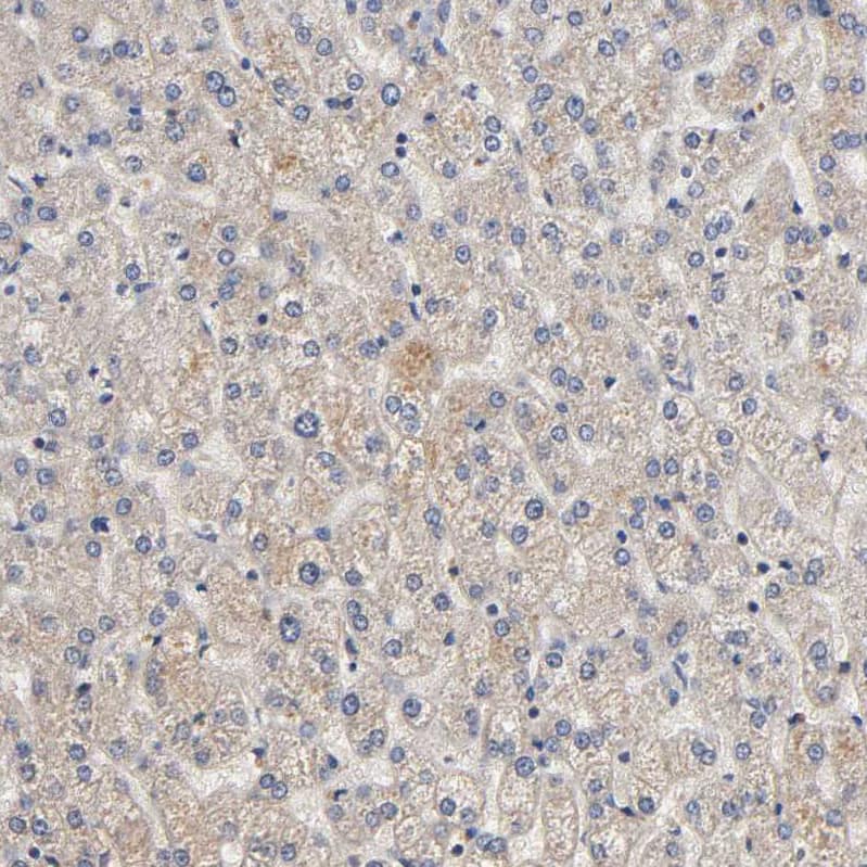
 at 0.1 µg/mL overnight at 4 °C. Tissue was stained using the Anti-Goat HRP-DAB Cell & Tissue Staining Kit (brown; Catalog # CTS008) and counterstained with hematoxylin (blue). Specific staining was localized to plasma membranes of keratinocytes. View our protocol for Chromogenic IHC Staining of Paraffin-embedded Tissue Sections.")
 tissue, loaded at 0.2 mg/mL. A specific band was detected for Cadherin-13 at approximately 114 kDa (as indicated) using 10 µg/mL of Goat Anti-Human Cadherin-13 Antigen Affinity-purified Polyclonal Antibody (Catalog # AF3264) followed by 1:50 dilution of HRP-conjugated Anti-Goat IgG Secondary Antibody (Catalog # HAF109). This experiment was conducted under reducing conditions and using the 12-230 kDa separation system.")
 Cell lysates from 46BR.1G1 and 7A3 cells were analyzed by Western blotting with anti-cadherin 13, anti-cadherin 4, and anti-alpha -tubulin antibodies. (B) Quantification of the assay was performed by densitometric analysis with NIH ImageJ 1.43 program. Bars show mean ± SEM of three independent experiments. Image collected and cropped by CiteAb from the following publication (//dx.plos.org/10.1371/journal.pone.0130561), licensed under a CC-BY license. Not internally tested by R&D Systems.")
, licensed under a CC-BY license. Not internally tested by R&D Systems.")
 Western blot analysis of total cell lysates. UV-F2 cells cultured in DMEM containing 5% serum from adiponectin knockout mice were treated with or without high molecular weight adiponectin (HMW-APN) (10 μg/mL) and roxadustat (Roxa) (50 μM) for 48 h (n = 3 for each group). (B) Western blot analysis of EVs isolated from cell culture medium by differential ultracentrifugation. UV-F2 cells cultured in FBS-free Advanced DMEM were treated with or without HMW-APN (20 μg/mL) and roxadustat (50 μM) for 48 h (n = 3 for each group). Alix, TSG101, and syntenin were evaluated as EV markers. (C) Western blot analysis of total cell lysates. UV-F2 cells transfected control (Cont) or T-cadherin (T-cad) siRNA were cultured in DMEM containing 5% serum from adiponectin knockout mice with or without HMW-APN (20 μg/mL) and roxadustat (50 μM) for 48 h (n = 3 for each group). (D) Western blot analysis of EVs isolated from cell culture medium. UV-F2 cells transfected Cont or T-cad siRNA were cultured in FBS-free Advanced DMEM with or without HMW-APN (20 μg/mL) and roxadustat (50 μM) for 48 h (n = 3). Data are means ± SEMs. *p AB-108-C, open histogram) followed by Allophycocyanin-conjugated Anti-Goat IgG Secondary Antibody (Catalog # F0108). View our protocol for Staining Membrane-associated Proteins.")
![Western Blot CD40/TNFRSF5 Antibody [Unconjugated]](https://images.novusbio.com/images/af632_human-cd40-tnfrsf5-affinity-purified-polyclonal-ab-822024950191.jpg)
![Western Blot CD40/TNFRSF5 Antibody [Unconjugated]](https://images.novusbio.com/images/af632_human-cd40-tnfrsf5-affinity-purified-polyclonal-ab-44202415425047.jpg)
![Simple Western CD40/TNFRSF5 Antibody [Unconjugated]](https://images.novusbio.com/images/antibody/af632_human-cd40-tnfrsf5-affinity-purified-polyclonal-ab-simple-western-58202111562.jpg)


![SDS-Page TNF-alpha [Unconjugated]](https://images.novusbio.com/images/protein/TNF-alpha_210-TA_256.jpg)
![Bioactivity TNF-alpha [Unconjugated]](https://images.novusbio.com/images/protein/TNFalpha_210TA_1658.jpg)
![SEC-MALS TNF-alpha [Unconjugated]](https://images.novusbio.com/images/210-ta_recombinant-human-tnf-alpha-protein-sec-mals-35202312244..jpg)
![Simple Western c-Rel Antibody [Unconjugated]](https://images.novusbio.com/images/antibody/cRel_AF2699_Simple_Western_16940.jpg)
![Western Blot c-Rel Antibody [Unconjugated]](https://images.novusbio.com/images/antibody/af2699_human-mouse-c-rel-affinity-purified-polyclonal-ab-western-blot-2102024121216..jpg)
![Western Blot WNK1 Antibody [Unconjugated]](https://images.novusbio.com/images/antibody/WNK1_AF2849_Western_Blot_5218.jpg)
![Immunocytochemistry Synaptotagmin 1 Antibody (ASV48) [Unconjugated]](https://images.novusbio.com/images/antibody/Synaptotagmin-1_MAB4364_Immunocytochemistry_11323.jpg)
![Knockout Validated Synaptotagmin 1 Antibody (ASV48) [Unconjugated]](https://images.novusbio.com/images/antibody/mab4364_rat-synaptotagmin-1-mab-clone-asv48-knockout-validated-2862024122433..png)
![Knockout Validated Synaptotagmin 1 Antibody (ASV48) [Unconjugated]](https://images.novusbio.com/images/antibody/mab4364_rat-synaptotagmin-1-mab-clone-asv48-knockout-validated-286202412275.jpg)


![Western Blot RB1 Antibody (607121) [Unconjugated]](https://images.novusbio.com/images/mab6495_human-rb1-mab-clone-607121-812025548577.jpg)
![Simple Western RB1 Antibody (607121) [Unconjugated]](https://images.novusbio.com/images/antibody/RB1_MAB6495_Simple_Western_16629.jpg)
![Western Blot RB1 Antibody (607121) [Unconjugated]](https://images.novusbio.com/images/antibody/RB1_MAB6495_Western_Blot_10216.jpg)
![N/A Adiponectin/Acrp30 [HRP]](https://images.novusbio.com/images/elisa/DATA_Adiponectin_DRP300_ELISA_812.jpg)
![N/A Adiponectin/Acrp30 [HRP]](https://images.novusbio.com/images/elisa/Adiponectin_DRP300_ELISA_173.jpg)
![N/A Adiponectin/Acrp30 [HRP]](https://images.novusbio.com/images/elisa/DATA_Adiponectin_DRP300_ELISA_813.jpg)


