DR555RB) and counterstained with DAPI (blue; Lunaphore Catalog #
DR100). Specific staining was localized to the cytoplasm of Kupffer cells. Protocol available in
COMET™ Panel Builder." class="big_lightbox">
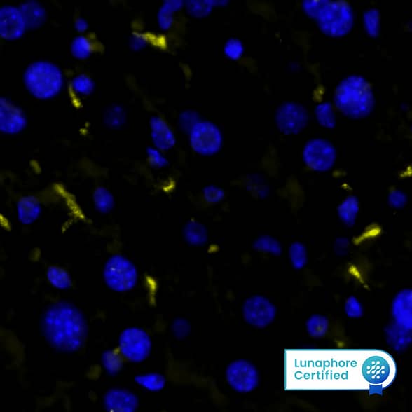
DR555RB) and counterstained with DAPI (blue; Lunaphore Catalog #
DR100). Specific staining was localized to the cytoplasm of Kupffer cells. Protocol available in
COMET™ Panel Builder." alt="CD68 was detected in immersion fixed paraffin-embedded sections of mouse Liver using Rabbit Anti-Mouse CD68, Monoclonal Antibody (Catalog #MAB101141) at 5ug/mL at 37 ° Celsius for 4 minutes. Before incubation with the primary antibody, tissue underwent an all-in-one dewaxing and antigen retrieval preprocessing using PreTreatment Module (PT Module) and Dewax and HIER Buffer H (pH 9; Epredia Catalog # TA-999-DHBH). Tissue was stained using the Alexa Fluor™ Plus 555 Goat anti-Rabbit IgG Secondary Antibody at 1:100 at 37 ° Celsius for 2 minutes. (Yellow; Lunaphore Catalog #
DR555RB) and counterstained with DAPI (blue; Lunaphore Catalog #
DR100). Specific staining was localized to the cytoplasm of Kupffer cells. Protocol available in
COMET™ Panel Builder." class="big_thumb" />
DR647RB) and counterstained with DAPI (blue; Lunaphore Catalog #
DR100). Specific staining was localized to the cytoplasm. Protocol available in
COMET™ Panel Builder." class="big_lightbox">

DR647RB) and counterstained with DAPI (blue; Lunaphore Catalog #
DR100). Specific staining was localized to the cytoplasm. Protocol available in
COMET™ Panel Builder." class="big_thumb" />
VC003). Before incubation with the primary antibody, tissue was subjected to heat-induced epitope retrieval using Antigen Retrieval Reagent-Basic (Catalog #
CTS013). Tissue was stained using DAB (brown) and counterstained with hematoxylin (blue). Specific staining was localized to Kupffer cells. View our protocol for
IHC Staining with VisUCyte HRP Polymer Detection Reagents." class="big_lightbox">
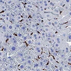
VC003). Before incubation with the primary antibody, tissue was subjected to heat-induced epitope retrieval using Antigen Retrieval Reagent-Basic (Catalog #
CTS013). Tissue was stained using DAB (brown) and counterstained with hematoxylin (blue). Specific staining was localized to Kupffer cells. View our protocol for
IHC Staining with VisUCyte HRP Polymer Detection Reagents." class="big_thumb" />
MAB1050, open histogram) followed by Phycoerythrin-conjugated Anti-Rabbit IgG Secondary Antibody (Catalog #
F0110). To facilitate intracellular staining, cells were fixed with Flow Cytometry Fixation Buffer (1x) (Catalog #
FC004) and permeabilized with ice-cold methanol." class="big_lightbox">
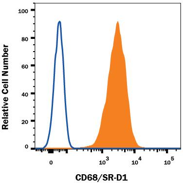
MAB1050, open histogram) followed by Phycoerythrin-conjugated Anti-Rabbit IgG Secondary Antibody (Catalog #
F0110). To facilitate intracellular staining, cells were fixed with Flow Cytometry Fixation Buffer (1x) (Catalog #
FC004) and permeabilized with ice-cold methanol." class="big_thumb" />
 VC001) or the HRP-conjugated Anti-Mouse IgG Secondary Antibody (Catalog # HAF007). Before incubation with the primary antibody, tissue was subjected to heat-induced epitope retrieval using VisUCyte Antigen Retrieval Reagent-Basic (Catalog # VCTS021). Tissue was stained using DAB (brown) and counterstained with hematoxylin (blue). Specific staining was localized to the cell surface of Kupffer cells. View our protocol for IHC Staining with VisUCyte HRP Polymer Detection Reagents." alt="CD68/SR‑D1 was detected in immersion fixed paraffin-embedded sections of human liver using Mouse Anti-Human CD68/SR‑D1 Monoclonal Antibody (Catalog # MAB20401) at 0.1 µg/ml for 1 hour at room temperature followed by incubation with the Anti-Mouse IgG VisUCyte™ HRP Polymer Antibody (Catalog # VC001) or the HRP-conjugated Anti-Mouse IgG Secondary Antibody (Catalog # HAF007). Before incubation with the primary antibody, tissue was subjected to heat-induced epitope retrieval using VisUCyte Antigen Retrieval Reagent-Basic (Catalog # VCTS021). Tissue was stained using DAB (brown) and counterstained with hematoxylin (blue). Specific staining was localized to the cell surface of Kupffer cells. View our protocol for IHC Staining with VisUCyte HRP Polymer Detection Reagents." class="big_thumb" />
 DR647MS) and counterstained with DAPI (blue; Lunaphore Catalog # DR100). Specific staining was localized to the cytoplasm. Protocol available in COMET™ Panel Builder." alt="CD68 was detected in immersion fixed paraffin-embedded sections of human tonsil using Mouse Anti-Human CD68 Monoclonal Antibody (Catalog # MAB2040) at 20 µg/mL at 37 ° Celsius for 4 minutes. Before incubation with the primary antibody, tissue underwent an all-in-one dewaxing and antigen retrieval preprocessing using PreTreatment Module (PT Module) and Dewax and HIER Buffer H (pH 9). Tissue was stained using the Alexa Fluor™ 647 Goat anti-Mouse IgG Secondary Antibody at 1:200 at 37 ° Celsius for 2 minutes. (Yellow; Lunaphore Catalog # DR647MS) and counterstained with DAPI (blue; Lunaphore Catalog # DR100). Specific staining was localized to the cytoplasm. Protocol available in COMET™ Panel Builder." class="big_thumb" />
 DR555RB) and counterstained with DAPI (blue; Lunaphore Catalog # DR100). Specific staining was localized to the cytoplasm of Kupffer cells. Protocol available in COMET™ Panel Builder." alt="CD68 was detected in immersion fixed paraffin-embedded sections of mouse Liver using Rabbit Anti-Mouse CD68, Monoclonal Antibody (Catalog #MAB101141) at 5ug/mL at 37 ° Celsius for 4 minutes. Before incubation with the primary antibody, tissue underwent an all-in-one dewaxing and antigen retrieval preprocessing using PreTreatment Module (PT Module) and Dewax and HIER Buffer H (pH 9; Epredia Catalog # TA-999-DHBH). Tissue was stained using the Alexa Fluor™ Plus 555 Goat anti-Rabbit IgG Secondary Antibody at 1:100 at 37 ° Celsius for 2 minutes. (Yellow; Lunaphore Catalog # DR555RB) and counterstained with DAPI (blue; Lunaphore Catalog # DR100). Specific staining was localized to the cytoplasm of Kupffer cells. Protocol available in COMET™ Panel Builder." class="big_thumb" />


![Flow Cytometry: CD68/SR-D1 Antibody (KP1) [NB100-683] - An intracellular stain was performed on THP-1 cells with CD68/SR-D1 Antibody NB100-683 (blue) and a matched isotype control (orange). Cells were fixed with 4% PFA and then permeabilized with 0.1% saponin. Cells were incubated in an antibody dilution of 1:50 for 30 minutes at room temperature, followed by Mouse IgG (H+L) Cross-Adsorbed Secondary Antibody.](http://images.novusbio.com/fullsize/CD68-SR-D1-Antibody-KP1-Flow-Cytometry-NB100-683-img0004.jpg)
![Immunocytochemistry/Immunofluorescence: CD68/SR-D1 Antibody (KP1) [NB100-683] - A431 cells were fixed in 4% paraformaldehyde for 10 minutes and permeabilized in 0.05% Triton X-100 in PBS for 5 minutes. The cells were incubated with CD68/SR-D1 Antibody [KP1] (NB100-683) at 1 ug/ml overnight at 4C and detected with an anti-mouse Dylight 488 (Green) at a 1:1000 dilution for 60 minutes. Nuclei were counterstained with DAPI (Blue). Cells were imaged using a 100X objective and digitally deconvolved.](http://images.novusbio.com/fullsize/CD68-SR-D1-Antibody-KP1-Immunocytochemistry-Immunofluorescence-NB100-683-img0012.jpg)
![Western Blot: CD68/SR-D1 Antibody (KP1) [NB100-683] - Total protein from mouse Raw cells was separated on a 7.5% gel by SDS-PAGE, transferred to PVDF membrane and blocked in 5% non-fat milk in TBST. The membrane was probed with 2.0 ug/ml anti-CD68 (NB100-683) in blocking buffer and detected with an anti-mouse HRP secondary antibody using NovaLume chemiluminescence detection reagent (NPB2-61915).](http://images.novusbio.com/fullsize/CD68-SR-D1-Antibody-KP1-Western-Blot-NB100-683-img0007.jpg)
![Flow Cytometry: CD68/SR-D1 Antibody (KP1) [NB100-683] - An intracellular stain was performed on A431 cells with CD68/SR-D1 [KP1] Antibody NB100-683AF647 (blue) and a matched isotype control (orange). Cells were fixed with 4% PFA and then permeabilized with 0.1% saponin. Cells were incubated in an antibody dilution of 2.5 ug/mL for 30 minutes at room temperature. Both antibodies were conjugated to Alexa Fluor 647.](http://images.novusbio.com/fullsize/CD68-SR-D1-Antibody-KP1-Flow-Cytometry-NB100-683-img0013.jpg)
![Immunocytochemistry/Immunofluorescence: CD68/SR-D1 Antibody (KP1) [NB100-683] - A431 cells were fixed in 4% paraformaldehyde for 10 minutes and permeabilized in 0.05% Triton X-100 in PBS for 5 minutes. The cells were incubated with CD68/SR-D1 Antibody [KP1] conjugated to Alexa Fluor 647 (NB100-683AF647) at 5 ug/ml for 1 hour at room temperature. Nuclei were counterstained with DAPI (Blue). Cells were imaged using a 40X objective.](http://images.novusbio.com/fullsize/CD68-SR-D1-Antibody-KP1-Immunocytochemistry-Immunofluorescence-NB100-683-img0011.jpg)
![Immunohistochemistry-Paraffin: CD68/SR-D1 Antibody (KP1) [NB100-683] - Analysis of a FFPE tissue section of human spleen using 1:200 dilution of CD68 antibody. The staining was developed using HRP labeled anti-rabbit secondary antibody and DAB reagent, and nuclei of cells were counter-stained with hematoxylin.](http://images.novusbio.com/fullsize/CD68-SR-D1-Antibody-KP1-Immunohistochemistry-Paraffin-NB100-683-img0006.jpg)
![Flow Cytometry: CD68/SR-D1 Antibody (KP1) [NB100-683] - An intracellular stain was performed on THP-1 cells with CD68 Antibody [KP1] NB100-683 (blue) and a matched isotype control (orange). Cells were fixed with 4% PFA and then permeabilized with 0.1% saponin. Cells were incubated in an antibody dilution of 1.0 ug/mL for 30 minutes at room temperature, followed by Mouse IgG (H+L) Cross-Adsorbed Secondary Antibody, Dylight 550 (35503, Thermo Fisher).](http://images.novusbio.com/fullsize/CD68-SR-D1-Antibody-KP1-Flow-Cytometry-NB100-683-img0005.jpg)
![Dual RNAscope ISH-IHC: CD68/SR-D1 Antibody (KP1) [NB100-683] - NOS2 mRNA (red) and CD68 protein (green) were detected in formalin-fixed paraffin-embedded tissue sections of human breast cancer. ACD's Integrated Co-Detection Workflow was performed using ACD RNAScope Probe Hs-NOS2 and CD68/SR-D1 antibody (KP1) at 1:100 dilution. Tissue was stained on Leica Bond RX using RNAscope (TM) 2.5 LS Reagent Kit-RED, BOND Polymer Refine Detection (DAB) and Hematoxylin, BOND Polymer Refine Red Detection and Hematoxylin and RNAscope (TM) 2.5 LS Green Accessory Pack. Tissue was counterstained with 50% hematoxylin (blue).](http://images.novusbio.com/fullsize/CD68-SR-D1-Antibody-KP1-Dual-RNAscope-ISH-IHC-NB100-683-img0010.jpg)


![Immunohistochemistry-Paraffin: CD68/SR-D1 Antibody (FA-11) [NBP2-33337] - Mouse spleen cryosection.](http://images.novusbio.com/fullsize/CD68-SR-D1-Antibody-FA-11-Immunohistochemistry-Paraffin-NBP2-33337-img0009.jpg "Immunohistochemistry-Paraffin: CD68/SR-D1 Antibody (FA-11) [NBP2-33337] - Mouse spleen cryosection.")
![Immunohistochemistry-Paraffin: CD68/SR-D1 Antibody (FA-11) [NBP2-33337] - Mouse spleen.](http://images.novusbio.com/fullsize/CD68-SR-D1-Antibody-FA-11-Immunohistochemistry-Paraffin-NBP2-33337-img0008.jpg)
![Immunocytochemistry/Immunofluorescence: CD68/SR-D1 Antibody (FA-11) [NBP2-33337] - Wehi-3 cells were fixed for 10 minutes using 10% formalin and then permeabilized for 5 minutes using 1X TBS + 0.5% Triton X-100. The cells were incubated with anti CD68 (FA-11) [NBP2-33337] at a 1:100 dilution overnight at 4C and detected with an anti-rat DyLight 488 (Green) at a 1:500 dilution. Actin was detected with Phalloidin 568 (Red) at a 1:200 dilution. Nuclei were counterstained with DAPI (Blue). Cells were imaged using a 40X objective.](http://images.novusbio.com/fullsize/CD68-SR-D1-Antibody-FA-11-Immunocytochemistry-Immunofluorescence-NBP2-33337-img0012.jpg)
![Flow (Intracellular): CD68/SR-D1 Antibody (FA-11) [NBP2-33337] - An intracellular stain was performed on Raw 246.7 cells with CD68/SR-D1 (FA-11) antibody NBP2-33337 (blue) and a matched isotype control MAB006 (orange). Cells were fixed with 4% PFA and then permeablized with 0.1% saponin. Cells were incubated in an antibody dilution of 1 ug/mL for 30 minutes at room temperature, followed by DyLight488-conjugated anti-rat secondary antibody.](http://images.novusbio.com/fullsize/CD68-SR-D1-Antibody-FA-11-Flow-Intracellular-NBP2-33337-img0011.jpg)
![Western Blot: CD68/SR-D1 Antibody (FA-11) [NBP2-33337] - Expression on J774 cells.](http://images.novusbio.com/fullsize/CD68-SR-D1-Antibody-FA-11-Western-Blot-NBP2-33337-img0010.jpg)
![Flow Cytometry: CD68/SR-D1 Antibody (FA-11) [NBP2-33337] - An intracellular stain was performed on Raw264.7 cells with CD68/SR-D1 Antibody (FA-11) NBP2-33337B (blue) and a matched isotype control (orange). Both antibodies were conjugated to Biotin. Cells were fixed with 4% PFA and then permeabilized with 0.1% saponin. Cells were incubated in an antibody dilution of 2.5 ug/mL for 30 minutes at room temperature, followed by Streptavidin - R-Phycoerythrin Protein (2012-1000, Novus Biologicals).](http://images.novusbio.com/fullsize/CD68-SR-D1-Antibody-FA-11-Flow-Cytometry-NBP2-33337-img0013.jpg)
![Flow Cytometry: CD68/SR-D1 Antibody (FA-11) [NBP2-33337] - Staining of permeabilised Mouse peritoneal Macrophages cells with Rat anti Mouse CD68 Antibody (Clone FA-11) visualised with F(ab')2 Goat Anti Rat IgG:FITC (Mouse Adsorbed).](http://images.novusbio.com/fullsize/CD68-SR-D1-Antibody-FA-11-Flow-Cytometry-NBP2-33337-img0007.jpg)


![Immunohistochemistry-Paraffin: CD68/SR-D1 Antibody (ED1) [NB600-985] - Cynomolgus monkey spleen. IHC-P image submitted by a verified customer review.](http://images.novusbio.com/fullsize/CD68-SR-D1-Antibody-ED1-Immunohistochemistry-Paraffin-NB600-985-img0012.jpg "Immunohistochemistry-Paraffin: CD68/SR-D1 Antibody (ED1) [NB600-985] - Cynomolgus monkey spleen. IHC-P image submitted by a verified customer review.")
![Flow Cytometry: CD68/SR-D1 Antibody (ED1) [NB600-985] - Staining of rat peritoneal macrophages cells.](http://images.novusbio.com/fullsize/CD68-SR-D1-Antibody-ED1-Flow-Cytometry-NB600-985-img0008.jpg)
![Flow Cytometry: CD68/SR-D1 Antibody (ED1) [NB600-985] - Staining of rat peritoneal macrophages.](http://images.novusbio.com/fullsize/CD68-SR-D1-Antibody-ED1-Flow-Cytometry-NB600-985-img0009.jpg)
![Flow Cytometry: CD68/SR-D1 Antibody (ED1) [NB600-985] - Staining of rat peritoneal macrophages](http://images.novusbio.com/fullsize/CD68-SR-D1-Antibody-ED1-Flow-Cytometry-NB600-985-img0010.jpg)
![Immunohistochemistry-Frozen: CD68/SR-D1 Antibody (ED1) [NB600-985] - The feline macrophage containing section was incubated with the antibody at 1:50 and the section was further stained with Alexa secondary antibody. Image was captured with an epifluorescence microscope. IHC-Fr image submitted by a verified customer review.](http://images.novusbio.com/fullsize/CD68-SR-D1-Antibody-ED1-Immunohistochemistry-Frozen-NB600-985-img0015.jpg)
![Flow Cytometry: CD68/SR-D1 Antibody (ED1) [NB600-985] - Decidual Macrophages were analyzed with a BD LSRFortessa. Antibody was diluted 1:100 in staining buffer before use. Isotype control is in Red. Sample with NB600-985AF700 is in Blue. Image from the Alexa Fluor 700 version of this antibody.](http://images.novusbio.com/fullsize/CD68-SR-D1-Antibody-ED1-Flow-Cytometry-NB600-985-img0016.jpg)
![Immunohistochemistry-Paraffin: CD68/SR-D1 Antibody (ED1) [NB600-985] - Staining of rat liver](http://images.novusbio.com/fullsize/CD68-SR-D1-Antibody-ED1-Immunohistochemistry-Paraffin-NB600-985-img0011.jpg)
![Flow Cytometry: CD68/SR-D1 Antibody (ED1) [NB600-985] - Staining of rat peritoneal macrophages cells.](http://images.novusbio.com/fullsize/CD68-SR-D1-Antibody-ED1-Flow-Cytometry-NB600-985-img0007.jpg)
![Flow Cytometry: CD68/SR-D1 Antibody (ED1) [NB600-985] - An intracellular stain was performed on Jurkat cells with BAK antibody (8B4) NBP1-74026PE (blue) and a matched isotype control NB600-985PE (orange). Cells were fixed with 4% PFA and then permeablized with 0.1% saponin. Cells were incubated in an antibody dilution of 1 ug/mL for 30 minutes at room temperature. Both antibodies were conjugated to phycoerythrin. Image using the PE form of this antibody.](http://images.novusbio.com/fullsize/CD68-SR-D1-Antibody-ED1-Flow-Cytometry-NB600-985-img0013.jpg)
![Flow (Intracellular): CD68/SR-D1 Antibody (ED1) [NB600-985] - An intracellular stain was performed on U-937 cells with CD68/SR-D1 Antibody (ED1) NB600-985PE (blue) and a matched isotype control (orange). Cells were fixed with 4% PFA and then permeabilized with 0.1% saponin. Cells were incubated in an antibody dilution of 5 ug/mL for 30 minutes at room temperature. Both antibodies were conjugated to phycoerythrin.](http://images.novusbio.com/fullsize/CD68-SR-D1-Antibody-ED1-Flow-Intracellular-NB600-985-img0014.jpg)
![Immunohistochemistry-Paraffin: CD68/SR-D1 Antibody (SPM130) [NBP2-32831] - Formalin-fixed, paraffin-embedded human tonsil (20X) stained with CD68 MAb (SPM130).](http://images.novusbio.com/fullsize/CD68-SR-D1-Antibody-SPM130-Immunohistochemistry-Paraffin-NBP2-32831-img0003.jpg "Immunohistochemistry-Paraffin: CD68/SR-D1 Antibody (SPM130) [NBP2-32831] - Formalin-fixed, paraffin-embedded human tonsil (20X) stained with CD68 MAb (SPM130).")
![Immunohistochemistry-Paraffin: CD68/SR-D1 Antibody (SPM130) [NBP2-32831] - Formalin-fixed, paraffin-embedded human tonsil (10X) stained with CD68 MAb (SPM130).](http://images.novusbio.com/fullsize/CD68-SR-D1-Antibody-SPM130-Immunohistochemistry-Paraffin-NBP2-32831-img0004.jpg)
![Immunohistochemistry-Frozen: CD68/SR-D1 Antibody (SPM130) [NBP2-32831] - The antibody was incubated with feline spleen section at 1:100 and the slide was further stained with Alexa secondary antibody. Image was captured with epifluorescence microscope. This image was submitted via customer review.](http://images.novusbio.com/fullsize/CD68-SR-D1-Antibody-SPM130-Immunohistochemistry-Frozen-NBP2-32831-img0007.jpg)
![Flow Cytometry: CD68/SR-D1 Antibody (SPM130) [NBP2-32831] - Flow Cytometry: CD68/SR-D1 Antibody (SPM130) [Biotin] [NBP2-34736B] - Mouse peripheral blood cells were unstained (left) or stained (right) with CD68/SR-D1 antibody and anti-biotin alexa-fluor 488 secondary antibody. Image from verified customer review.](http://images.novusbio.com/fullsize/CD68-SR-D1-Antibody-SPM130-Flow-Cytometry-NBP2-32831-img0008.jpg)

![Immunohistochemistry-Paraffin: CD68/SR-D1 Antibody (C68/684) [NBP2-32832] - Formalin-fixed, paraffin-embedded human Tonsil stained with .](http://images.novusbio.com/fullsize/CD68-SR-D1-Antibody-C68-684-Immunohistochemistry-Paraffin-NBP2-32832-img0006.jpg "Immunohistochemistry-Paraffin: CD68/SR-D1 Antibody (C68/684) [NBP2-32832] - Formalin-fixed, paraffin-embedded human Tonsil stained with .")
![Western Blot: CD68/SR-D1 Antibody (C68/684) [NBP2-32832] - Western Blot Analysis of human Spleen tissue lysate using CD68/SR-D1 Antibody (C68/684).](http://images.novusbio.com/fullsize/CD68-SR-D1-Antibody-C68-684-Western-Blot-NBP2-32832-img0007.jpg)
![Immunocytochemistry/Immunofluorescence: CD68/SR-D1 Antibody (C68/684) [NBP2-32832] - Immunofluorescence staining of U87MG cells using CD68/SR-D1 Antibody (C68/684) followed by goat anti-Mouse IgG conjugated to CF488 (green). Nuclei are stained with Red Dot](http://images.novusbio.com/fullsize/CD68-SR-D1-Antibody-C68-684-Immunocytochemistry-Immunofluorescence-NBP2-32832-img0004.jpg)
![Flow Cytometry: CD68/SR-D1 Antibody (C68/684) [NBP2-32832] - Flow Cytometric Analysis of U87MG cells using CD68/SR-D1 Antibody (C68/684).followed by goat anti-Mouse IgG-CF488 (Blue); Isotype Control (Red).](http://images.novusbio.com/fullsize/CD68-SR-D1-Antibody-C68-684-Flow-Cytometry-NBP2-32832-img0008.jpg)
![Immunohistochemistry-Paraffin: CD68/SR-D1 Antibody (C68/684) [NBP2-32832] - Formalin-fixed, paraffin-embedded human Histiocytoma stained with CD68/SR-D1 Antibody (C68/684).](http://images.novusbio.com/fullsize/CD68-SR-D1-Antibody-C68-684-Immunohistochemistry-Paraffin-NBP2-32832-img0005.jpg)
![Immunocytochemistry/Immunofluorescence: CD68/SR-D1 Antibody (3F7D3) [NBP2-37265] - Immunofluorescence analysis of Hela cells using CD68 mouse mAb (green). Blue: DRAQ5 fluorescent DNA dye.](http://images.novusbio.com/fullsize/CD68-SR-D1-Antibody-3F7D3-Immunocytochemistry-Immunofluorescence-NBP2-37265-img0005.jpg "Immunocytochemistry/Immunofluorescence: CD68/SR-D1 Antibody (3F7D3) [NBP2-37265] - Immunofluorescence analysis of Hela cells using CD68 mouse mAb (green). Blue: DRAQ5 fluorescent DNA dye.")
![Western Blot: CD68/SR-D1 Antibody (3F7D3) [NBP2-37265] - Western blot analysis using CD68 mouse mAb against U937 (1), Hela (2), HepG2 (3), Jurkat (4) cell lysate.](http://images.novusbio.com/fullsize/CD68-SR-D1-Antibody-3F7D3-Western-Blot-NBP2-37265-img0004.jpg)
![Immunohistochemistry-Paraffin: CD68/SR-D1 Antibody (3F7D3) [NBP2-37265] - Analysis of bladder cancer tissues using CD68 mouse mAb with DAB staining.](http://images.novusbio.com/fullsize/CD68-SR-D1-Antibody-3F7D3-Immunohistochemistry-NBP2-37265-img0008.jpg)
![Flow Cytometry: CD68/SR-D1 Antibody (3F7D3) [NBP2-37265] - Flow cytometric analysis of Hela cells using CD68 mouse mAb (green) and negative control (red).](http://images.novusbio.com/fullsize/CD68-SR-D1-Antibody-3F7D3-Flow-Cytometry-NBP2-37265-img0006.jpg)
![Western Blot: CD68/SR-D1 Antibody (3F7D3) [NBP2-37265] - Western blot analysis using CD68 mAb against human CD68 (AA: 42-155) recombinant protein. (Expected MW is 37.4 kDa)](http://images.novusbio.com/fullsize/CD68-SR-D1-Antibody-3F7D3-Western-Blot-NBP2-37265-img0002.jpg)
![Western Blot: CD68/SR-D1 Antibody (3F7D3) [NBP2-37265] - Western blot analysis using CD68 mAb against HEK293 (1) and CD68 (AA: 42-155)-hIgGFc transfected HEK293 (2) cell lysate.](http://images.novusbio.com/fullsize/CD68-SR-D1-Antibody-3F7D3-Western-Blot-NBP2-37265-img0003.jpg)
![Immunohistochemistry-Paraffin: CD68/SR-D1 Antibody (3F7D3) [NBP2-37265] - Analysis of endometrial cancer tissues using CD68 mouse mAb with DAB staining.](http://images.novusbio.com/fullsize/CD68-SR-D1-Antibody-3F7D3-Immunohistochemistry-NBP2-37265-img0007.jpg)
![ELISA: CD68/SR-D1 Antibody (3F7D3) [NBP2-37265] - Red: Control Antigen (100ng); Purple: Antigen (10ng); Green: Antigen (50ng); Blue: Antigen (100ng);](http://images.novusbio.com/fullsize/CD68-SR-D1-Antibody-3F7D3-ELISA-NBP2-37265-img0001.jpg)
![Immunohistochemistry-Paraffin: CD68/SR-D1 Antibody (514H12) [NB100-65101] - Detection of Mouse anti Human CD68 (NB100-65101) in an immunohistochemical analysis of formalin fixed, paraffin embedded tonsil section following heat mediated antigen retrieval with TRIS-EDTA buffer, pH9.0 using the Histar Detection Kit.](http://images.novusbio.com/fullsize/CD68-SR-D1-Antibody-514H12-Immunohistochemistry-Paraffin-NB100-65101-img0001.jpg "Immunohistochemistry-Paraffin: CD68/SR-D1 Antibody (514H12) [NB100-65101] - Detection of Mouse anti Human CD68 (NB100-65101) in an immunohistochemical analysis of formalin fixed, paraffin embedded tonsil section following heat mediated antigen retrieval with TRIS-EDTA buffer, pH9.0 using the Histar Detection Kit.")
![Immunohistochemistry-Paraffin: CD68/SR-D1 Antibody (001) [NBP2-90914] - Staining of rat CD68/SR-D1 in rat liver with rabbit monoclonal antibody (1:2000).](http://images.novusbio.com/fullsize/CD68-SR-D1-Antibody-001-Immunohistochemistry-Paraffin-NBP2-90914-img0002.jpg "Immunohistochemistry-Paraffin: CD68/SR-D1 Antibody (001) [NBP2-90914] - Staining of rat CD68/SR-D1 in rat liver with rabbit monoclonal antibody (1:2000).")
![Immunohistochemistry-Paraffin: CD68/SR-D1 Antibody (001) [NBP2-90914] - Staining of rat CD68/SR-D1 in rat lung with rabbit monoclonal antibody (1:2000).](http://images.novusbio.com/fullsize/CD68-SR-D1-Antibody-001-Immunohistochemistry-Paraffin-NBP2-90914-img0001.jpg)
 VC001). Before incubation with the primary antibody, tissue was subjected to heat-induced epitope retrieval using Antigen Retrieval Reagent-Basic (Catalog #
  VC001). Tissue was stained using ACD RNAscope® 2.5 HD Duplex Detection Reagents (Catalog # 322500). Specific staining was localized to cytoplasm in lymphocytes." class="big_thumb" />
 DR647RB) and counterstained with DAPI (blue; Lunaphore Catalog #
  VC003). Before incubation with the primary antibody, tissue was subjected to heat-induced epitope retrieval using Antigen Retrieval Reagent-Basic (Catalog #
  MAB1050, open histogram) followed by Phycoerythrin-conjugated Anti-Rabbit IgG Secondary Antibody (Catalog #
 ![Western Blot: CD68/SR-D1 Antibody [NBP2-48923] - Analysis in U-87MG ATCC cells transfected with control siRNA, target specific siRNA probe #1 and #2. Remaining relative intensity is presented. Loading control: Anti-GAPDH.](http://images.novusbio.com/fullsize/CD68-SR-D1-Antibody-Western-Blot-NBP2-48923-img0008.jpg "<b>Genetic Strategies Validation. </b>Western Blot: CD68/SR-D1 Antibody [NBP2-48923] - Analysis in U-87MG ATCC cells transfected with control siRNA, target specific siRNA probe #1 and #2. Remaining relative intensity is presented. Loading control: Anti-GAPDH.")
![<b>Genetic Strategies Validation. </b>Immunohistochemistry-Paraffin: CD68/SR-D1 Antibody [NBP2-48923] - Immunohistochemical staining of human lung shows strong cytoplasmic positivity in macrophages.](http://images.novusbio.com/fullsize/CD68-SR-D1-Antibody-Immunohistochemistry-Paraffin-NBP2-48923-img0005.jpg)
![<b>Orthogonal Strategies Validation. </b>Immunohistochemistry-Paraffin: CD68/SR-D1 Antibody [NBP2-48923] - Staining in human tonsil and skeletal muscle tissues . Corresponding CD68 RNA-seq data are presented for the same tissues.](http://images.novusbio.com/fullsize/CD68-SR-D1-Antibody-Immunohistochemistry-Paraffin-NBP2-48923-img0009.jpg)
![Immunohistochemistry-Paraffin: CD68/SR-D1 Antibody [NBP2-48923] - Immunohistochemical staining of human tonsil shows strong cytoplasmic positivity in a subset of non-germinal center cells.](http://images.novusbio.com/fullsize/CD68-SR-D1-Antibody-Immunohistochemistry-Paraffin-NBP2-48923-img0003.jpg)
![Immunohistochemistry-Paraffin: CD68/SR-D1 Antibody [NBP2-48923] - Immunohistochemical staining of human rectum shows strong cytoplasmic positivity in tissue macrophages.](http://images.novusbio.com/fullsize/CD68-SR-D1-Antibody-Immunohistochemistry-Paraffin-NBP2-48923-img0004.jpg)
![Immunohistochemistry-Paraffin: CD68/SR-D1 Antibody [NBP2-48923] - Immunohistochemical staining of human skeletal muscle shows no positivity as expected.](http://images.novusbio.com/fullsize/CD68-SR-D1-Antibody-Immunohistochemistry-Paraffin-NBP2-48923-img0002.jpg)


