DR555RT) and counterstained with DAPI (blue; Lunaphore Catalog #
DR100). Specific staining was localized to the cell membrane. Protocol available in
COMET™ Panel Builder." class="big_lightbox">
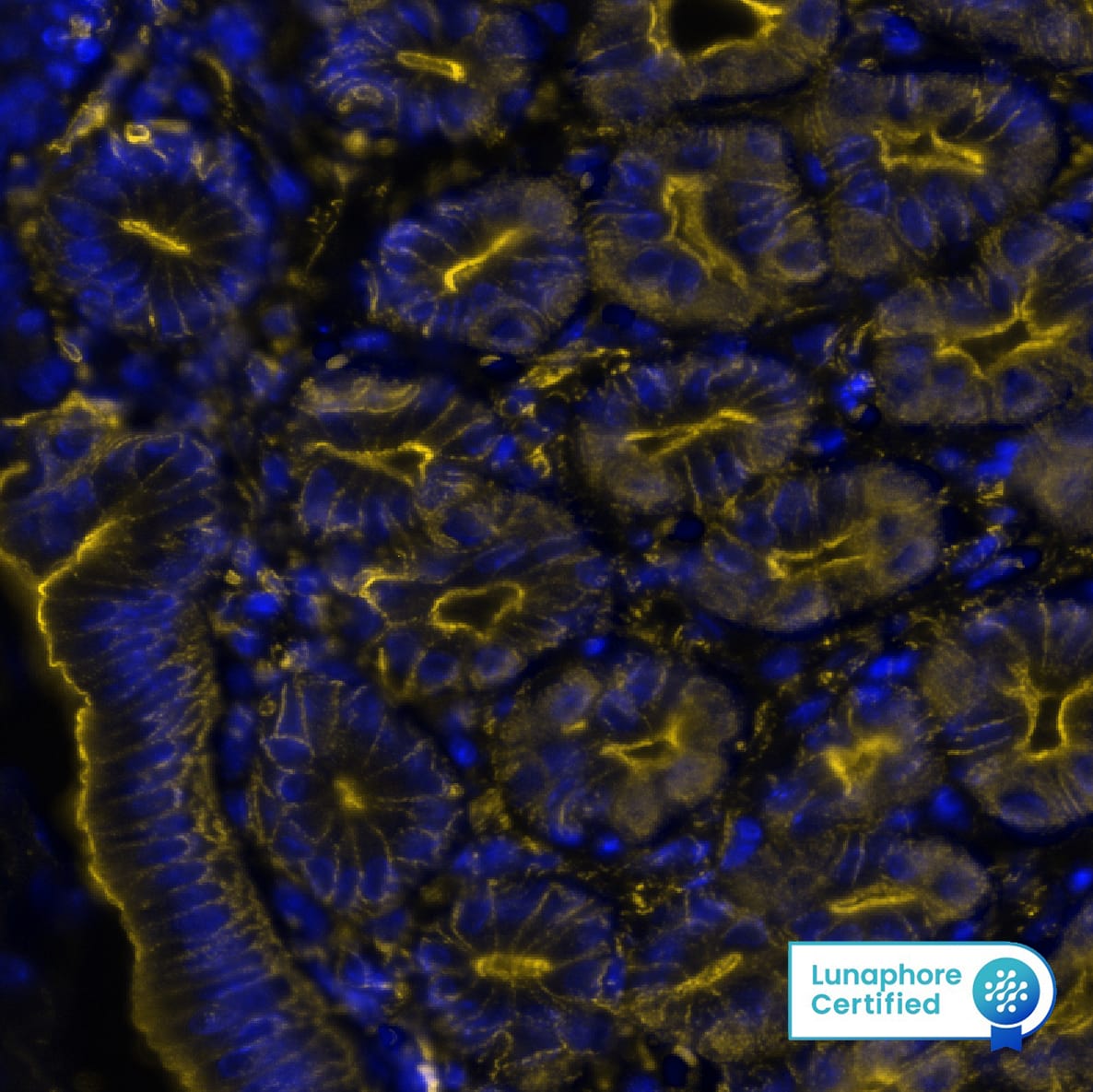
DR555RT) and counterstained with DAPI (blue; Lunaphore Catalog #
DR100). Specific staining was localized to the cell membrane. Protocol available in
COMET™ Panel Builder." alt=" beta -Actin was detected in perfusion fixed paraffin-embedded sections of mouse Stomach using Rat Anti-Mouse beta -Actin, Monoclonal Antibody (Catalog #MAB11702) at 0.25ug/mL at 37° Celsius for 2 minutes.Before incubation with the primary antibody, tissue underwent an all-in-one dewaxing and antigen retrieval preprocessing using PreTreatment Module (PT Module) and Dewax and HIER Buffer H (pH 9; Epredia Catalog #TA-999-DHBH). Tissue was stained using the Alexa Fluor™ 555 Goat anti-Rat IgG Secondary Antibody at 1:100 at 37 ° Celsius for 2 minutes. (Yellow; Lunaphore Catalog #
DR555RT) and counterstained with DAPI (blue; Lunaphore Catalog #
DR100). Specific staining was localized to the cell membrane. Protocol available in
COMET™ Panel Builder." class="big_thumb" />
DR555RT) and counterstained with DAPI (blue; Lunaphore Catalog #
DR100). Specific staining was localized to the cytoplasm. Protocol available in
COMET™ Panel Builder." class="big_lightbox">
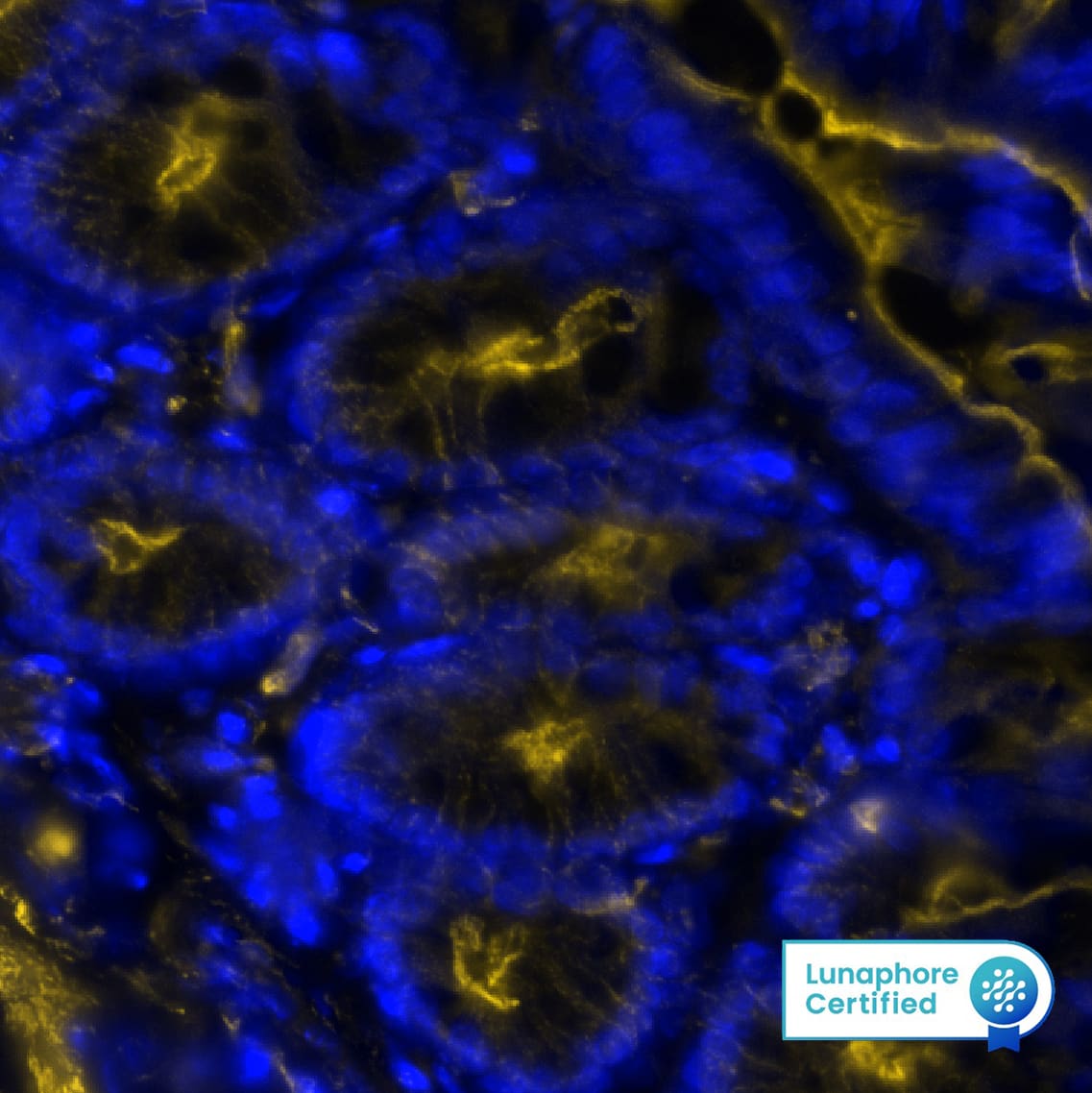
DR555RT) and counterstained with DAPI (blue; Lunaphore Catalog #
DR100). Specific staining was localized to the cytoplasm. Protocol available in
COMET™ Panel Builder." class="big_thumb" />
DR555RT) and counterstained with DAPI (blue; Lunaphore Catalog #
DR100). Specific staining was localized to the cytoplasm. Protocol available in
COMET™ Panel Builder." class="big_lightbox">
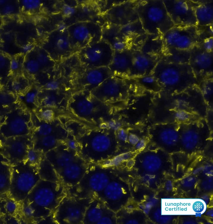
DR555RT) and counterstained with DAPI (blue; Lunaphore Catalog #
DR100). Specific staining was localized to the cytoplasm. Protocol available in
COMET™ Panel Builder." class="big_thumb" />
DR647RT) and counterstained with DAPI (blue; Lunaphore Catalog #
DR100). Specific staining was localized to the cytoplasm. Protocol available in
COMET™ Panel Builder." class="big_lightbox">
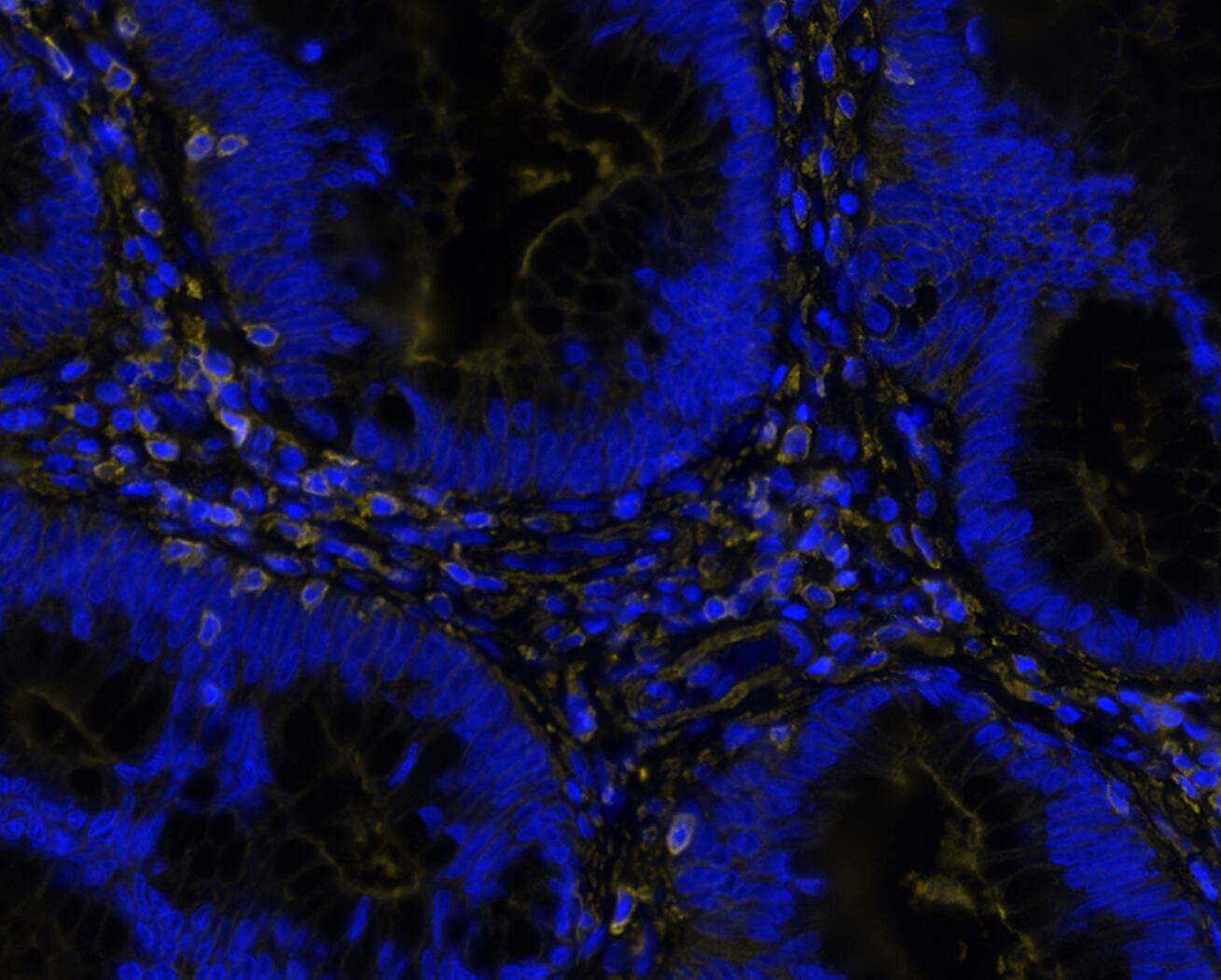
DR647RT) and counterstained with DAPI (blue; Lunaphore Catalog #
DR100). Specific staining was localized to the cytoplasm. Protocol available in
COMET™ Panel Builder." class="big_thumb" />
HAF005). A specific band was detected for beta ‑Actin at approximately 45 kDa (as indicated). This experiment was conducted under reducing conditions and using
Western Blot Buffer Group 1." class="big_lightbox">
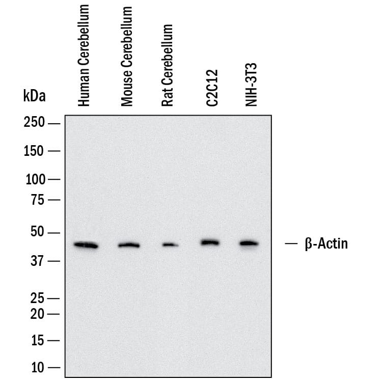
HAF005). A specific band was detected for beta ‑Actin at approximately 45 kDa (as indicated). This experiment was conducted under reducing conditions and using
Western Blot Buffer Group 1." class="big_thumb" />
HAF005) in Milk-free Antibody Diluent (Catalog # 043-524). This experiment was conducted under reducing conditions and using the 12-230kDa separation system.
Right: Simple Western electropherogram showing the same Rat Anti-Mouse beta ‑Actin Monoclonal Antibody (Catalog # MAB11702) tested at 10 µg/ml (blue line) and 50 µg/ml (green line) in the C2C12 mouse myoblast cell line." class="big_lightbox">
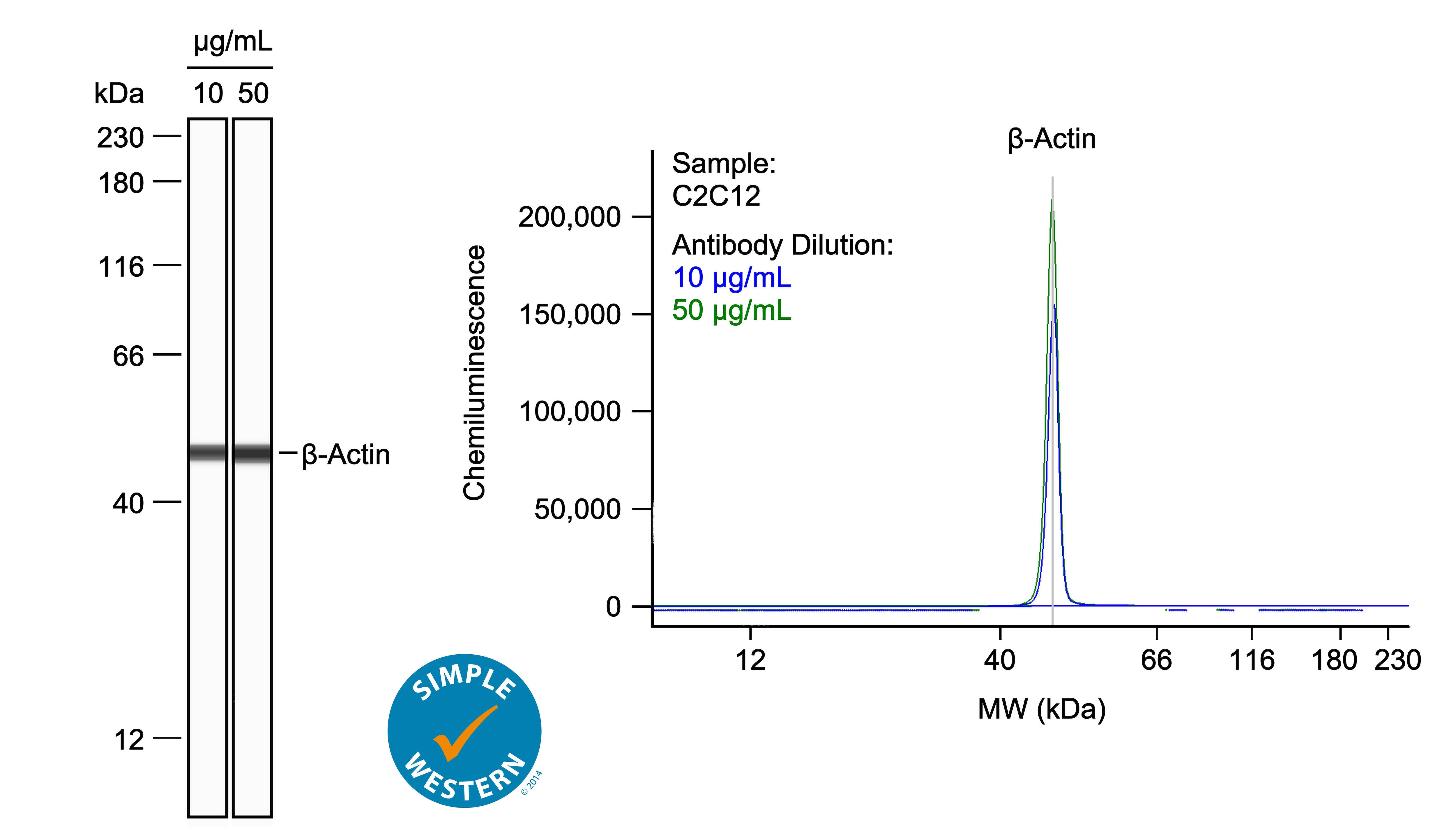
HAF005) in Milk-free Antibody Diluent (Catalog # 043-524). This experiment was conducted under reducing conditions and using the 12-230kDa separation system.
Right: Simple Western electropherogram showing the same Rat Anti-Mouse beta ‑Actin Monoclonal Antibody (Catalog # MAB11702) tested at 10 µg/ml (blue line) and 50 µg/ml (green line) in the C2C12 mouse myoblast cell line." class="big_thumb" />
 HAF018). A specific band was detected for beta -Actin at approximately 45 kDa (as indicated). This experiment was conducted under reducing conditions and using Immunoblot Buffer Group 1." alt="Western blot shows lysates of A431 human epithelial carcinoma cell line, C2C12 mouse myoblast cell line, and Rat-2 rat embryonic fibroblast cell line. PVDF membrane was probed with 0.01 µg/mL of Mouse Anti-Human/Mouse/Rat beta -Actin Monoclonal Antibody (Catalog # MAB8929) followed by HRP-conjugated Anti-Mouse IgG Secondary Antibody (Catalog # HAF018). A specific band was detected for beta -Actin at approximately 45 kDa (as indicated). This experiment was conducted under reducing conditions and using Immunoblot Buffer Group 1." class="big_thumb" />
 NL007) and counterstained with DAPI (blue). Specific staining was localized to cytoplasm. View our protocol for Fluorescent ICC Staining of Cells on Coverslips." alt=" beta -Actin was detected in immersion fixed HeLa human cervical epithelial carcinoma cell line using Mouse Anti-Human beta -Actin Monoclonal Antibody (Catalog # MAB8969) at 10 µg/mL for 3 hours at room temperature. Cells were stained using the NorthernLights™ 557-conjugated Anti-Mouse IgG Secondary Antibody (red; Catalog # NL007) and counterstained with DAPI (blue). Specific staining was localized to cytoplasm. View our protocol for Fluorescent ICC Staining of Cells on Coverslips." class="big_thumb" />
 DR555RT) and counterstained with DAPI (blue; Lunaphore Catalog # DR100). Specific staining was localized to the cell membrane. Protocol available in COMET™ Panel Builder." alt=" beta -Actin was detected in perfusion fixed paraffin-embedded sections of mouse Stomach using Rat Anti-Mouse beta -Actin, Monoclonal Antibody (Catalog #MAB11702) at 0.25ug/mL at 37° Celsius for 2 minutes.Before incubation with the primary antibody, tissue underwent an all-in-one dewaxing and antigen retrieval preprocessing using PreTreatment Module (PT Module) and Dewax and HIER Buffer H (pH 9; Epredia Catalog #TA-999-DHBH). Tissue was stained using the Alexa Fluor™ 555 Goat anti-Rat IgG Secondary Antibody at 1:100 at 37 ° Celsius for 2 minutes. (Yellow; Lunaphore Catalog # DR555RT) and counterstained with DAPI (blue; Lunaphore Catalog # DR100). Specific staining was localized to the cell membrane. Protocol available in COMET™ Panel Builder." class="big_thumb" />


![Western Blot: beta-Actin Antibody (AC-15) [NB600-501] - MCDK cells induced with C22H24N2O8 to control the expression of the gene of interest. Beta-actin blocking confirms the albumin assay showing that an equal amount of lysate was loaded in each lane.](http://images.novusbio.com/fullsize/beta-Actin-Antibody-AC-15-Western-Blot-NB600-501-img0023.jpg)
![Western Blot: beta-Actin Antibody (AC-15) [NB600-501] - Beta-Actin expression in Fibroblast Hs68 cell lysate using anti-beta-Actin antibody. Image from verified customer review.](http://images.novusbio.com/fullsize/beta-Actin-Antibody-AC-15-Western-Blot-NB600-501-img0026.jpg)
![Western Blot: beta-Actin Antibody (AC-15) [NB600-501] - Cell line lysates were separated on SDS-PAGE and probed with 1:5000 Monoclonal Anti-beta-Actin Clone: AC-15 . The antibody was developed using Goat Anti-Mouse IgG-Peroxidase and a chemiluminescent substrate.Lanes: 1. HeLa, 2. JURKAT, 3. COS7, 4. NIH-3T3, 5. PC-12, 6. RAT2, 7.CHO, 8. MDBK, 9. MDCK](http://images.novusbio.com/fullsize/beta-Actin-Antibody-AC-15-Western-Blot-NB600-501-img0040.jpg)
![Immunocytochemistry/Immunofluorescence: beta-Actin Antibody (AC-15) [NB600-501] - Analysis of beta-Actin antibody on mouse fibroblasts. Image from verified customer review.](http://images.novusbio.com/fullsize/beta-Actin-Antibody-AC-15-Immunocytochemistry-Immunofluorescence-NB600-501-img0038.jpg)
![Immunohistochemistry: beta-Actin Antibody (AC-15) [NB600-501] - Staining of formalin-fixed, paraffin-embedded human heart sections with 1:1000 Monoclonal Anti-beta-Actin Clone: AC-15.](http://images.novusbio.com/fullsize/beta-Actin-Antibody-AC-15-Immunohistochemistry-NB600-501-img0039.jpg)
![Western Blot: beta-Actin Antibody (AC-15) [NB600-501] - Analysis of beta-Actin in bovine whole skeletal muscle lysate. Image from verified customer review.](http://images.novusbio.com/fullsize/beta-Actin-Antibody-AC-15-Western-Blot-NB600-501-img0016.jpg)
![Western Blot: beta-Actin Antibody (AC-15) [NB600-501] - Whole cell extract of human fibroblasts was separated on SDS-PAGE and blotted with Monoclonal Anti-beta-Actin. The antibody was developed with Goat Anti-Mouse IgG, Peroxidase conjugate and AEC substrate. Lanes A: Antibody dilution 1:5,000 B: Negative control (only secondary antibody).](http://images.novusbio.com/fullsize/beta-Actin-Antibody-AC-15-Western-Blot-NB600-501-img0022.jpg)
![Western Blot: beta-Actin Antibody (AC-15) [NB600-501] - beta-Actin expression in human glioma cells using anti-beta-Actin antibody. Image from verified customer review.](http://images.novusbio.com/fullsize/beta-Actin-Antibody-AC-15-Western-Blot-NB600-501-img0027.jpg)
![Western Blot: beta-Actin Antibody (AC-15) [NB600-501] - HEK293T cells transfected with Niaph virus Matrix Protein, expression 24h later. This image was submitted via customer review.](http://images.novusbio.com/fullsize/beta-Actin-Antibody-AC-15-Western-Blot-NB600-501-img0033.jpg)


![Immunocytochemistry/Immunofluorescence: beta-Actin Antibody (AC-15) [NB600-501] - Cultured FS-11 human fibroblast cell line stained using Monoclonal Anti-Beta-Actin, Clone No. AC-15 (NB600-501).](http://images.novusbio.com/fullsize/beta-Actin-Antibody-AC-15-Immunocytochemistry-Immunofluorescence-NB600-501-img0019.jpg)
![Immunocytochemistry/Immunofluorescence: beta-Actin Antibody (AC-15) [NB600-501] - HS-68 cells.](http://images.novusbio.com/fullsize/beta-Actin-Antibody-AC-15-Immunocytochemistry-Immunofluorescence-NB600-501-img0024.jpg)
![Simple Western: beta-Actin Antibody (AC-15) [NB600-501] - Analysis using the HRP conjugate of NB600-501. Electropherogram image(s) of corresponding Simple Western lane view. Beta-Actin antibody was used at 1:500 dilution on Hela, MCF-7, SH-SY5Y, & Jurkat lysate(s).](http://images.novusbio.com/fullsize/beta-Actin-Antibody-AC-15-Simple-Western-NB600-501-img0029.jpg)
![Simple Western: beta-Actin Antibody (AC-15) [NB600-501] - Analysis using the HRP conjugate of NB600-501. Image shows a specific band for Beta-Actin in 0.2 mg/mL of Hela, MCF-7, SH-SY5Y, & Jurkat lysate(s). This experiment was performed under reducing conditions using the 12-230 kDa separation system. * Non-specific interaction with the 230 kDa Simple Western standard may be seen with this antibody.](http://images.novusbio.com/fullsize/beta-Actin-Antibody-AC-15-Simple-Western-NB600-501-img0041.jpg)


![The hypoxia-activated pro-drug Tirapazamine (TPZ) reduces stem-like Aldefluorbright population in vitro & in liver metastases(a) Human colonospheres cultured in hypoxia (0.1%) & normoxia (21%) for 24 hrs in absence or presence of TPZ for 4 hrs. Cells processed for IF analysis of gamma H2AX (green). DAPI (blue) used to visualize cell nuclei. (b) Bar graphs showing quantification of gamma H2AX foci observed in (a), shown as % of mean gamma H2AX intensity at normoxia (n=3). (c) Representative confocal pictures of comet assay of colonospheres cultured as in (a). (d) Bar graphs showing quantification of DNA damage observed with the comet assay (Extend tail moment) (n=77). (e) Experiment was performed as in (a), but cells were either treated or untreated for 16 hrs. Cells were lysed & analyzed by WB for the indicated markers (cropped). (f) Colonospheres cultured in hypoxia & treated with TPZ for 16 hrs. The cells were FACS sorted into Aldefluorbright & Aldefluordim populations & analyzed by WB for the indicated markers. (g) Human colonospheres cultured in hypoxia (0.1%) for 24 hrs in absence or presence of TPZ for 4 hrs & FACS sorted into Aldefluorbright & Aldefluordim populations. Bar graph shows the fold change Aldefluordim & Aldefluorbright cells upon TPZ treatment (n=3). (h) C26 tumor cells were injected into the liver parenchyma of immune-deficient mice. Following tumor initiation, tumor-bearing liver lobes were subjected to a vascular clamping protocol [24] to induce hypoxia & treated with saline (control) or TPZ (treatment) for 10 days. At endpoint, livers were excised & expression of ALDH1 was examined by IHC. Magnification 20x, T=tumor. (i) Bar graphs showing quantification of ALDH1 (n=28) observed in (h) (n=14). * = significant (p>0.05). ns = not significant. Image collected & cropped by CiteAb from the following publication (//www.oncotarget.com/lookup/doi/10.18632/oncotarget.21145), licensed under a CC-BY license. Not internally tested by Novus Biologicals.](http://images.novusbio.com/fullsize/nb600-501_mouse-monoclonal-beta-actin-antibody-ac-15-210202423454879.jpg)
![Western Blot: beta-Actin Antibody (AC-15) [NB600-501] - Effect of IFN-gamma pretreatment on carfilzomib sensitivity of H727 (a) & H23 (b) cells. IFN-gamma (150 U⋅ml−1) pretreatment for 24 h led to increased expression (left panel) & activity (middle panel) of proteasome immuno-subunits, & sensitized H727 cells to Cfz, while desensitizing H23 cells towards Cfz. The IC50 values displayed a statistically significant difference between IFN-gamma -pretreated cells & vehicle control (P value < 0.01, n = 3, Student’s t-test comparing the log transformed IC50 values obtained from three independent runs). Image collected & cropped by CiteAb from the following publication (//pubmed.ncbi.nlm.nih.gov/30858500), licensed under a CC-BY license. Not internally tested by Novus Biologicals.](http://images.novusbio.com/fullsize/nb600-501_mouse-monoclonal-beta-actin-antibody-ac-15-310202415392582.jpg)
![Western Blot: beta-Actin Antibody (AC-15) [NB600-501] - RARA overexpression increases chemosensitivity to AM80(A, B) Overexpression of either RARAwt or RARAR394Q increases chemosensitivity of (A) HuT78 & (B) Karpas 299 cells to AM80. The presence of the R394Q mutation does not reduce chemosensitivity significantly. (C) RARAwt overexpression increases retinoic acid response element (RARE) activity (indicated by luciferase) in HuT78 cells treated with AM80. (D) RARAwt overexpression increases the degree of CDK2, CDK4 & CDK6 inhibition caused by AM80. wt, wild-type; CDK, cyclin-dependent kinase. Image collected & cropped by CiteAb from the following publication (//www.oncotarget.com/lookup/doi/10.18632/oncotarget.15441), licensed under a CC-BY license. Not internally tested by Novus Biologicals.](http://images.novusbio.com/fullsize/nb600-501_mouse-monoclonal-beta-actin-antibody-ac-15-310202415293342.jpg)
![Western Blot: beta-Actin Antibody (AC-15) [NB600-501] - RARA drives expression of cyclin-dependent kinases(A) Knockdown of RARA causes G1 cell cycle arrest (P = 0.004) in Mac-1 cells. (B) Expression of the regulators of cell cycle progression, CDK6, CDK4, & to a lesser extent, CDK2, is inhibited by RARA knockdown in Mac-1 cells. (C) Expression of CDK6, CDK4, & CDK2 is increased following overexpression of RARAwt & RARAR394Q in HuT78 cells. RARA, retinoic acid receptor alpha; wt, wild-type; siRNA, small interfering RNA; CDK, cyclin-dependent kinase. Image collected & cropped by CiteAb from the following publication (//www.oncotarget.com/lookup/doi/10.18632/oncotarget.15441), licensed under a CC-BY license. Not internally tested by Novus Biologicals.](http://images.novusbio.com/fullsize/nb600-501_mouse-monoclonal-beta-actin-antibody-ac-15-310202415363726.jpg)
![Western Blot: beta-Actin Antibody (AC-15) [NB600-501] - (A) Protein expression level of CCL2 with WARS silencing under control & IFN gamma stimulation conditions, performed on human primary PBMC-derived macrophages. *p<0.05, Student’s t-test(B) Western blot analysis showing the protein levels of phosphorylation of STAT1 with WARS silencing under control & IFN gamma stimulation conditions, performed on human primary PBMC-derived macrophages. (C) Relative mRNA & protein expression levels for CCL2, TNF alpha , STAT1, JAK2 & CD36 with GBP1 silencing under control & IFN gamma stimulation conditions, performed on THP-1-differentiated macrophage-like cells. (D) Western blot analysis showing the protein levels of JAK2 & phospho-STAT1 with GBP1 silencing in THP-1 macrophage-like cells at 3 & 6 hr after IFN gamma stimulation. Image collected & cropped by CiteAb from the following publication (//pubmed.ncbi.nlm.nih.gov/30303482), licensed under a CC-BY license. Not internally tested by Novus Biologicals.](http://images.novusbio.com/fullsize/nb600-501_mouse-monoclonal-beta-actin-antibody-ac-15-31020241534361.jpg)
![Western Blot: beta-Actin Antibody (AC-15) [NB600-501] - Sortilin forms homodimers on the cell surface of HEK293 cells.A, schematic of FLAG-sortilin & His6-sortilin. FLAG tag & His6 tag were inserted following propeptide & 3 amino acids (Ser78-Ala79-Pro80) in sortilin. SP, signal peptide; PP, propeptide. B, overexpression of FLAG-sortilin & His6-sortilin in HEK293 cells was validated by Western blotting. C & D, detection of binding of FLAG-sortilin & His6-sortilin on the cell surface of HEK293 in TR-FRET assay (C) & HTRF assay (D). Change of FRET signal by expression of His6-sortilin is indicated by percent change (mean ± S.D., three independent experiments). Error bars represent S.D. *, p < 0.05; **, p < 0.01 by t test. IB, immunoblotting. Image collected & cropped by CiteAb from the following publication (//pubmed.ncbi.nlm.nih.gov/29382723), licensed under a CC-BY license. Not internally tested by Novus Biologicals.](http://images.novusbio.com/fullsize/nb600-501_mouse-monoclonal-beta-actin-antibody-ac-15-310202415382440.jpg)
![Western Blot: beta-Actin Antibody (AC-15) [NB600-501] - Western blot analysis of neurofilament proteins. Protein levels of NF-H, phosphorylated NF-H, NF-M & phosphorylated NF-M are enhanced in pEGFP-Peripherin cells, especially at 8 days of NGF induction. However, protein level of NF-L is not influenced in pEGFP-Peripherin cells. The EGFP-Peripherin fusion protein is constantly expressed in pEGFP-Peripherin cells, while not in PC12 cells. The protein level of endogenous peripherin is similar between control PC12 cells & pEGFP-Peripherin cells. Image collected & cropped by CiteAb from the following publication (//jbiomedsci.biomedcentral.com/articles/10.1186/1423-0127-19-8), licensed under a CC-BY license. Not internally tested by Novus Biologicals.](http://images.novusbio.com/fullsize/nb600-501_mouse-monoclonal-beta-actin-antibody-ac-15-310202415293324.jpg)
![Western Blot: beta-Actin Antibody (AC-15) [NB600-501] - Activation of NF-kappa B through NRAGE is specific to the non-canonical BMP pathway. A: 293 cells were transfected with full length NRAGE:EGFP or stimulated with BMP-4 & incubated with or without IKK VII inhibitor, & NF-kappa B activation assessed by luciferase assay. B: 293 cells were transfected with full length NRAGE:EGFP or stimulated with BMP-4 & incubated with or without Tak1 or Tab1 morpholinos, & NF-kappa B activation assessed by luciferase assay. C: Endogenous phosphorylation of IKK-alpha / beta in 293 cells transfected with Tak1 or Tab1 morpholinos prior to 10 ng/ml BMP-4 stimulation for one hour. D: 293 cells were transfected with full length NRAGE:EGFP, incubated with or without Tak1 or Tab1 morpholinos immunoprecipitated with rabbit anti-GFP & western blotted for IKK alpha / beta -phosphorylation & total IKK alpha . E: Western blot of Tak1 & Tab1 proteins in 293 cells with & without morpholinos. Luciferase assays were performed in triplicate & presented as fold increase over renilla. * P < 0.05; vs NRAGE construct or BMP-4 treatment by the two-tailed unpaired Student's t-test. Image collected & cropped by CiteAb from the following publication (//pubmed.ncbi.nlm.nih.gov/20100315), licensed under a CC-BY license. Not internally tested by Novus Biologicals.](http://images.novusbio.com/fullsize/nb600-501_mouse-monoclonal-beta-actin-antibody-ac-15-310202416175218.jpg)
![Western Blot: beta-Actin Antibody (AC-15) [NB600-501] - 16:4(n-3) stimulation increases cPLA2 activity in splenic macrophages. A) Schematic overview of hydrolysis sites of PC to yield metabolites, such as LPC. Splenic macrophages were harvested from murine BALB/c spleens by magnetic bead sorting using F4/80 Abs. Macrophages were incubated with either vehicle or 16:4(n-3) (25 nM) for indicated times. B, C) Fluorescent-based analysis of PLA1 & cPLA2 enzymatic activity revealed a decrease in PLA1 activity (B) & a significant time-dependent increase in cPLA2 activity (C) upon stimulation with 16:4(n-3). Preincubation of splenic macrophages with the GPR120-specific antagonist AH-7614 did not affect 16:4(n-3)-mediated changes in PLA1 activity but was able to prevent the 16:4(n-3)-induced increase in cPLA2 activity. D) In addition, splenic macrophage cell lysates were analyzed by Western blot for phospho-cPLA2. Actin was used as a loading control. E) Treatment of tumor-bearing BALB/c mice with cisplatin (cis) & sCM derived from splenocytes that were incubated with 16:4(n-3) & AACOCF3 did not induce chemoresistance, whereas cotreatment with cisplatin & sCM+ 16:4(n-3) did. Panel B shows grouped results of 2 independent experiments with similar outcomes. Panels C & D show results of 4 independent experiments. Panel E shows grouped results of 2 independent experiments with similar outcomes (n = 8 per group). Data are presented as means ± sem. Statistical significance was determined by 2-tailed Student’s t test (B, C) or 1-way ANOVA (E); all compared with vehicle control (B, C) or with cisplatin alone (E) unless indicated otherwise. *P < 0.05; **P < 0.01. Image collected & cropped by CiteAb from the following publication (//pubmed.ncbi.nlm.nih.gov/28183801), licensed under a CC-BY license. Not internally tested by Novus Biologicals.](http://images.novusbio.com/fullsize/nb600-501_mouse-monoclonal-beta-actin-antibody-ac-15-31020241616048.jpg)
![Western Blot: beta-Actin Antibody (AC-15) [NB600-501] - WEGL reduces serum LPS & TLR4-related signalling pathways in HFD mice.Effects of WEGL treatment on serum endotoxin (a) TLR4 protein production (b,c) JNK phosphorylation (d,e) & I kappa B-alpha production (f,g) were examined in the liver & epididymal adipose tissues of chow- & HFD-fed mice as described in Fig. 1. Serum endotoxin (EU ml−1) was determined as mean±s.e.m. using the limulus amebocyte lysate assay kit. Representative immunoblots for target proteins in b-g are shown. Molecular weight markers were indicated as kilodaltons (kDa). Protein levels were normalized to internal controls ( beta -actin or total JNK, T-JNK) & the relative ratio to the Chow group was labelled on the top of immunoblots. Graph bars in a with different letters on top represent statistically significant results (P<0.05) based on Newman–Keuls post hoc one-way ANOVA analysis, whereas bars labelled with the same letter correspond to results that show no statistically significant differences. Where two letters are present on top of the bar, each letter should be compared separately with the letters of other bars to determine whether the results show statistically significant differences. Image collected & cropped by CiteAb from the following publication (//www.nature.com/articles/ncomms8489), licensed under a CC-BY license. Not internally tested by Novus Biologicals.](http://images.novusbio.com/fullsize/nb600-501_mouse-monoclonal-beta-actin-antibody-ac-15-31020241616029.jpg)
![Western Blot: beta-Actin Antibody (AC-15) [NB600-501] - Mutation of Cys783 abolishes dimerization of sortilin.A, schematic of His6-sortilin 10CC+TMD, FLAG-sortilin WT & C783A, & His6-sortilin ICD+TMD WT & C783A. Cysteine 783 was replaced by alanine. SP, signal peptide; PP, propeptide. B, expression vector of His6-sortilin 10CC+TMD was transfected in HEK293 cells. Dimerization of His6-sortilin 10CC+TMD was detected in non-reducing Western blotting with anti-His6 antibody (n = 3). C, sortilin ICD+TMD C783A did not form homodimers in HEK293 cells in the non-reducing Western blotting (n = 3). D & E, C783A decreased sortilin homodimers of low molecular weight in the cells (D) & extracellular vesicles (E) of HEK293 cells in non-reducing Western blotting (n = 3). F & G, 24-h incubation with 2-FPA, an inhibitor of palmitoylation, increased sortilin homodimers of low molecular weight in HEK293 cells stably overexpressing FLAG-sortilin (F) & their extracellular vesicles (G) (n = 3). Monomers & homodimers of high & low molecular weight are abbreviated as MO, D(HMW), & D(LMW), respectively. IB, immunoblotting. Image collected & cropped by CiteAb from the following publication (//pubmed.ncbi.nlm.nih.gov/29382723), licensed under a CC-BY license. Not internally tested by Novus Biologicals.](http://images.novusbio.com/fullsize/nb600-501_mouse-monoclonal-beta-actin-antibody-ac-15-310202416175237.jpg)
![Western Blot: beta-Actin Antibody (AC-15) [NB600-501] - (a) Cell viability (IC50 values) for a panel of established cancer cell lines as measured by MTS assay following incubation with carfilzomib (Cfz) for 72 h. H727 cells are most resistant to Cfz among 21 tested cell lines. (b) Representative images of H727 cells growing in the presence of Cfz (250 nM) assessed via light microscopy. On day 3 of Cfz treatment, live cell not stained with trypan blue were counted (expressed as % cell viability relative to the vehicle control; no statistically significant difference between Cfz-treated H727 cells & the vehicle control by Student’s t-test, P value > 0.1, n = 3). (c) Immunoblotting results showing no detectable expression of P-glycoprotein (P-gp) in H727 cells. DLD-1 cells with acquired Cfz resistance via P-gp upregulation (DLD-1/CfzR) were used as a positive control. (d) The co-treatment of reversin-121 (7.5 µM, P-gp inhibitor) did not affect the sensitivity of H727 cells to Cfz. The IC50 values did not show statistically significant difference between in the presence & absence of reversin-121 (Student’s t-test). (e) The levels of remaining Cfz in culture media were comparable between H727 & H23 cells (no statistically significant differences, t-tests the Holm-Sidak method to correct for multiple comparisons with alpha = 0.05). Image collected & cropped by CiteAb from the following publication (//pubmed.ncbi.nlm.nih.gov/30858500), licensed under a CC-BY license. Not internally tested by Novus Biologicals.](http://images.novusbio.com/fullsize/nb600-501_mouse-monoclonal-beta-actin-antibody-ac-15-3102024161895.jpg)
![Western Blot: beta-Actin Antibody (AC-15) [NB600-501] - (a) Immunoblots showing the expression of cP & iP catalytic subunits in H727 & H23 cells. Immunoblots for additional cell lines displaying differential expression of proteasome catalytic subunits are presented in Supplementary Fig. S2. (b) Heat map displaying differential proteasome activity profiles in H727 & H23 cell lines. Purified human 20S cP & iP were used as controls for individual subunits: 20S cP for beta 5 & beta 1 & 20S iP for beta 5i & beta 1i. The numbers represent hydrolysis rates of respective substrates (RFU/min, mean values derived from three technical replicates) & were converted to color format & clustered by using the program “R” (//www.R-project.org). (c) Remaining catalytic activities of individual proteasome subunits in H727 & H23 cells 4 h after treatment with 20 nM of carfilzomib (left panel) or 20 nM bortezomib (right panel). Data are presented as mean ± SD derived from three technical replicates. Image collected & cropped by CiteAb from the following publication (//pubmed.ncbi.nlm.nih.gov/30858500), licensed under a CC-BY license. Not internally tested by Novus Biologicals.](http://images.novusbio.com/fullsize/nb600-501_mouse-monoclonal-beta-actin-antibody-ac-15-31020241616080.jpg)
![Western Blot: beta-Actin Antibody (AC-15) [NB600-501] - Effects of insulin on AKT & ERK phosphorylation & cell viability in primary human pancreatic duct cells. Phosphorylated AKT & ERK were measured in primary pancreatic exocrine cultures treated with the indicated concentrations of insulin for 5 minutes (A, B) & 24 hours (C, D) (n =3-4) Fold refers to the fold change of sample relative to control at the same time point. (E) Quantification of automated cell-counting studies employing live-cell imaging of Hoechst-labeled cell cultures over 60 hours. (n =3). (F) Quantification of proliferation by BrdU staining of treated relative to untreated over 3 days (n =4). (G) Quantification of the average number of dying/dead treated cells, propidium iodide (PI) labeled, over 60 hours relative to non-treated cells. (n =3). (H) Human exocrine cells were exposed to 0, 0.2, 2, 20, 200 nM insulin for 3 days. Bright-field images are representative of 3 cultures. (I) Effects of inhibition of RAF1/ERK signalling on PI incorporation with 10 μM GW5074 or AKT signalling with 100 nM Akti1/2 on human primary pancreatic exocrine cell viability (n =3). SF denotes serum free. Repeated Measures ANOVA analyses with Bonferroni’s post-test were performed. *Represents statistical significance of p < 0.05 when compared to DMSO control. Image collected & cropped by CiteAb from the following publication (//pubmed.ncbi.nlm.nih.gov/25373319), licensed under a CC-BY license. Not internally tested by Novus Biologicals.](http://images.novusbio.com/fullsize/nb600-501_mouse-monoclonal-beta-actin-antibody-ac-15-310202416175219.jpg)
![Western Blot: beta-Actin Antibody (AC-15) [NB600-501] - Liver injury induces intra-nuclear metabolic changes & histone hyper-acetylation. Nuclear (A) acetyl-CoA, (B) lactate, & (C) nHDAC activity in livers harvested 6 h after the injection of either saline or CD95-Ab (n = 3 per group). (D) Western blotting for acetylated histone H3 (H3K9) & H4 (H4K5) on livers harvested 6 h after the injections of either saline or CD95-Ab. Histone H3 & beta -actin were used as loading controls. Each line corresponds to an independent mouse. (E) Affinity purification-liquid chromatography tandem-mass spectrometry of liver proteins enriched by immunoprecipitation with acetyl lysine-specific antibody revealed 13 proteins with fold-change >2 in CD95-Ab vs. saline liver samples (n = 5 per group). (F) Venn diagram including the 751 genes upregulated in livers of mice injected with CD95-Ab & the H3K9Ac ChIP-seq data on eight-week-old mouse livers in ENCODE showing a high degree of overlap (Hypergeometric test; p value: 9.643501e−58). (G) Chart generated using custom annotation scripts shows that response to external stimulus including inflammatory & defense response processes, & cytokine-cytokine receptor interaction pathways were enriched among the 467 genes overlapping between the two groups according to KEGG pathways. Means ± SDs are shown; t test; *p <0.05. Ab, antibody; nHDAC, nuclear histone deacetylase. (This figure appears in colour on the web.) Image collected & cropped by CiteAb from the following publication (//pubmed.ncbi.nlm.nih.gov/29580866), licensed under a CC-BY license. Not internally tested by Novus Biologicals.](http://images.novusbio.com/fullsize/nb600-501_mouse-monoclonal-beta-actin-antibody-ac-15-31020241616055.jpg)
![Western Blot: beta-Actin Antibody (AC-15) [NB600-501] - In vivo inhibition of histone acetylation by garcinol improves survival & reduces injury in acute liver failure. (A) Kaplan-Meier survival curves of mice receiving 20 mg/kg/day of garcinol (n = 8) or saline (n = 5) for 5 days prior to the injection of the CD95-Ab. Means ± SDs are shown. Long-rank test p = 0.0473. (B) Western blotting for histone H3 (H3) & acetylated histone H3 (H3K9) on livers harvested 6 h after the administrations of CD95-Ab. beta -actin was used as a loading control. (C) Densitometric quantifications of H3K9 over H3 from the Western blotting shown in B; t test; *p <0.05. (D) Representative TUNEL (upper panels) & H&E (lower panels) staining of livers harvested 6 h after the administrations of CD95-Ab or saline. (E) Quantifications of TUNEL-positive cells & (F) Necrotic areas on livers of at least three sections from each mouse (at least n = 4 mice per group). Means ± SDs are shown. Likelihood ratio test for generalized linear model: *p = 2.2 × 10⁻¹⁶. ANOVA plus Tukey’s post hoc test; for necrotic area: *p = 2.45 × 10⁻⁸. Enzyme activities of PDHC (G) & LDH (H) in nuclear fractions (nPDHC & nLDH) of livers harvested 6 h after CD95-Ab injections in mice treated with garcinol (n = 5) or saline (n = 4). Means ± SDs are shown. ANOVA plus Tukey’s post hoc test: p = 0.11 for nPDHC; p = 0.1 for nLDH. Ab, antibody; A.U., arbitrary units; nLDH, nuclear lactate dehydrogenase; nPDHC, nuclear pyruvate dehydrogenase complex; NS, not statistically significant; TUNEL, terminal deoxynucleotidyl transferase dUTP nick end labeling. (This figure appears in colour on the web.) Image collected & cropped by CiteAb from the following publication (//pubmed.ncbi.nlm.nih.gov/29580866), licensed under a CC-BY license. Not internally tested by Novus Biologicals.](http://images.novusbio.com/fullsize/nb600-501_mouse-monoclonal-beta-actin-antibody-ac-15-310202416171468.jpg)
![Western Blot: beta-Actin Antibody (AC-15) [NB600-501] - Activation of the mTOR signaling pathway during spheroid formation on chitosan film. (a, Left panel) After 48 hr of seeding on monolayer or with chitosan coating (Spheroid), cell lysates were analyzed using the Human Phospho-MAPK Array kit. (a, Right panel) the spots of phospho-mTOR (S2448) were analyzed by an image analysis system. (b & c) Whole-cell lysates were analyzed by western blotting with specific antibodies against indicated molecules. beta -tubulin & actin were used as the loading control. (d) Immunostaining of p-mTOR (S2448), p-S6 (S235), & p-4EBP1 in 3D spheres. DAPI merged images are shown in the low panel. Scale bar = 50 μM. The results are expressed as the mean ± standard deviation of three independent experiments, which is representative of MSCs from two individuals. **p < 0.01 compared with monolayer cells. Image collected & cropped by CiteAb from the following publication (//www.nature.com/articles/s41598-017-10708-0), licensed under a CC-BY license. Not internally tested by Novus Biologicals.](http://images.novusbio.com/fullsize/nb600-501_mouse-monoclonal-beta-actin-antibody-ac-15-31020241616077.jpg)
![Western Blot: beta-Actin Antibody (AC-15) [NB600-501] - Triple AAV Vectors Drive Full-Length Protein Expression in the Pig Retina(A) Western blot (WB) analysis of lysates from Large White pig retina 2 months following subretinal injection of either single or triple AAV2/8 vectors encoding for ED under the control of the PR-specific promoter IRBP. The arrow indicates the full-length protein. (B) WB analysis of truncated products in lysates from Large White pig retina 2 months following subretinal injection of the combinations of triple ED-AAVs. Lysates of HEK293 cells infected with an ED-AAV 1 + 2 + 3 are shown on the left as positive control. The arrows indicate the following products: #A, ED full-length protein; #E, probably from AAV 3. alpha -3xflag, WB with anti-3xflag antibodies; alpha -beta -Actin, WB with anti-beta -Actin antibodies, used as loading control. Neg, lysates of retinas following injection with PBS. The molecular weight ladder is depicted on the left; 200 μg of proteins were loaded. Image collected & cropped by CiteAb from the following publication (//pubmed.ncbi.nlm.nih.gov/29292161), licensed under a CC-BY license. Not internally tested by Novus Biologicals.](http://images.novusbio.com/fullsize/nb600-501_mouse-monoclonal-beta-actin-antibody-ac-15-310202416173521.jpg)
![Western Blot: beta-Actin Antibody (AC-15) [NB600-501] - (a) Immunoblots of proteasome catalytic subunits in H727 cells transfected with siRNA targeting beta 5, beta 2 or beta 5i. (b) The catalytic activity of beta 5 subunit was decreased in H727 cells transfected with siRNA targeting beta 5 compared with H727 cells transfected with scrambled siRNA. (c) Effects of siRNA knockdown of beta 5, beta 5i, or beta 2 on Cfz sensitivity (IC50 values) in H23 & H727 cells. Data are shown as mean ± SD derived from a single non-linear regression based on n = 3 replicates per compound per concentration. (d) Heat map showing proteasome catalytic subunit activity profiles of 6 PI-naïve patient MM samples purchased from Conversant Biologics & AllCells (left). The numbers represent hydrolysis rates of respective substrates (RFU/min, mean values derived from three technical replicates) & were converted to color format & clustered by using the program “R” (//www.R-project.org) (left panel). Carfilzomib (Cfz) cell viability of the same 6 patient MM cells was measured via CellTiter-Glo Luminescent Cell Viability Assay (right panel). Image collected & cropped by CiteAb from the following publication (//pubmed.ncbi.nlm.nih.gov/30858500), licensed under a CC-BY license. Not internally tested by Novus Biologicals.](http://images.novusbio.com/fullsize/nb600-501_mouse-monoclonal-beta-actin-antibody-ac-15-31020241617528.jpg)
![Western Blot: beta-Actin Antibody (AC-15) [NB600-501] - 14-3-3 zeta suppresses hedgehog signalling to control the expression of p27Kip1.(a) Measurement of relative luciferase activity from various lengths of the Cdkn1b promoter in control or 14-3-3 zeta -depleted NIH-3T3 cells (n=7–8 per group, *P<0.05 when compared with control cells as assessed by Student's t-test). (b) Transcription factor binding sites within the −939–554 promoter region of Cdkn1b. (c) Hedgehog signalling (Gli-dependent) transcriptional activity in control or si14-3-3 zeta -transfected Shh-light2 cells treated with 100 nM SAG. (n=4 per group, *P<0.05 when compared to control cells, as assessed by Student's t-test). (d) Expression levels of various components of the hedgehog signalling pathway in differentiating control & 14-3-3 zeta -depleted 3T3-L1 cells, as measured by RNA-seq (n=4 per group, *P<0.05 when compared to control cells, as assessed by Student's t-test). (e) Immunoblotting of full length (∼170 kDa) & processed (∼80 kDa) Gli3 in 14-3-3 zeta -depleted 3T3-L1 cells treated with MDI (representative of n=3 experiments). (f,g) Co-immunoprecipitation of endogenous (f) 14-3-3 zeta or (g) Gli3 from lysates of 3T3-L1 cells treated with MDI for 24 h (representative of n=3 experiments) (h) ChIP-qPCR analysis of Gli3 occupancy on the Cdkn1b promoter in 3T3-L1 cells treated with MDI for 24 h (n=4 per group, *P<0.05, as assessed by Student's t-test). (i,j) Co-transfection of siRNA against 14-3-3 zeta & Gli3 was used to examine whether knockdown of both proteins could restore differentiation, as determined by Ppar gamma expression (i) or Oil Red-O staining (j) (n=4 independent experiments). Error bars represent s.e.m. Image collected & cropped by CiteAb from the following publication (//pubmed.ncbi.nlm.nih.gov/26220403), licensed under a CC-BY license. Not internally tested by Novus Biologicals.](http://images.novusbio.com/fullsize/nb600-501_mouse-monoclonal-beta-actin-antibody-ac-15-310202416175264.jpg)
![Western Blot: beta-Actin Antibody (AC-15) [NB600-501] - Silencing MFN1 or MFN2 reveals their distinct roles in DENV infection.(A & B) RT-qPCR (A) & western blot (B) validation of A549 cells stably expressing shRNA targeting LacZ, MFN1 or MFN2. Data in panels A & C-F are mean ± SD (n = 3 per group) & were compared by two-tailed Student’s t test. Quantification in panel B is the relative ratio of the indicated protein to actin expression. (C) LDH release assay of shLacZ-, shMFN1- or shMFN2-expressing A549 cells infected with DENV serotype 2 (moi 10) for the indicated time. (D) DENV plaque-forming assay of shLacZ-, shMFN1- or shMFN2-expressing A549 cells infected with DENV (moi 0.1) for 24 or 48 h. (E) Analysis of antiviral activity in culture media from indicated cells with DENV infection (moi 5, 48 h) against dSinF-Luc/2A virus. (F) RT-qPCR analysis of IFN beta mRNA level in DENV infected (moi 10) A549-shLacZ, -shMFN1, & -shMFN2 cells for the indicated time. (G) Western blot analysis of DENV infected (moi 10) A549 cells expressing shLacZ, shMFN1 or shMFN2. The relative ratios of band intensity were quantified as indicated. (H) Flow cytometry with JC-1 staining of shLacZ-, shMFN1 or shMFN2-expressing A549 cells infected with DENV (moi 5) for 30 h. Decreased red/green fluorescence ratio of JC-1 represents disrupted MMP. CCCP, carbonyl cyanide m-chlorophenyl hydrazone, an ionophore that disrupts MMP. (I) Plaque forming assay of vesicular stomatitis virus (VSV) by using A549 cells expressing shLacZ, shMFN1 or shMFN2 as indicated. pfu, plaque-forming unit. Image collected & cropped by CiteAb from the following publication (//dx.plos.org/10.1371/journal.ppat.1005350), licensed under a CC-BY license. Not internally tested by Novus Biologicals.](http://images.novusbio.com/fullsize/nb600-501_mouse-monoclonal-beta-actin-antibody-ac-15-310202416165632.jpg)
![Western Blot: beta-Actin Antibody (AC-15) [NB600-501] - Effects of HSM on IL-18 gene expression & secretion in human macrophages.Cells were pre-treated with various concentrations (1 or 2%) of HSM extract for 20 h, followed by treatment with LPS (0.5 μg/ml) for 3 h & ATP (5 mM) for 1 h. (a) The mRNA expression levels of IL-18 were determined by RT-PCR analysis. (b) IL-18 mRNAs were quantified using real-time PCR. beta -actin gene expression was used for normalization. The results are expressed as fold changes, considering one as the value of untreated cells. (c) The amount of IL-18 in cell culture supernatants was detected by ELISA. (d) The presence of IL-18 in cell lysates & cell culture supernatants were analyzed by Western blot analysis. Data are presented as means ± SE of three experiments performed in duplicate. #P < 0.01 versus untreated cells. *P < 0.01 versus HSM-untreated control (ethanol) cells. †P < 0.05 versus HSM (1%) treated cells. Image collected & cropped by CiteAb from the following publication (//pubmed.ncbi.nlm.nih.gov/23459183), licensed under a CC-BY license. Not internally tested by Novus Biologicals.](http://images.novusbio.com/fullsize/nb600-501_mouse-monoclonal-beta-actin-antibody-ac-15-31020241616018.jpg)
![Western Blot: beta-Actin Antibody (AC-15) [NB600-501] - Wnt effect on AChR clustering requires interaction with MuSK. A, Wnt9a & Wnt11, but not Wnt7a, bind MuSK in vitro. Flag-Wnts immobilized beads were incubated with MuSKect-Myc. Bound proteins were isolated, resolved by SDS-PAGE & blotted with antibodies against Myc & Flag. B, Wnt11 binds CRD of MuSK. Flag-Wnt11 immobilized beads were incubated with MuSKect, MuSKect delta CRD or MuSKCRD. Interactions were assayed as in A. *, p < 0.05, n = 3. C, Wnt-induced AChR clustering is rescued by MuSK, but not MuSK delta CRD, in MuSK-/- cells. MuSK-/- cells were transfected without or with respective constructs & resulting myotubes (identified by GFP encoded by the expression construct) were assayed for AChR cluster formation in response to agrin or Wnts as in Figure 1. Arrows, AChR clusters. *, p < 0.01, Student's t test. D, Wnt9a & Wnt11 induce MuSK tyrosine phosphorylation. C2C12 myotubes were treated by agrin or Wnts for 1 h. MuSK was purified by immunoprecipitation & probed with the 4G10 antibody. E, Wnt9a & Wnt11 induce MuSK dimerization. Flag-MuSK & MuSK-Myc were transfected into C2C12 myoblasts & resulting myotubes were treated by Wnts, agrin or control media for 1 h. Flag-MuSK was precipitated by Flag antibody & associated MuSK-Myc was determined by anti-Myc antibody. Band intensity of immunoblot was analyzed by ImageJ software. *, p < 0.01, one-way ANOVA with Student's t test. Image collected & cropped by CiteAb from the following publication (//pubmed.ncbi.nlm.nih.gov/22309736), licensed under a CC-BY license. Not internally tested by Novus Biologicals.](http://images.novusbio.com/fullsize/nb600-501_mouse-monoclonal-beta-actin-antibody-ac-15-31020241616082.jpg)
![Western Blot: beta-Actin Antibody (AC-15) [NB600-501] - Amiodarone enhanced liver growth & hepatocyte proliferation in the liver regeneration after PHx.Wild-type mice were intraperitoneally injected with vehicle (Veh), amiodarone (AD), or chloroquine (CQ) at 0.5 h before PHx or sham operation & then once per day until 168 h. Liver tissues were harvested at 0–168 h after surgery. Liver-to-body-weight ratios were calculated (A). Liver sections at 24 h after the sham operation, PHx with Veh, AD, or CQ were stained with H&E; original magnification, 400X (B). Representative immunohistochemical staining of Ki67 is shown (C). The percentage of Ki67-positive nuclei in hepatocyte was counted under low-power fields (200 ×) in 15 random sections from at least six different mice (D). Liver tissues were harvested at 0 h, 12 h, 24 h, or 48 h after surgery, & the tissue extracts were analyzed for PCNA, cyclin A, B, D1, E, p21, TGF-beta 1, & beta -actin protein by Western blotting (E,F). The values are shown as the mean ± SD in the bar graph & compared by Student’s t test. #P < 0.05 versus vehicle-treated PHx. Scale bar, 25 μm in B, 50 μm in C (n = 6). Image collected & cropped by CiteAb from the following publication (//www.nature.com/articles/srep15807), licensed under a CC-BY license. Not internally tested by Novus Biologicals.](http://images.novusbio.com/fullsize/nb600-501_mouse-monoclonal-beta-actin-antibody-ac-15-310202416171486.jpg)
![Western Blot: beta-Actin Antibody (AC-15) [NB600-501] - Cell death & DNA damage in response to treatment in A549/p53 dominant-negative cells(A) Confirmation of stable clones was done by western blot analysis using p53 specific antibody. (B) MTT assay for cisplatin shows that the mutant cell line is resistant to the drug as compared p54-wt A549 cells. (C) Confocal images of A549/p53 dominant negative cells shows an elevated level of DNA damage in combination than single agent treatment. Increased accumulation of gamma -H2AX indicates DNA damage. Nucleus= Blue (DAPI), F-actin= Red (TRITC), gamma -H2AX= Green (FITC). (D) Western blot analysis shows a caspase mediated cell death in A549/p53 mut cells in response to treatment. (E) AnnexinV-PI dual staining & flow cytometry analysis shows significantly higher apoptosis in combination treatment than low dose cisplatin & scriptaid. Q1=necrotic population, Q2=late phase apoptosis, Q3=live cells, Q4= early phase apoptosis. C= control, L=2μg/ml cisplatin, S1= 1μg/ml scriptaid. * indicates p < 0.05. Image collected & cropped by CiteAb from the following publication (//www.oncotarget.com/lookup/doi/10.18632/oncotarget.12378), licensed under a CC-BY license. Not internally tested by Novus Biologicals.](http://images.novusbio.com/fullsize/nb600-501_mouse-monoclonal-beta-actin-antibody-ac-15-310202416171425.jpg)
![Western Blot: beta-Actin Antibody (AC-15) [NB600-501] - Sortilin forms homodimers in the extracellular & intracellular domains with intermolecular disulfide bonds in HEK293 cells.A, schematic of FLAG-sortilin Full, ECD+TMD, & ICD+TMD. SP, signal peptide; PP, propeptide. B & C, protein expression of FLAG-sortilin Full, ECD+TMD, & ICD+TMD was validated in reducing (B) & non-reducing (C) Western blotting using anti-FLAG antibody. FLAG-sortilin Full & ECD+TMD form homodimers & multimers. Empty vector was used as a control. D, HEK293 cells transiently overexpressing FLAG-sortilin Full or ECD+TMD were treated with a cross-linker, BS3, & the cell lysates were used for reducing Western blotting with anti-FLAG antibody, showing dimerization of sortilin Full & ECD+TMD (n = 3). E, HEK293 cells stably overexpressing FLAG-sortilin ICD+TMD (FLAG-sortilin ICD+TMD HEK293 cells) were incubated with DMSO (Control), 20 μmol/liter MG-132 (MG) or 10 μmol/liter chloroquine (Chlo) for 7 h, & then reducing Western blotting was performed using anti-sortilin antibody. MG-132 increased the protein expression of FLAG-sortilin ICD+TMD, but chloroquine did not (n = 3). F, FLAG-sortilin ICD+TMD HEK293 cells were incubated with DMSO or MG-132 (2–20 μmol/liter) for 7 or 24 h. MG-132 increased FLAG-sortilin ICD+TMD in a time- & concentration-dependent manner (n = 3). G, following 16-h incubation of HEK293 cells (Control) or FLAG-sortilin ICD+TMD HEK293 cells (ICD+TMD) with MG-132 (5 μmol/liter) & immunoprecipitation with anti-FLAG antibody, non-reducing Western blotting showed dimerization of sortilin ICD+TMD using anti-sortilin antibody (n = 3). Monomers, homodimers, & multimers are abbreviated as MO, D, & MU, respectively. IB, immunoblotting. Image collected & cropped by CiteAb from the following publication (//pubmed.ncbi.nlm.nih.gov/29382723), licensed under a CC-BY license. Not internally tested by Novus Biologicals.](http://images.novusbio.com/fullsize/nb600-501_mouse-monoclonal-beta-actin-antibody-ac-15-310202416182613.jpg)
![Western Blot: beta-Actin Antibody (AC-15) [NB600-501] - Defects in adipogenesis due to 14-3-3 zeta depletion are due to cell cycle arrest & up-regulation of Cdkn1b/p27Kip1.(a–d) Analysis by flow cytometry (a) of control or si14-3-3 zeta -transfected 3T3-L1 cells at different stages of the cell cycle at 0 (b), 24 (c) & 48 (d) hours following differentiation (n=4 independent experiments; *P<0.05 when compared with siCon-transfected cells, assessed by Student's t-test). (e) Analysis of various cell cycle regulatory genes by RNA-Seq from siCon- or si14-3-3 zeta transfected 3T3-L1 adipocytes (n=4 per goup, *P<0.05 when compared with siCon cells at the same time point, assessed by Student's t-test). (f) Immunoblotting of p27Kip1 from lysates of differentiating of siCon- or si14-3-3 zeta -3T3-L1 adipocytes (n=4 experiments). (g,h) Co-transfection of siRNA against 14-3-3 zeta & p27Kip1 was used to examine whether knockdown of both proteins could restore differentiation, as determined by Ppar gamma expression (g) or Oil Red-O staining (h). (n=4 independent experiments). Error bars represent s.e.m. Image collected & cropped by CiteAb from the following publication (//pubmed.ncbi.nlm.nih.gov/26220403), licensed under a CC-BY license. Not internally tested by Novus Biologicals.](http://images.novusbio.com/fullsize/nb600-501_mouse-monoclonal-beta-actin-antibody-ac-15-310202416175214.jpg)
![Western Blot: beta-Actin Antibody (AC-15) [NB600-501] - Effects of HSM on IL-1 beta gene expression & secretion in human macrophages.Cells were pre-treated with either 1 or 2% of HSM extract for 20 h, followed by treatment with LPS (0.5 μg/ml) for 3 h & with ATP (5 mM) for 1 h. (a) The mRNA expression levels of IL-1 beta were determined by RT-PCR analysis. (b) IL-1 beta mRNAs were quantified using real-time PCR. beta -actin gene expression was used for normalization. The results are expressed as fold changes, considering one as the value of untreated cells. (c) The amount of IL-1 beta in cell culture supernatants was detected by ELISA. (d) The presence of IL-1 beta in cell lysates & cell culture supernatants were analyzed by Western blot analysis. Data are presented as means ± SE of three experiments performed in duplicate. #P < 0.01 versus untreated cells. *P < 0.01 versus HSM-untreated (ethanol-treated) control cells. †P < 0.05 versus HSM (1%) treated cells. Image collected & cropped by CiteAb from the following publication (//pubmed.ncbi.nlm.nih.gov/23459183), licensed under a CC-BY license. Not internally tested by Novus Biologicals.](http://images.novusbio.com/fullsize/nb600-501_mouse-monoclonal-beta-actin-antibody-ac-15-310202416165621.jpg)
![Western Blot: beta-Actin Antibody (AC-15) [NB600-501] - (A) shows western blots of nuclear extracts of mCCD cells treated with 100 μmol/L CoCl2. Treatment with CoCl2 increased stabilization of HIF1 alpha in the nucleus of the cells. GAPDH levels are shown as loading controls. (B) shows western blots of whole cell extracts of mCCD cells treated with 100 μmol/L CoCl2 for 4 h. As shown in the images, treatment with CoCl2 increased the EPO production by cells. Beta actin levels are shown as loading controls. (C) shows western blots of whole cell extracts of mCCD cells treated with 100 μmol/L CoCl2 for 4 h & chronic fluid flow. As shown in the images, treatments decreased ZO‐1 production by cells independently of fluid flow stimulation. Beta actin levels are shown as loading controls. (D) shows western blots of whole cell extracts of mCCD cells treated with 100 μmol/L CoCl2 for 4 h & chronic fluid flow. As shown in the images, treatments decreased NaKATPase production by cells independently of fluid flow stimulation. Beta actin levels are shown as loading controls. (E) Densitometric analysis of Western blots presented in (A–D). HIF1 alpha level: P = 0.004 (N = 3), t‐test, showing significant nuclear stabilization of HIF1 alpha . EPO level: increases slightly though not statistically significant (N = 3, P > 0.05, One‐way ANOVA). ZO‐1 level: P < 0.0001 (N = 4), One‐way ANOVA, showing significant decrease in ZO‐1 levels, independent of fluid flow. NaKATPase alpha 1 subunit level: P = 0.001 (N = 3), One‐way ANOVA, showing significant decrease in NaKATPase alpha 1 subunit levels, largely independent of fluid flow. Image collected & cropped by CiteAb from the following publication (//pubmed.ncbi.nlm.nih.gov/29263117), licensed under a CC-BY license. Not internally tested by Novus Biologicals.](http://images.novusbio.com/fullsize/nb600-501_mouse-monoclonal-beta-actin-antibody-ac-15-310202416171461.jpg)
![Western Blot: beta-Actin Antibody (AC-15) [NB600-501] - MFN1 & MFN2 manipulate MAVS-triggered signaling differently.(A & B) Western blot (A) & RT-qPCR (B) analysis of A549+on/myc-MAVS cells with stably expressing shRNA targeting the indicated genes after 48 h of Dox treatment. RT-qPCR data are mean ± SD (n = 3 per group) & were compared by two-tailed Student’s t test. (C) Dual-luciferase activity analysis of A549 cells cotransfected with the indicated MFNs (cloned in pcDNA3.1 vector; 0.5 μg), stimulator or control (Flag-MAVS/pcDNA3 or Flag-GFP/pcDNA3; 0.4 μg), IRF3/pCR3.1 (0.3 μg), p125-Luc (0.2 μg) & pRL-TK (0.1 μg) for 24 h. The firefly luciferase activity (p125-Luc) was normalized to that of renilla luciferase (pRL-TK) & the relative luciferase activities are shown. Data are mean ± SD (n = 3 per group) & were compared by two-tailed Student’s t test. Image collected & cropped by CiteAb from the following publication (//dx.plos.org/10.1371/journal.ppat.1005350), licensed under a CC-BY license. Not internally tested by Novus Biologicals.](http://images.novusbio.com/fullsize/nb600-501_mouse-monoclonal-beta-actin-antibody-ac-15-31020241616021.jpg)
![Western Blot: beta-Actin Antibody (AC-15) [NB600-501] - Mutation of Cys783 abolishes dimerization of sortilin.A, schematic of His6-sortilin 10CC+TMD, FLAG-sortilin WT & C783A, & His6-sortilin ICD+TMD WT & C783A. Cysteine 783 was replaced by alanine. SP, signal peptide; PP, propeptide. B, expression vector of His6-sortilin 10CC+TMD was transfected in HEK293 cells. Dimerization of His6-sortilin 10CC+TMD was detected in non-reducing Western blotting with anti-His6 antibody (n = 3). C, sortilin ICD+TMD C783A did not form homodimers in HEK293 cells in the non-reducing Western blotting (n = 3). D & E, C783A decreased sortilin homodimers of low molecular weight in the cells (D) & extracellular vesicles (E) of HEK293 cells in non-reducing Western blotting (n = 3). F & G, 24-h incubation with 2-FPA, an inhibitor of palmitoylation, increased sortilin homodimers of low molecular weight in HEK293 cells stably overexpressing FLAG-sortilin (F) & their extracellular vesicles (G) (n = 3). Monomers & homodimers of high & low molecular weight are abbreviated as MO, D(HMW), & D(LMW), respectively. IB, immunoblotting. Image collected & cropped by CiteAb from the following publication (//pubmed.ncbi.nlm.nih.gov/29382723), licensed under a CC-BY license. Not internally tested by Novus Biologicals.](http://images.novusbio.com/fullsize/nb600-501_mouse-monoclonal-beta-actin-antibody-ac-15-310202416182626.jpg)
![Western Blot: beta-Actin Antibody (AC-15) [NB600-501] - Effects of HSM on inflammasome components & caspase-4 activation in human macrophages.Cells were pretreated with various concentrations (1 or 2%) of HSM extract for 20 h, followed by treatment with LPS (0.5 μg/ml) for 3 h & ATP (5 mM) for 1 h. (a) The mRNA expression levels of ASC, NLRP1, NLRP3 & caspase-4 were determined by RT-PCR, using beta -actin as the internal control. (b) ASC, NLRP1, NLRP3 & caspase-4 mRNAs were quantified using real-time PCR. beta -actin gene expression was used for normalization. The results are expressed as fold changes, considering one as the value of untreated cells. (c) Cell lysates were analyzed by Western blot analysis using specific anti-ASC, anti-NLRP1 & anti-NLRP3 antibodies. (d) Cell lysates were analyzed for protein levels of caspase-4 by Western blot analysis. beta -actin was used as an internal control. Data are presented as means ± SE of three experiments preformed in duplicate. #P < 0.01 versus untreated cells. versus HSM-untreated control (ethanol) cells. *P < 0.01 versus HSM-untreated control (ethanol) cells. Image collected & cropped by CiteAb from the following publication (//pubmed.ncbi.nlm.nih.gov/23459183), licensed under a CC-BY license. Not internally tested by Novus Biologicals.](http://images.novusbio.com/fullsize/nb600-501_mouse-monoclonal-beta-actin-antibody-ac-15-310202416154643.jpg)
![Western Blot: beta-Actin Antibody (AC-15) [NB600-501] - Proteasome inhibitors restored the expression of MFN2 & induced apoptosis under hypoxic conditions. HPASMCs were exposed to hypoxic conditions & were treated with bortezomib & marizomib for 24 h. The expression of MFN2, pro-PARP1, & cleaved PARP1 were detected by Western blotting. The expression of actin was used as a loading control. Bortezomib (A) & marizomib (B) restored the expression of MFN2-inhibited by hypoxia, inhibited the levels of pro-PARP1, & induced the level of cleaved PARP1. One-way ANOVA was used to determine the differences between the experimental & vehicle control (DMSO) groups & the Newman–Keuls Multiple Comparison Test was used as a post hoc test following ANOVA. The data represent means ±standard error of three independent experiments. * p < 0.05 versus normoxic group. # p < 0.05 versus hypoxic control group. Image collected & cropped by CiteAb from the following publication (//pubmed.ncbi.nlm.nih.gov/35453623), licensed under a CC-BY license. Not internally tested by Novus Biologicals.](http://images.novusbio.com/fullsize/nb600-501_mouse-monoclonal-beta-actin-antibody-ac-15-310202416171437.jpg)
![Western Blot: beta-Actin Antibody (AC-15) [NB600-501] - Ad.5-TCTV induces ER stress & apoptosis in cancer cells. (A) Breast cancer cells (MDA-MB-231 & SUM-159) treated with 25 pfu of Ad.5-E1A or the indicated pfu of Ad.5-CTV or Ad.5-TCTV for 72 h. Cells were then collected & Western blotting analysis was performed for signaling molecules using specific antibodies & beta -actin served as a loading control. (B) Breast cancer cells were cultured in 8-well chamber slides & infected with 25 pfu of Ad.5-E1A, Ad.5-CTV, or Ad.5-TCTV for 72 h. Cells were fixed & TUNEL assays were performed. Data presented as TUNEL positive cells in a defined microscopic field as compared with un-treated control cells. (C) Prostate cancer cells (DU-145 & PC-3) were treated as described in A, cells were then collected & Western blotting analysis was performed for signaling molecules using specific antibodies & beta -actin served as a loading control. (D) Prostate cancer cells were treated as described in B & cells were fixed & TUNEL assays were performed. Data presented as TUNEL positive cells in a defined microscopic field as compared with un-treated control cells. Scale bar: 200 μM. Image collected & cropped by CiteAb from the following publication (//pubmed.ncbi.nlm.nih.gov/33670594), licensed under a CC-BY license. Not internally tested by Novus Biologicals.](http://images.novusbio.com/fullsize/nb600-501_mouse-monoclonal-beta-actin-antibody-ac-15-310202416161837.jpg)
![Western Blot: beta-Actin Antibody (AC-15) [NB600-501] - Pyruvate dehydrogenase kinase 1 (PDK 1) regulates DRP1 mediated mitochondrial fission & glycolytic phenotype in BRAFV600E cells. (A, B) Western blot & representative confocal micrographs (2.5x zoom, 60x original magnification) of si-PDK1 silenced BRAFV600E CRC cells, showing a reduction in both pDRP1S616 levels as well as mitochondrial fission after PDK1 knockdown, 30-40 cells were quantified (n=3, mean± SEM), red= mitochondria, CMX Ros stained & blue= Nuclei, DAPI stained, statistical analysis was done by two way ANOVA, followed by Bonferroni post-tests comparing replicate means by column with corresponding p values shown. (C, D) Genetic & pharmacologic knockdown of PDK1 in BRAFV600E CRC cells leads to a reduction in cell proliferation (n= 4, mean± SEM) & clonogenic rates in soft agar (n=3, mean± SEM) respectively; statistical analysis for proliferation was done by 2-way ANOVA followed by Bonferroni post-tests comparing replicate means by row with corresponding p values shown, & statistical analysis for soft agar clonogenic assay was done by unpaired t-test followed by Welch’s correction assuming unequal variance in means, corresponding p values shown. Image collected & cropped by CiteAb from the following publication (//pubmed.ncbi.nlm.nih.gov/33738242), licensed under a CC-BY license. Not internally tested by Novus Biologicals.](http://images.novusbio.com/fullsize/nb600-501_mouse-monoclonal-beta-actin-antibody-ac-15-310202416163797.jpg)
![Western Blot: beta-Actin Antibody (AC-15) [NB600-501] - Expression of Secretagogin in different regions of human & rat brain. Tissues from different brain areas were analyzed for relative Secretagogin gene & protein expression by RT-qPCR & Western blotting. (A) Human post-mortem tissues from three individuals were analyzed with regard to the following specific brain regions: cerebellum, frontal cortex, parietal cortex, hippocampus, thalamus, & olfactory bulb. Western blot analysis was performed from tissue of white matter, thalamus, hypothalamus, hippocampus, stem ganglia, cerebellum, frontal cortex, parietal cortex, & occipital cortex. Equal protein loading was verified by staining with beta -actin antibody. (B) Rat brain tissues from three adult rats were dissected & analyzed from cerebellum, frontal cortex, parietal cortex, hippocampus, striatum, & olfactory bulb. A representative Western blot of equal amounts of protein from cerebellum, frontal cortex, parietal cortex, hippocampus, striatum, & olfactory bulb is shown on the right. beta -actin immunostaining was again used as loading control. Image collected & cropped by CiteAb from the following publication (//journal.frontiersin.org/article/10.3389/fnmol.2012.00084/abstract), licensed under a CC-BY license. Not internally tested by Novus Biologicals.](http://images.novusbio.com/fullsize/nb600-501_mouse-monoclonal-beta-actin-antibody-ac-15-3102024161892.jpg)
![Western Blot: beta-Actin Antibody (AC-15) [NB600-501] - Effects of insulin on AKT & ERK phosphorylation & cell viability in primary human pancreatic duct cells. Phosphorylated AKT & ERK were measured in primary pancreatic exocrine cultures treated with the indicated concentrations of insulin for 5 minutes (A, B) & 24 hours (C, D) (n =3-4) Fold refers to the fold change of sample relative to control at the same time point. (E) Quantification of automated cell-counting studies employing live-cell imaging of Hoechst-labeled cell cultures over 60 hours. (n =3). (F) Quantification of proliferation by BrdU staining of treated relative to untreated over 3 days (n =4). (G) Quantification of the average number of dying/dead treated cells, propidium iodide (PI) labeled, over 60 hours relative to non-treated cells. (n =3). (H) Human exocrine cells were exposed to 0, 0.2, 2, 20, 200 nM insulin for 3 days. Bright-field images are representative of 3 cultures. (I) Effects of inhibition of RAF1/ERK signalling on PI incorporation with 10 μM GW5074 or AKT signalling with 100 nM Akti1/2 on human primary pancreatic exocrine cell viability (n =3). SF denotes serum free. Repeated Measures ANOVA analyses with Bonferroni’s post-test were performed. *Represents statistical significance of p < 0.05 when compared to DMSO control. Image collected & cropped by CiteAb from the following publication (//pubmed.ncbi.nlm.nih.gov/25373319), licensed under a CC-BY license. Not internally tested by Novus Biologicals.](http://images.novusbio.com/fullsize/nb600-501_mouse-monoclonal-beta-actin-antibody-ac-15-310202416161811.jpg)
![Western Blot: beta-Actin Antibody (AC-15) [NB600-501] - Lack of HIF-P4H-1 increases the amount of p53. (a) Western blot analysis of p53 in wt & Hif-p4h-1−/− MEFs cultured in 21% or 1% O2 for 24 h. (b,c) Western blot analysis of p53 in Hif-p4h-1 & scrambled (Scrm) siRNA transfected HEK293 cells (b) & wt MEFs treated with 50 µM FG4497 for 24 h (c). (d) qPCR analysis of p53 mRNA in wt & Hif-p4h-1−/− MEFs cultured in 21% or 1% O2 for 24 h. (e) Analysis of p53 protein turnover rate. Hif-p4h-1−/− & wt MEFs were treated with 200 μg/ml of cycloheximide for the indicated time points & whole cell lysates were blotted for p53. (f) Western blot analysis of MDM2 in wt & Hif-p4h-1−/− MEFs cultured in 21% or 1% O2 for 24 h. (g) Western blot analysis of p53 & MDM2 in wt MEFs treated with 10 μM nutlin-3a for 24 h with or without overexpression of human HIF-P4H-1 (OE). (h,i) Western blot analysis of HIF1 alpha in wt & Hif-p4h-1−/− MEFs cultured in 21% or 1% O2 for 24 h (h) & in scrambled & Hif-p4h-1 siRNA transfected HEK293 cells (i). (j) Western blot analysis of ubiquitination of p53 in Hif-p4h-1 & scrambled siRNA transfected HEK293 cells. The cells were transfected with HA-ubiquitin & endogenous p53 was immunoprecipitated followed by Western blotting with anti-HA & anti-P53 antibodies. (k) Western blot analysis of p53 in Hif-p4h-1−/− & wt MEFs were treated with or without 10 μM MG132 for 5 h. Data are presented as representative Western blots & as mean ± s.d., n = at least 3 individual MEF isolates or experiments. *P < 0.05, **P < 0.01 & ***P < 0.001, two-tailed Student’s t-test. Unprocessed original scans of blots are shown in Supplementary Fig. 5. Image collected & cropped by CiteAb from the following publication (//www.nature.com/articles/s41598-017-17376-0), licensed under a CC-BY license. Not internally tested by Novus Biologicals.](http://images.novusbio.com/fullsize/nb600-501_mouse-monoclonal-beta-actin-antibody-ac-15-310202416165646.jpg)
![Western Blot: beta-Actin Antibody (AC-15) [NB600-501] - Lack of HIF-P4H-1 suppresses NF-kappa B activity. (a) Western blot analysis of Rel-A in wt & Hif-p4h-1−/− MEFs subjected to 21% or 1% O2 for 24 h. (b–d) Analysis of NF-kappa B luciferase reporter activity in wt & Hif-p4h-1−/− MEFs (b), in Hif-p4h-1 & scrambled (Scrm) siRNA transfected HEK293 cells (c) & in MDA-MB-231 cells treated with 50 µM FG4497 for 6 h (d). Cells were harvested 48 h after the NF-kappa B luciferase reporter plasmid transfection. siRNA transfection was performed 24 h before the NF-kappa B luciferase reporter plasmid transfection. FG4497 was added 6 h before the cell harvest. (e) Western blot analysis of Rel-A, p-Rel-A, I kappa B alpha , p105 & p50 in wt & Hif-p4h-1−/− MEFs treated with or without 200 ng/ml LPS for 12 h. (f,g) Analysis of Rel-A & p-Rel-A by Western blotting (f) & NF-kappa B luciferase reporter activity (g) in wt MEFs treated with or without 200 ng/ml LPS & 50 µM FG4497 for 6 h. (h,i) Analysis of Rel-A & p-Rel-A by Western blotting (h) & NF-kappa B luciferase reporter activity (i) in Hif-p4h-1−/− MEFs transfected with empty vector (EV) or a vector encoding V5-tagged human recombinant HIF-P4H-1 (OE) & treated with or without 200 ng/ml LPS for 12 h. (j) qPCR analysis of Rel-A mRNA in Hif-p4h-1−/− & wt MEFs treated with 200 ng/ml of LPS for 12 h. (k) Western blot analysis of Rel-A & p-Rel-A in cytosolic & nuclear fractions of Hif-p4h-1−/− & wt MEFs. (l) NF-kappa B luciferase reporter activity in HIF1A−/− HCT116 cells treated with FG4497 or transfected with HIF-P4H-1 siRNA. Data are presented as representative Western blots & as mean ± s.d., n = 3–4 individual MEF isolates or experiments. *P < 0.05, **P < 0.01 & ***P < 0.001, two-tailed Student’s t-test. Unprocessed original scans of blots are shown in Supplementary Fig. 5. Image collected & cropped by CiteAb from the following publication (//www.nature.com/articles/s41598-017-17376-0), licensed under a CC-BY license. Not internally tested by Novus Biologicals.](http://images.novusbio.com/fullsize/nb600-501_mouse-monoclonal-beta-actin-antibody-ac-15-310202416154632.jpg)
![Western Blot: beta-Actin Antibody (AC-15) [NB600-501] - The cleaved MFN fragments were further degraded by host proteasome machinery.(A) Western blot analysis of A549+on/MFN1 (lane 1–6) & /MFN2 (lane 7–12) cells with Dox treatment (all lanes) & DENV infection (lane 3–6 & 9–12). The cells were infected with DENV serotype 2 (moi 5) for 24 h, then incubated in culture media containing Dox (1 μg/ml) with (lanes 5–6 & 11–12) or without MG132 (0.5 μM) for 16 h. (B) Western blot analysis of four different serotypes DENV-infected A549 cells incubated with MG132-containing medium. (C) Western blot analysis of endogenous MFN1 & 2 cleavage in A549 stable cells overexpressing WT or protease-dead mutant (S135A) of DENV NS2B3 with or without 18-h treatment with MG132 (0.5 μM). Filled arrow: full-length; open arrow: cleaved product; star: non-specific band. The longer exposure images for endogenous MFN1 & 2 signals are also shown in panels B & C. Image collected & cropped by CiteAb from the following publication (//dx.plos.org/10.1371/journal.ppat.1005350), licensed under a CC-BY license. Not internally tested by Novus Biologicals.](http://images.novusbio.com/fullsize/nb600-501_mouse-monoclonal-beta-actin-antibody-ac-15-310202416171432.jpg)
![Western Blot: beta-Actin Antibody (AC-15) [NB600-501] - Zinc-finger motifs of ZAP are required for JEV RNA binding & its antiviral activity.(A) Schematic representation of human ZAP isoforms (ZAP-L, 902 a.a. & ZAP-S, 699 a.a.). The four tandem CCCH-type zinc-finger (ZF) motifs within the N-terminus of ZAP are shown as solid black boxes. Deletion of the four ZFs (a.a. 73–193) are indicated with a dashed line. (B) 293T/17 cells infected with JEV (MOI = 5) for 16 h were transfected with plasmids expressing EGFP, WT or ZF-deleted ZAP-L-V5 & ZAP-S-V5 for additional 24 h. The viral RNA bound with V5-tagged proteins was pulled down by anti-V5 agarose affinity gel & amplified by RT-PCR with JEV 3′-UTR specific primers (middle panel). RT-PCR of input viral RNA in JEV-infected cells (lower panel). Western blot analysis of the immunoprecipitated ZAP-L & ZAP-S (WT & del4ZFs) (upper panel). (C-E) The indicated cells were infected with JEV (MOI = 5) for 10 h. Cell lysates, total RNA, & culture supernatants were determined for the indicated proteins by western blot (C), viral RNA by RT-PCR (D), & viral titer by plaque assay (E). Representative data from three independent experiments shown as mean ± SD (n = 3) were analyzed by two-tailed Student’s t test. *** P≤0.001. Image collected & cropped by CiteAb from the following publication (//pubmed.ncbi.nlm.nih.gov/30016363), licensed under a CC-BY license. Not internally tested by Novus Biologicals.](http://images.novusbio.com/fullsize/nb600-501_mouse-monoclonal-beta-actin-antibody-ac-15-310202416173543.jpg)
![Western Blot: beta-Actin Antibody (AC-15) [NB600-501] - CD95-induced senescence is mediated by low-level canonical caspase signaling. (a) CRC29 colonospheres were exposed once (cycle 1) or chronically exposed to CD95L (10 ng/ml) for 2 weeks (cycle 2), & collected at the indicated time points. Cells were collected & analyzed for the presence of activated (cleaved) caspase-3 by western blotting. (b–c) CRC29 colonospheres were exposed to CD95L (5 ng/ml) in the presence or absence of zVAD (25 μM) for 1 or 7 days & were then processed for imunofluorescence analysis of caspase-3 activation (b) & DNA damage ( gamma -H2AX) (c) An overview of the significance of all comparisons is provided in Supplementary Table S1. (d) Colonspheres were exposed to CD95L (10 ng/ml) for 24 h in the presence or absence of zVAD (25 μM) followed by western blot analysis of caspase cleavage & iCAD processing. (e & f) CRC29 colonospheres were exposed to increasing concentrations of CD95L as indicated in the presence or absence of zVAD (25 μM), after which SA-beta GAL (e) & clone-forming capacity (f) were assessed at 7 or 14 days, respectively. The asterisks indicate significant differences (ordinary one-way ANOVA) (****P<0.00001; **P<0.001). An overview of the significance of all comparisons is provided in Supplementary Table S1 Image collected & cropped by CiteAb from the following publication (//pubmed.ncbi.nlm.nih.gov/28300842), licensed under a CC-BY license. Not internally tested by Novus Biologicals.](http://images.novusbio.com/fullsize/nb600-501_mouse-monoclonal-beta-actin-antibody-ac-15-310202416171496.jpg)
![Western Blot: beta-Actin Antibody (AC-15) [NB600-501] - mRNA & protein expression in Subject 1 & control lymphoblast cells. A. Scatter graph of mRNA expression of EXOC6, RPJB and HES1in lymphoblasts. Values for EXOC6, RPJB and HES1 mRNA expression were normalized to the corresponding beta -actin mRNA levels. The results derived from at least three sets of samples & the mean level of each sample is represented in the graph, horizontal bar is an average value between three controls. The difference between the study sample & the average of the controls has been evaluated by student t-test (*, p < 0.05; **, P < 0.01). B. RBPJ protein expression. C. HES1 protein expression. C1-C5 indicate control samples from the lymphoblasts, S1 indicate study sample from Subject1. Cell lysates were analyzed by SDS-PAGE & immunoblotting with membrane probed for RBPJ, HES1 & beta -actin, Values for RBPJ & HES1 expression were normalized to the corresponding beta -actin levels. The results derived from at least three sets of samples & the mean value of each sample is represented in the bar graph. The difference between Subject1 & the average of three controls has been evaluated by student t-test (*, p < 0.05). Image collected & cropped by CiteAb from the following publication (//pubmed.ncbi.nlm.nih.gov/23837398), licensed under a CC-BY license. Not internally tested by Novus Biologicals.](http://images.novusbio.com/fullsize/nb600-501_mouse-monoclonal-beta-actin-antibody-ac-15-310202416175275.jpg)
![Western Blot: beta-Actin Antibody (AC-15) [NB600-501] - Ketogenic diet induces anti-inflammatory IL-4 & Arginase-1 expression in D2 retina & ON. a Bar graph showing Il-4 mRNA normalized to Hprt mRNA by qRT-PCR in untreated (Unt) & keto D2 retina, *p = 0.0264; n = 8 samples per group. b, c IL-4 protein levels in the retina (b), **p = 0.0014, & ON (c), *p = 0.0121, analyzed by ELISA; n = 8 samples per group. dArg1 mRNA levels in retina normalized by Hprt mRNA levels, *p = 0.0368; n = 8 samples per group. e Immunohistochemical analysis of Arginase-1 (green) in the untreated & keto D2 retina & ON. Iba1 (red) labels microglia & DAPI stains nuclei. f, g Percentage of mean fluorescence of Arginase-1 in inner retina (f), ***p = 0.0001, & in the ROI of the proximal ON (g), ***p = 0.0001; five retinas & ON from each group were analyzed, with five sections from each individual retina & ON. h Western blot analysis of Arginase-1 protein in the retina. i Quantification by densitometry of Arginase-1 levels normalized to beta -actin levels, ***p = 0.0001; n = 3 blots per group, each with independent samples. j Arginase-1 protein in the ON normalized to beta -actin levels, as determined by capillary electrophoresis, **p = 0.0006; n = 3 ON per group. All bar graphs are presented as the mean ± SEM, analyzed by two-tailed unpaired t test. Scale bar, 20 μm Image collected & cropped by CiteAb from the following publication (//pubmed.ncbi.nlm.nih.gov/30424795), licensed under a CC-BY license. Not internally tested by Novus Biologicals.](http://images.novusbio.com/fullsize/nb600-501_mouse-monoclonal-beta-actin-antibody-ac-15-310202416175293.jpg)
![Western Blot: beta-Actin Antibody (AC-15) [NB600-501] - Substituting the transmembrane domain of sortilin with the corresponding domain of CD43 does not decrease the dimeric form of sortilin.A, schematic of FLAG-sortilin wildtype (WT) & FLAG-sortilin CD43-TMD. The transmembrane domain of sortilin was replaced with that of CD43. SP, signal peptide; PP, propeptide. B, FLAG-sortilin WT & FLAG-sortilin CD43-TMD were transiently overexpressed in HEK293 cells, & non-reducing Western blotting was carried out using cell lysate with anti-FLAG antibody (n = 3). Monomers, homodimers, & multimers are abbreviated as MO, D, & MU, respectively. C & D, His6-sortilin WT or His6-sortilin CD43-TMD was transiently overexpressed in HEK293 cells stably overexpressing FLAG-sortilin, & immunoprecipitation was performed using anti-FLAG M2 antibody. Western blotting was carried out using whole-cell lysates (C) & immunoprecipitants (D). His6-sortilin CD43-TMD coprecipitated with FLAG-sortilin as well as His6-sortilin WT. Arrows, sortilin wildtype or sortilin CD43-TMD (n = 3). E, in FLAG-sortilin HEK293 cells or HEK293 cells, His6-sortilin CD43-TMD was overexpressed. The cells were subjected to TR-FRET assay. Change of FRET signal by expression of His6-sortilin WT or CD43-TMD is indicated by percent change (mean ± S.D., n = 4, one independent experiment). Error bars represent S.D. *, p < 0.05; **, p < 0.01 by t test. F & G, in FLAG-sortilin HEK293 cells, His6-sortilin WT or His6-sortilin CD43-TMD were overexpressed. The cell lysates were subjected to non-reducing Western blotting with anti-FLAG antibody (F) & anti-His6 antibody (G), demonstrating that substituting the transmembrane domain of sortilin with that of CD43 did not decrease dimerization (n = 3). IB, immunoblotting. Image collected & cropped by CiteAb from the following publication (//pubmed.ncbi.nlm.nih.gov/29382723), licensed under a CC-BY license. Not internally tested by Novus Biologicals.](http://images.novusbio.com/fullsize/nb600-501_mouse-monoclonal-beta-actin-antibody-ac-15-31020241616023.jpg)
![Western Blot: beta-Actin Antibody (AC-15) [NB600-501] - The expression of HO-1 is higher in ALI/ARDS-developing mice compared to HP-developing mice. (a) Representative images of lung sections subject to immunohistochemistry for detection of HO-1 protein (brown), counterstained with hematoxylin. The graph represents the quantification of protein expression of HO-1 by immunohistochemistry on the 7th day after infection (DAI). Dashed line represents the average of values from noninfected mice. (Mann–Whitney test, n = 10, ∗∗p ≤ 0.01). (b) Immunoblot of HO-1 & beta actin control (left). Protein levels of HO-1 measured by immunoblot densitometry (right). Values are expressed in HO-1 band densities adjusted by the beta actin control. (c) Expression of HO-1 mRNA levels in lungs of ALI/ARDS-developing mice & HP-developing mice (unpaired t-test, n = 28, ∗p ≤ 0.05). (d) Values of HO-1 in lung cell lysates of ALI/ARDS versus HP-developing mice (Mann–Whitney test, n = 8, ∗p ≤ 0.05). (e) Protein levels of HO-1 in the serum of ALI/ARDS-developing mice & HP-developing mice (Mann–Whitney test, n = 9, ∗p ≤ 0.05). (f) Bilirubin levels in the serum of ALI/ARDS & HP infected mice. Bilirubin levels are significantly higher in ALI/ARDS than in noninfected mice (one-way ANOVA with Bonferroni's multiple comparison test n = 38, #p ≤ 0.05). The dashed lines represent the average values of noninfected mice (minimum n = 3). In graphs ((b) & (e)) the values of noninfected mice were equal or less than 0. In graphs with fold increase, the values of ALI/ARDS-developing & HP-developing mice are compared to the average values of noninfected mice. Image collected & cropped by CiteAb from the following publication (//pubmed.ncbi.nlm.nih.gov/27974865), licensed under a CC-BY license. Not internally tested by Novus Biologicals.](http://images.novusbio.com/fullsize/nb600-501_mouse-monoclonal-beta-actin-antibody-ac-15-310202416154638.jpg)
![Western Blot: beta-Actin Antibody (AC-15) [NB600-501] - Analysis of NLRP3, IL-1 beta , & ARRB2 expression in untreated (Unt) & Keto D2 & D2G retina & ON. a, bNlrp3 (a), *p = 0.0219, & Il-1 beta (b), *p = 0.0151 mRNA levels normalized to Hprt mRNA levels determined by qRT-PCR; n = 8 samples per group. c Western blot analysis of NLRP3 & IL-1 beta protein levels in the retina. d, e Quantification by densitometry of NLRP3 (d), ***p = 0.0001, ***p = 0.0001, & mature IL-1 beta (e), ***p = 0.0001, ***p = 0.0001 protein levels normalized to beta -actin levels; n = 3 blots per group, each with independent samples. f IL-1 beta protein levels in the ON analyzed by ELISA, **p = 0.0092; n = 8 samples per group. g Western blot analysis of ARRB2 protein levels in the retina. h Quantification by densitometry of ARRB2 protein normalized to beta -actin; n = 3 blots per group, each with independent samples. i Capillary tube electrophoresis of ARRB2 protein levels in the optic nerve normalized to beta -actin ***p = 0.0001, ***p = 0.0001; n = 3 ON per group. All bar graphs are presented as the mean ± SEM, n = 5–9, analyzed by two-tailed unpaired t test Image collected & cropped by CiteAb from the following publication (//pubmed.ncbi.nlm.nih.gov/30424795), licensed under a CC-BY license. Not internally tested by Novus Biologicals.](http://images.novusbio.com/fullsize/nb600-501_mouse-monoclonal-beta-actin-antibody-ac-15-31020241618916.jpg)
![Western Blot: beta-Actin Antibody (AC-15) [NB600-501] - Sortilin S316E & sortilin wp increase dimerization in HEK293 cells, & the addition of propeptide decreases dimerization in the extracellular vesicles of FLAG-sortilin HEK293 cells.A, schematic of FLAG-sortilin WT, S316E, & wp. Serine 316 was replaced by glutamic acid in FLAG-sortilin S316E. Propeptide was removed in FLAG-sortilin wp. SP, signal peptide; PP, propeptide. B, S316E increased dimerization of sortilin in HEK293 cells (n = 3). C, removal of propeptide increased dimerization of sortilin in HEK293 cells (n = 3). D & E, addition of propeptide (100 nmol/liter) decreased dimerization of sortilin in the extracellular vesicles of FLAG-sortilin HEK293 cells (E), whereas a decrease in the cells was not observed (D) (n = 2). Monomers & homodimers of high & low molecular weight are abbreviated as MO, D(HMW), & D(LMW), respectively. Vec, vector; IB, immunoblotting. Image collected & cropped by CiteAb from the following publication (//pubmed.ncbi.nlm.nih.gov/29382723), licensed under a CC-BY license. Not internally tested by Novus Biologicals.](http://images.novusbio.com/fullsize/nb600-501_mouse-monoclonal-beta-actin-antibody-ac-15-310202416163765.jpg)
![Western Blot: beta-Actin Antibody (AC-15) [NB600-501] - Chronic CD95 stimulation reduces colony-forming capacity in human colonospheres. (a) Human colonosphere cultures isolated from the tumors of nine distinct colon cancer patients were pretreated with FC control or FC-CD95L (10 ng/ml) for 2 weeks, seeded as single cells in Matrigel & cultured continuously in the absence or presence of FC-CD95L. Colony formation was scored after 2 weeks. The experiments were performed multiple times with similar results (L145: n=4, L146: n=4, L167: n=3, CRC47 n=4, CRC29 n=3, L193 n=2, CRC26 n=1, L169 n=4, CR16 n=3). One representative experiment is shown (triplicate values). (b) The expression of CD95 was measured with FACS (CH11, left panel) & with western blotting (middle panel; asterisks indicate non-specific bands). The FACS results are plotted as the percentage CH11-positive cells over control for each cell line. Averaged FACS values were then plotted against the percentage growth inhibition (right panel). The Pearson correlation & the accompanying P-value are shown in the inset. (c) CRC29 cells were cultured in the presence of FC control or FC-CD95L for 2 weeks & were then processed for FACS analysis of DNA content using PI. (d) CRC29 cells were cultured in the presence of control or CD95L for 2 weeks, labeled with CFSE, & subsequently plated in Matrigel as single cells. Cells were collected from the Matrigel & analyzed for maintenance of the fluorescent signal by FACS at days 2, 4, & 7 after plating. (e) The experiment was performed as in a, comparing a 2-day treatment to chronic stimulation with FC-CD95L during colony formation Image collected & cropped by CiteAb from the following publication (//pubmed.ncbi.nlm.nih.gov/28300842), licensed under a CC-BY license. Not internally tested by Novus Biologicals.](http://images.novusbio.com/fullsize/nb600-501_mouse-monoclonal-beta-actin-antibody-ac-15-310202416163722.jpg)
![Western Blot: beta-Actin Antibody (AC-15) [NB600-501] - (A) shows western blots of nuclear extracts of mCCD cells treated with 100 μmol/L CoCl2. Treatment with CoCl2 increased stabilization of HIF1 alpha in the nucleus of the cells. GAPDH levels are shown as loading controls. (B) shows western blots of whole cell extracts of mCCD cells treated with 100 μmol/L CoCl2 for 4 h. As shown in the images, treatment with CoCl2 increased the EPO production by cells. Beta actin levels are shown as loading controls. (C) shows western blots of whole cell extracts of mCCD cells treated with 100 μmol/L CoCl2 for 4 h & chronic fluid flow. As shown in the images, treatments decreased ZO‐1 production by cells independently of fluid flow stimulation. Beta actin levels are shown as loading controls. (D) shows western blots of whole cell extracts of mCCD cells treated with 100 μmol/L CoCl2 for 4 h & chronic fluid flow. As shown in the images, treatments decreased NaKATPase production by cells independently of fluid flow stimulation. Beta actin levels are shown as loading controls. (E) Densitometric analysis of Western blots presented in (A–D). HIF1 alpha level: P = 0.004 (N = 3), t‐test, showing significant nuclear stabilization of HIF1 alpha . EPO level: increases slightly though not statistically significant (N = 3, P > 0.05, One‐way ANOVA). ZO‐1 level: P < 0.0001 (N = 4), One‐way ANOVA, showing significant decrease in ZO‐1 levels, independent of fluid flow. NaKATPase alpha 1 subunit level: P = 0.001 (N = 3), One‐way ANOVA, showing significant decrease in NaKATPase alpha 1 subunit levels, largely independent of fluid flow. Image collected & cropped by CiteAb from the following publication (//pubmed.ncbi.nlm.nih.gov/29263117), licensed under a CC-BY license. Not internally tested by Novus Biologicals.](http://images.novusbio.com/fullsize/nb600-501_mouse-monoclonal-beta-actin-antibody-ac-15-310202416154613.jpg)
![Western Blot: beta-Actin Antibody (AC-15) [NB600-501] - JEV NS5 interacts with mitochondrial trifunctional protein (MTP), the enzyme complex involved in LCFA beta -oxidation.(A) Western blot analysis of protein levels of the indicated proteins in A549 cells with JEV infection (MOI = 10) or NS5-Flag overexpression. (B) Western blot analysis of V5-tag, Flag-tag, & actin in HEK293T cells co-transfected with HADH alpha -V5-His plus the indicated plasmids expressing Flag-tagged JEV viral proteins for 24 h, then immunoprecipitated with anti-Flag affinity gel. WCL, whole-cell lysates. (C) Immunoprecipitation (IP) analysis with control IgG or anti-Flag affinity gel in A549, GFP-A549 & NS5-Flag-A549 cells. The protein bands identified as HADH alpha & HADH beta are indicated by arrows. (D) IP with anti-Flag affinity gel & Western blot analysis with the indicated antibodies in HEK293T cells co-transfected with vector control or NS5-Flag plus HADH alpha -V5-His or HADH beta -HA for 24 h. (E) IP analysis with nickel beads & Western blot analysis with the indicated antibodies in HEK293T cells adsorbed with JEV for 3 h, then transfected with vector control, HADH alpha -V5-His or HADH beta -V5-His for 24 h. Image collected & cropped by CiteAb from the following publication (//dx.plos.org/10.1371/journal.ppat.1004750), licensed under a CC-BY license. Not internally tested by Novus Biologicals.](http://images.novusbio.com/fullsize/nb600-501_mouse-monoclonal-beta-actin-antibody-ac-15-310202416175277.jpg)
![Western Blot: beta-Actin Antibody (AC-15) [NB600-501] - Lack of HIF-P4H-1 increases the amount of p53. (a) Western blot analysis of p53 in wt & Hif-p4h-1−/− MEFs cultured in 21% or 1% O2 for 24 h. (b,c) Western blot analysis of p53 in Hif-p4h-1 & scrambled (Scrm) siRNA transfected HEK293 cells (b) & wt MEFs treated with 50 µM FG4497 for 24 h (c). (d) qPCR analysis of p53 mRNA in wt & Hif-p4h-1−/− MEFs cultured in 21% or 1% O2 for 24 h. (e) Analysis of p53 protein turnover rate. Hif-p4h-1−/− & wt MEFs were treated with 200 μg/ml of cycloheximide for the indicated time points & whole cell lysates were blotted for p53. (f) Western blot analysis of MDM2 in wt & Hif-p4h-1−/− MEFs cultured in 21% or 1% O2 for 24 h. (g) Western blot analysis of p53 & MDM2 in wt MEFs treated with 10 μM nutlin-3a for 24 h with or without overexpression of human HIF-P4H-1 (OE). (h,i) Western blot analysis of HIF1 alpha in wt & Hif-p4h-1−/− MEFs cultured in 21% or 1% O2 for 24 h (h) & in scrambled & Hif-p4h-1 siRNA transfected HEK293 cells (i). (j) Western blot analysis of ubiquitination of p53 in Hif-p4h-1 & scrambled siRNA transfected HEK293 cells. The cells were transfected with HA-ubiquitin & endogenous p53 was immunoprecipitated followed by Western blotting with anti-HA & anti-P53 antibodies. (k) Western blot analysis of p53 in Hif-p4h-1−/− & wt MEFs were treated with or without 10 μM MG132 for 5 h. Data are presented as representative Western blots & as mean ± s.d., n = at least 3 individual MEF isolates or experiments. *P < 0.05, **P < 0.01 & ***P < 0.001, two-tailed Student’s t-test. Unprocessed original scans of blots are shown in Supplementary Fig. 5. Image collected & cropped by CiteAb from the following publication (//www.nature.com/articles/s41598-017-17376-0), licensed under a CC-BY license. Not internally tested by Novus Biologicals.](http://images.novusbio.com/fullsize/nb600-501_mouse-monoclonal-beta-actin-antibody-ac-15-310202416175265.jpg)
![Western Blot: beta-Actin Antibody (AC-15) [NB600-501] - Celecoxib induced cell cycle arrest at the G2 phase in CGCCA cells.CGCCA cells were treated with the indicated concentration of celecoxib for 1 day, & the cell cycle distribution was analysed. (A & B) After 1 day of celecoxib treatment, CGCCA cells were analysed by flow cytometry to determine cell cycle distribution. (C) Treated CGCCA cells were stained with PI to show condensed chromatin (white arrow) in M-phase cells. In the right lower panel, the (+) & (–) are examples of cells with & without condensed chromatin, respectively. (D) Ratio of CGCCA cells at M-phase. (E) Expression levels of Cdc25C & CDK1 in CGCCA cells after 1 day of treatment with the indicated concentrations of celecoxib as measured using western blot. Results are presented as % of the control. Each value is a mean ± SD of 3 to 5 determinations. *p<0.05, **p<0.001 (versus control). Image collected & cropped by CiteAb from the following publication (//dx.plos.org/10.1371/journal.pone.0069928), licensed under a CC-BY license. Not internally tested by Novus Biologicals.](http://images.novusbio.com/fullsize/nb600-501_mouse-monoclonal-beta-actin-antibody-ac-15-310202416171464.jpg)
![Western Blot: beta-Actin Antibody (AC-15) [NB600-501] - Effects of MFN1 or MFN2 overexpression on DENV infection.(A) Western blot analysis of A549+on/HA-MFN1 cells induced with Dox (1 μg/ml) or not for 18 h, then infected with DENV serotype 2 for 24 or 48 h by the indicated moi. The relative ratios of band intensity were quantified by ImageJ. p.i. (h): hours post infection. (B) DENV plaque-forming assay of culture supernatants from A549+on/HA-MFN1 cells cultured with or without Dox (1 μg/ml) for 18 h, then infected with DENV (moi 0.1 or 10) as indicated. Data in panels B-D & F-G are mean ± SD (n = 3 per group) & were compared by two-tailed Student’s t test. (C) RT-qPCR analysis of IFN beta mRNA expression at indicated time point in DENV-infected (moi 10) A549+on/HA-MFN1 cells with or without an 18 h-Dox-pretreatment. (D) Analysis of antiviral activity against dSinF-Luc/2A virus in culture media from A549+on/HA-MFN1 cells with DENV infection (moi 5, 48 h) with or without 18-h Dox pretreatment. (E) Western blot analysis of A549+on/HA-MFN2 cells induced with Dox (1 μg/ml, 18 h) or not, then infected with DENV (moi 10) for the indicated time. (F & G) Analysis of the culture supernatants derived from panel E for DENV titer (F) & LDH release (G). (H) Flow cytometry of A549+on/HA-MFN2 cells double stained with JC-1 & annexin V. Cells were infected with DENV (moi 5) for 48 h with or without 18-h Dox pre-treatment. Decreased red/green fluorescence ratio of JC-1 represents disrupted MMP. Image collected & cropped by CiteAb from the following publication (//dx.plos.org/10.1371/journal.ppat.1005350), licensed under a CC-BY license. Not internally tested by Novus Biologicals.](http://images.novusbio.com/fullsize/nb600-501_mouse-monoclonal-beta-actin-antibody-ac-15-310202416173518.jpg)
![Western Blot: beta-Actin Antibody (AC-15) [NB600-501] - HSP90 & HSP70 interact with PABPN1.(A-C) A17-PABPN1 induced INIs in C2C12 cells. Mouse muscle C2C12 myoblasts were transfected with 0.5 μg GFP-tagged A10-PABPN1 or A17-PABPN1 constructs by lipofectamine 2000. Twenty-four hours post-transfection, myoblasts were fixed with 4% PFA, & the nuclei were stained with DAPI (blue). A10-PABPN1 formed small inclusions in nuclear speckles (arrows) whereas A17-PABPN1 formed large INIs (A). Scale bar, 10 μm. Myoblasts were switched to differentiation medium 24 hr after transfection. Fully differentiated myotubes were fixed & stained with DAPI. INIs formed by A10-PABPN1 & A17-PABPN1 are indicated by arrows (B). Cells in 10 random fields with INIs in the nucleus were scored (C). Data are shown as the mean ± SEM, n = 5; *, P < 0.01. (D) Interaction of PABPN1 with HSP90. Lysates from HEK293 cells transfected with Flag-HSP90 & GFP-tagged A17-PABPN1 were immunoprecipitated with an anti-Flag antibody. Immunoprecipitates (IP) were analyzed by Western blot (IB). (E) Interaction of PABPN1 with HSP70. Lysates from HEK293 cells transfected with Flag-HSP70 & GFP-tagged A17-PABPN1 were immunoprecipitated with an anti-Flag antibody. (F) HSP90 associates with the PABPN1 aggregates. C2C12 myoblasts were transfected with A17-PABPN1 constructs, & the cells were processed for immunofluorescence staining using a HSP90 antibody 48 hr after transfection. Arrows indicate the recruitment of HSP90 to the PABPN1 aggregates. Scale bar, 10 μm. Image collected & cropped by CiteAb from the following publication (//dx.plos.org/10.1371/journal.pone.0138936), licensed under a CC-BY license. Not internally tested by Novus Biologicals.](http://images.novusbio.com/fullsize/nb600-501_mouse-monoclonal-beta-actin-antibody-ac-15-310202416154620.jpg)
![Western Blot: beta-Actin Antibody (AC-15) [NB600-501] - Intralipid enhances the biodistribution of paclitaxel in mice organs. (A–E) Determination by LC/MS of biodistribution of paclitaxel in mice organs at 24 or 48 hrs after Abraxane treatment. The organs include liver (A), lung (B), kidney (C), spleen (D), & heart (E). The number above the box denotes the median amount of remaining paclitaxel in mice organs. n.d., not determined. (F) Western blot analysis of the apoptosis signal of the cleaved caspase-3 in the liver at 24 & 48 hrs after treatment with 45 or 90 mg/kg Abraxane with or without Intralipid. The blots of caspase-3 & beta -actin are cropped from different parts of the same gel. (G) The intensities of the apoptosis signals normalized with beta -actin are quantified, & the values summarized in the right box-and-whisker plot, denote the relative intensities compared with those in the control group. (H) Immunohistochemistry of the cleaved caspase-3 in liver sections. Immunohistochemistry was performed on liver tissues in each group, & one case is shown. Original: 200x magnification; Inserts: 400 magnification. Image collected & cropped by CiteAb from the following publication (//pubmed.ncbi.nlm.nih.gov/32071352), licensed under a CC-BY license. Not internally tested by Novus Biologicals.](http://images.novusbio.com/fullsize/nb600-501_mouse-monoclonal-beta-actin-antibody-ac-15-310202416163733.jpg)
![Western Blot: beta-Actin Antibody (AC-15) [NB600-501] - Effects of HSM on ATP-mediated caspase-1 gene expression & activation in human macrophages.Cells were pretreated with HSM extracts (1 or 2%) for 20 h, followed by treatment with LPS (0.5 μg/ml) for 3 h & subsequently ATP (5 mM) for 1 h. (a) The mRNA expression levels of caspase-1 were determined by RT-PCR analysis. (b) Caspase-1 mRNAs were quantified using real-time PCR. beta -actin gene expression was used for normalization. The results are expressed as fold changes, considering one as the value of untreated cells. (c) The secretion of caspase-1 subunit p20 into the supernatants of THP-1 macrophages was assessed by ELISA. (d) Cell lysates & culture supernatants were Western-blotted to detect pro-caspase-1 p45 & caspase-1 subunit p20. Data are presented as means ± SE of three experiments preformed in duplicate. #P < 0.01 versus untreated cells. *P < 0.01 versus HSM-untreated control (ethanol) cells. †P < 0.05 versus HSM (1%) treated cells. Image collected & cropped by CiteAb from the following publication (//pubmed.ncbi.nlm.nih.gov/23459183), licensed under a CC-BY license. Not internally tested by Novus Biologicals.](http://images.novusbio.com/fullsize/nb600-501_mouse-monoclonal-beta-actin-antibody-ac-15-310202416161828.jpg)
![Western Blot: beta-Actin Antibody (AC-15) [NB600-501] - Effects of HSM on ATP-mediated caspase-1 gene expression & activation in human macrophages.Cells were pretreated with HSM extracts (1 or 2%) for 20 h, followed by treatment with LPS (0.5 μg/ml) for 3 h & subsequently ATP (5 mM) for 1 h. (a) The mRNA expression levels of caspase-1 were determined by RT-PCR analysis. (b) Caspase-1 mRNAs were quantified using real-time PCR. beta -actin gene expression was used for normalization. The results are expressed as fold changes, considering one as the value of untreated cells. (c) The secretion of caspase-1 subunit p20 into the supernatants of THP-1 macrophages was assessed by ELISA. (d) Cell lysates & culture supernatants were Western-blotted to detect pro-caspase-1 p45 & caspase-1 subunit p20. Data are presented as means ± SE of three experiments preformed in duplicate. #P < 0.01 versus untreated cells. *P < 0.01 versus HSM-untreated control (ethanol) cells. †P < 0.05 versus HSM (1%) treated cells. Image collected & cropped by CiteAb from the following publication (//pubmed.ncbi.nlm.nih.gov/23459183), licensed under a CC-BY license. Not internally tested by Novus Biologicals.](http://images.novusbio.com/fullsize/nb600-501_mouse-monoclonal-beta-actin-antibody-ac-15-31020241618911.jpg)
![Western Blot: beta-Actin Antibody (AC-15) [NB600-501] - Effects of androgen & anti-androgen on cell proliferation, cell cycle, & Cdk2 activity in LNCaP 104-R1 cells.(A) Gene expression of CDKN1B was determined in LNCaP 104-R1 cells treated with 0, 0.1, or 10 nM R1881 for 96 h using qRT-PCR. LNCaP 104-R1 cells treated with 10 nm R1881 in the presence or absence of 5 µm anti-androgen Casodex for 96 h. Asterisk * denotes significant difference p<0.05 of the treated cells as compared to control cells. (B) Relative cell number was determined by fluorometric DNA assay. (C) Percentage of cell population in S phase was determined by flow cytometry analysis. Asterisk * denotes a significant difference (p<0.05) of the treated cells as compared to control cells. (D) Histone H1 phosphorylation was assayed using Cdk2 immunoprecipitated from cell lysate containing 2 mg protein. Relative radioactivity was determined by scanning with a Storm 860 phosphoimager (Molecular Dynamics, Sunnyvale, CA, U.S.A.). Protein expression level of Cdk2, p27Kip1, & beta -actin were determined by Western blotting from the same cell lysates. Abundance of beta -actin protein was used as loading control. Image collected & cropped by CiteAb from the following publication (//pubmed.ncbi.nlm.nih.gov/25271736), licensed under a CC-BY license. Not internally tested by Novus Biologicals.](http://images.novusbio.com/fullsize/nb600-501_mouse-monoclonal-beta-actin-antibody-ac-15-31020241617526.jpg)
![Western Blot: beta-Actin Antibody (AC-15) [NB600-501] - ENERGI-F704 attenuates LPS-induced nuclear translocation & production of NF-kappa B in BV2 cells. BV2 cells were stimulated with 200 ng/mL LPS & subsequently treated with ENERGI-F704 incorporating with or without compound C for 1 h. (a) After treatments, cells were fixed for immunocytochemical staining. NF-kappa B & nuclei were visualized using Alexa Fluor 488 (green) & DAPI (blue), respectively. (b) Ratio of nuclear: cytoplasmic immunofluorescence of NF-kappa B was assessed by microscopy image & quantified using ImageJ software. Ratio < 1 indicates brighter cytoplasmic staining for NF-kappa B, whereas ratios > 1 indicate brighter nuclear staining for NF-kappa B. (c) BV2 cells were incubated with 200 ng/mL LPS in the presence of ENERGI-F704 for 24 h. After treatments, cell lysates were used to determine the levels of NF-kappa B using Western blotting. Data are presented as the mean ± SEM of three independent experiments (one-way ANOVA; * & **, P < 0.05 & P < 0.01; specific comparison to LPS-treated control). Representative images of three independent experiments are shown. Image collected & cropped by CiteAb from the following publication (//pubmed.ncbi.nlm.nih.gov/25025067), licensed under a CC-BY license. Not internally tested by Novus Biologicals.](http://images.novusbio.com/fullsize/nb600-501_mouse-monoclonal-beta-actin-antibody-ac-15-310202416175284.jpg)
![Western Blot: beta-Actin Antibody (AC-15) [NB600-501] - Sortilin forms homodimers in the extracellular & intracellular domains with intermolecular disulfide bonds in HEK293 cells.A, schematic of FLAG-sortilin Full, ECD+TMD, & ICD+TMD. SP, signal peptide; PP, propeptide. B & C, protein expression of FLAG-sortilin Full, ECD+TMD, & ICD+TMD was validated in reducing (B) & non-reducing (C) Western blotting using anti-FLAG antibody. FLAG-sortilin Full & ECD+TMD form homodimers & multimers. Empty vector was used as a control. D, HEK293 cells transiently overexpressing FLAG-sortilin Full or ECD+TMD were treated with a cross-linker, BS3, & the cell lysates were used for reducing Western blotting with anti-FLAG antibody, showing dimerization of sortilin Full & ECD+TMD (n = 3). E, HEK293 cells stably overexpressing FLAG-sortilin ICD+TMD (FLAG-sortilin ICD+TMD HEK293 cells) were incubated with DMSO (Control), 20 μmol/liter MG-132 (MG) or 10 μmol/liter chloroquine (Chlo) for 7 h, & then reducing Western blotting was performed using anti-sortilin antibody. MG-132 increased the protein expression of FLAG-sortilin ICD+TMD, but chloroquine did not (n = 3). F, FLAG-sortilin ICD+TMD HEK293 cells were incubated with DMSO or MG-132 (2–20 μmol/liter) for 7 or 24 h. MG-132 increased FLAG-sortilin ICD+TMD in a time- & concentration-dependent manner (n = 3). G, following 16-h incubation of HEK293 cells (Control) or FLAG-sortilin ICD+TMD HEK293 cells (ICD+TMD) with MG-132 (5 μmol/liter) & immunoprecipitation with anti-FLAG antibody, non-reducing Western blotting showed dimerization of sortilin ICD+TMD using anti-sortilin antibody (n = 3). Monomers, homodimers, & multimers are abbreviated as MO, D, & MU, respectively. IB, immunoblotting. Image collected & cropped by CiteAb from the following publication (//pubmed.ncbi.nlm.nih.gov/29382723), licensed under a CC-BY license. Not internally tested by Novus Biologicals.](http://images.novusbio.com/fullsize/nb600-501_mouse-monoclonal-beta-actin-antibody-ac-15-310202416161825.jpg)
![Western Blot: beta-Actin Antibody (AC-15) [NB600-501] - RAF1/ERK signalling is preferentially required for PANC1 cell survival in the absence of exogenous insulin. (A) Effects of different small molecule inhibitors on propidium iodide (PI) incorporation (PI) in PANC1 cells were tracked & expressed as the fold change in the percent of PI & Hoechst co-positive cells over total Hoechst positive cells at that hour relative to t =0 hour. Kinetic data were analyzed relative to serum-free control by two-way ANOVA (n =3) Data points that have been shaded solid black represent statistical significance when compared to non-treated conditions at that time point. # Indicates statistical significance in cells treated with Akti1/2 when compared to control at that time point. (B) Average number of PI positive cells over time of each treated group in Figure 5A is shown as a histogram expressed in arbitrary units (AU). GW5074 exhibited statistical significance, where as other treatments did not yield significance. U0126 p = 0.38, GW5074 *p = 0.0005, Akti-1/2 p = 0.395, Wort. p = 0.292 (n = 3). (C) The effect of 24 hours treatment with inhibitors on cleaved caspase 3 protein levels in PANC1 cells. This is a representative immunoblot of three independent biological replicates (n =3). (D-E) PANC1 cells were serum starved & treated with either DMSO, 10 μM GW5074, 10 μM U0126, 200 nM Akti-1/2 & 1 mM wortmannin (wort.) for 24 hours & 120 hours (n =4-5). Cell viability of PANC1 cells was expressed as the fold change of the treated relative to control. (C-E) One-way ANOVA analysis with Bonferroni post-test was performed. *Represents statistical significance of p < 0.05 where treated groups are compared to control (-) in the post-hoc test. Image collected & cropped by CiteAb from the following publication (//pubmed.ncbi.nlm.nih.gov/25373319), licensed under a CC-BY license. Not internally tested by Novus Biologicals.](http://images.novusbio.com/fullsize/nb600-501_mouse-monoclonal-beta-actin-antibody-ac-15-310202416175255.jpg)
![Western Blot: beta-Actin Antibody (AC-15) [NB600-501] - Analysis of HCAR expression in D2 & D2G retina & ON. a, b Bar graphs showing Hcar1 (a), ***p = 0.0001, & Hcar2 (b) mRNA normalized to Hprt mRNA by qRT-PCR; n = 8 ON per group. c Immunohistochemical analysis of HCAR1 (red) expression in the untreated (Unt) & keto D2 retina & ON. GFAP (green) labels Müller glia & astrocytes, Iba1 (cyan) labels microglia, & DAPI (blue) stains nuclei. Arrows indicate colocalization of HCAR1 with Iba1+ microglia & GFAP+ astrocytes. d, e Percentage of mean fluorescence of HCAR1 in the inner retina (d), **p = 0.0036, & in the ROI of the proximal ON (e), ***p = 0.0001; five retinae per group, with five sections/retina. f Western blot analysis of HCAR1 protein level in retina. g Quantification by densitometry of HCAR1 protein normalized to beta -actin, ***p = 0.0008; n = 3 blots per group, each with independent samples. h HCAR1 protein in the ON normalized to beta -actin levels, as determined by capillary electrophoresis, **p = 0.0025; n = 3 ON per group. All bar graphs are presented as the mean ± SEM, n = 5, analyzed by two-tailed unpaired t test. Scale bar, 20 μm Image collected & cropped by CiteAb from the following publication (//pubmed.ncbi.nlm.nih.gov/30424795), licensed under a CC-BY license. Not internally tested by Novus Biologicals.](http://images.novusbio.com/fullsize/nb600-501_mouse-monoclonal-beta-actin-antibody-ac-15-31020241616028.jpg)
![Simple Western: beta-Actin Antibody (AC-15) [NB600-501] - iPSC-RPE from an ARB donor show reduced levels of Best1 expression compared to five different iPSC-RPE control lines. (A) To compare Best1 protein expression levels between different iPSC-RPE lines, beta -actin was used as a loading control. (B) While iPSCs displayed no detectable Best1 protein expression, all shown iPSC-RPE lines – regardless of donor origin – displayed detectable Best1 protein expression. (C) The three control iPSC-RPE lines derived from unrelated, unaffected donors all exhibited similar levels of Best1 protein expression. The dad exhibited reduced levels compared to the unrelated controls & the mom showed levels that were about 50% that of the dad. This was expected as the antibody used cannot detect the mutant Best1I366fsX18 which both the mom & the ARB patient are heterozygous for. Best1 expression levels in the ARB iPSC-RPE line were about a quarter of the levels observed in the Mom 214 iPSC-RPE clone. (D) Widefield immunofluorescent staining of Best1 reveals basolateral staining in each of the iPSC-RPE lines, indicating that Best1 protein is properly localized regardless if Best1R141H and/or Best1I366fsX18 are present. Immunofluorescent signal was notably weaker in the ARB iPSC-RPE clones than in the control clones. Image collected & cropped by CiteAb from the following publication (//www.nature.com/articles/s41598-018-21651-z), licensed under a CC-BY license. Not internally tested by Novus Biologicals.](http://images.novusbio.com/fullsize/nb600-501_mouse-monoclonal-beta-actin-antibody-ac-15-310202416163795.jpg)
![Western Blot: beta-Actin Antibody (AC-15) [NB600-501] - Nifedipine & the expression p-AMPK, SREBP-1/2(nuclear), PPAR-alpha (nuclear) & lipin-1 (nuclear) in 48 h. (a) The AMPK/actin was upregulated to 107.1%, 112.6%, & 116.4%, significantly at doses of 7.5, 15, & 30 μM (p < 0.05), the ratio p-AMPK/AMPK was downregulated to 82.1%, 58.7%, & 49.9%, respectively. (b,c) SREBP-1 was activated in control. The SREBP-1/HDAC was upregulated to 109.9% & 132.0% by nifedipine at doses of 7.5 & 30 μM, respectively (p < 0.01); The SREBP-2 were elevated to 127.5%, 146.7% & 161.0% by nifedipine at doses of 7.5, 15 & 30 μM, respectively (p < 0.05 in 7.5 μM, p < 0.01 in 15 & 30 μM) (d) nifedipine downregulated PPAR-alpha . The PPAR alpha /Lamin B was suppressed from 80.2%, 85.6%, to 96.4% compared to control (100%) upon administration of nifedipine at doses 7.5, 15 & 30 μM, respectively. (p < 0.05 in 15 & 30 μM) (e) Lipin-1 was activated in control. The lipin-1/lamin B was upregulated after nifedipine treatment to 102.5%, 115.8% & 121.1%, respectively, by nifedipine at doses of 7.5, 15, & 30 μM, respectively (p < 0.05 in 15 & 30 μM). p-values ≤ 0.05 (marked as *) were considered statistically significant. In addition, p-values ≤ 0.01 are marked as **. Image collected & cropped by CiteAb from the following publication (//pubmed.ncbi.nlm.nih.gov/30934807), licensed under a CC-BY license. Not internally tested by Novus Biologicals.](http://images.novusbio.com/fullsize/nb600-501_mouse-monoclonal-beta-actin-antibody-ac-15-310202416173528.jpg)
![Western Blot: beta-Actin Antibody (AC-15) [NB600-501] - JEV NS5 interacts with mitochondrial trifunctional protein (MTP), the enzyme complex involved in LCFA beta -oxidation.(A) Western blot analysis of protein levels of the indicated proteins in A549 cells with JEV infection (MOI = 10) or NS5-Flag overexpression. (B) Western blot analysis of V5-tag, Flag-tag, & actin in HEK293T cells co-transfected with HADH alpha -V5-His plus the indicated plasmids expressing Flag-tagged JEV viral proteins for 24 h, then immunoprecipitated with anti-Flag affinity gel. WCL, whole-cell lysates. (C) Immunoprecipitation (IP) analysis with control IgG or anti-Flag affinity gel in A549, GFP-A549 & NS5-Flag-A549 cells. The protein bands identified as HADH alpha & HADH beta are indicated by arrows. (D) IP with anti-Flag affinity gel & Western blot analysis with the indicated antibodies in HEK293T cells co-transfected with vector control or NS5-Flag plus HADH alpha -V5-His or HADH beta -HA for 24 h. (E) IP analysis with nickel beads & Western blot analysis with the indicated antibodies in HEK293T cells adsorbed with JEV for 3 h, then transfected with vector control, HADH alpha -V5-His or HADH beta -V5-His for 24 h. Image collected & cropped by CiteAb from the following publication (//dx.plos.org/10.1371/journal.ppat.1004750), licensed under a CC-BY license. Not internally tested by Novus Biologicals.](http://images.novusbio.com/fullsize/nb600-501_mouse-monoclonal-beta-actin-antibody-ac-15-310202416163768.jpg)
![Western Blot: beta-Actin Antibody (AC-15) [NB600-501] - The effect of AM1241, SB203580, & PDTC on the thermal threshold & the expression of P2Y12 & P2Y13 receptors. Data are presented as TWL. All values represent mean ± standard deviation. a Comparison of thermal stimulation pain threshold in the ipsilateral hind paw of rats (n = 8). Spinal ADPbetaS injection reduced TWL from days 3 to 7 after ADPbetaS injection. *P < 0.05 compared w/ control group. Pretreated w/ AM1241 (100 PM, 20 min), SB203580 (50 μM), or PDTC (5 μg/10 μl) significantly inhibited the reduced w/drawal threshold days 3 to 7 in ADPbetaS-treated rats, respectively (compared w/ ADPbetaS-treated rats, AM1241: P < 0.05; SB203580: P < 0.05; PDTC: P < 0.05). b RT-PCR results show the expression of P2Y12 mRNA expression in the dorsal spinal cord of the rats in every groups (n = 8 per group). *P < 0.05 means comparison w/ the control group; +P < 0.05 means comparison w/ the ADPbetaS-treated rats. c RT-PCR results show the expression of P2Y13 mRNA expression in the dorsal spinal cord of the rats in every group (n = 8 per group). *P < 0.05 means comparison w/ the control group; +P < 0.05 means comparison w/ the ADPbetaS-treated rats. d Western blotting image of P2Y12R expression. The top panel the target band, P2Y12R, & the bottom one for the loading control beta-actin. e Western blotting quantitative analysis of the P2Y12R expression in the dorsal spinal cord of the rats. *P < 0.05 means comparison w/ the control; +P < 0.05 means comparison w/ the ADPbetaS-treated rats. f Western blotting quantitative analysis of the P2Y13R expression in the dorsal spinal cord of the rats. *P < 0.05 means comparison w/ the control; +P < 0.05 means comparison w/ the ADPbetaS-treated rats Image collected & cropped by CiteAb from the following publication (//jneuroinflammation.biomedcentral.com/articles/10.1186/s12974-017-0960-0), licensed under a CC-BY license. Not internally tested by Novus Biologicals.](http://images.novusbio.com/fullsize/nb600-501_mouse-monoclonal-beta-actin-antibody-ac-15-31020241617357.jpg)
![Western Blot: beta-Actin Antibody (AC-15) [NB600-501] - Effects of insulin on AKT & ERK phosphorylation & cell viability in primary human pancreatic duct cells. Phosphorylated AKT & ERK were measured in primary pancreatic exocrine cultures treated with the indicated concentrations of insulin for 5 minutes (A, B) & 24 hours (C, D) (n =3-4) Fold refers to the fold change of sample relative to control at the same time point. (E) Quantification of automated cell-counting studies employing live-cell imaging of Hoechst-labeled cell cultures over 60 hours. (n =3). (F) Quantification of proliferation by BrdU staining of treated relative to untreated over 3 days (n =4). (G) Quantification of the average number of dying/dead treated cells, propidium iodide (PI) labeled, over 60 hours relative to non-treated cells. (n =3). (H) Human exocrine cells were exposed to 0, 0.2, 2, 20, 200 nM insulin for 3 days. Bright-field images are representative of 3 cultures. (I) Effects of inhibition of RAF1/ERK signalling on PI incorporation with 10 μM GW5074 or AKT signalling with 100 nM Akti1/2 on human primary pancreatic exocrine cell viability (n =3). SF denotes serum free. Repeated Measures ANOVA analyses with Bonferroni’s post-test were performed. *Represents statistical significance of p < 0.05 when compared to DMSO control. Image collected & cropped by CiteAb from the following publication (//pubmed.ncbi.nlm.nih.gov/25373319), licensed under a CC-BY license. Not internally tested by Novus Biologicals.](http://images.novusbio.com/fullsize/nb600-501_mouse-monoclonal-beta-actin-antibody-ac-15-31020241616087.jpg)
![Western Blot: beta-Actin Antibody (AC-15) [NB600-501] - Intrathecal injection of SB203580 (50 μM) or PDTC (5 μg/10 ml) suppressed the increased expression of P2Y12 & P2Y13R in the dorsal spinal cord of CCI rats. a Comparison of thermal stimulation pain threshold in the ipsilateral hind paw of the rats (n = 8). Data are presented as TWL. CCI reduced TWL from days 1 to 7 after nerve injury. *P < 0.05 compared with control group. Pretreated with SB203580 (50 μM) or PDTC (5 μg/10 ml) significantly inhibited the reduced withdrawal threshold days 1 to 7 in CCI rats, respectively (compared with vehicle-treated CCI group, SB203580: +P < 0.05; PDTC: +P < 0.05). b RT-PCR results show the expression of P2Y12 mRNA expression in the rat dorsal spinal cord (n = 8 per group). *P < 0.05 means comparison with the control group; +P < 0.05 means comparison with the vehicle-treated CCI group. c RT-PCR results show the expression of P2Y13 mRNA expression in the rat dorsal spinal cord (n = 8 per group). *P < 0.05 means comparison with the control group; +P < 0.05 means comparison with the vehicle-treated CCI group. d Western blotting image of P2Y12R & P2Y13R expression. The top panel was the target band, & the bottom one was for the loading control beta-actin. e Western blotting quantitative analysis of the P2Y12R expression in the rat dorsal spinal cord. *P < 0.05 means comparison with the sham group; +P < 0.05 means comparison with the vehicle-treated CCI group. f Western blotting quantitative analysis of the P2Y13R expression in the rat dorsal spinal cord. *P < 0.05 means comparison with the sham group; +P < 0.05 means comparison with the vehicle-treated CCI group Image collected & cropped by CiteAb from the following publication (//jneuroinflammation.biomedcentral.com/articles/10.1186/s12974-017-0960-0), licensed under a CC-BY license. Not internally tested by Novus Biologicals.](http://images.novusbio.com/fullsize/nb600-501_mouse-monoclonal-beta-actin-antibody-ac-15-310202416173522.jpg)
![Western Blot: beta-Actin Antibody (AC-15) [NB600-501] - Effects of HSM on the expression of P2X4R & P2X7R & ROS production in human macrophages.Cells were pretreated with various concentrations (1 or 2%) of HSM extract for 20 h, followed by treatment with LPS (0.5 μg/ml) for 3 h & ATP (5 mM) for 1 h. (a) The mRNA expression levels of P2X4R & P2X7R were determined by RT-PCR, using beta -actin as the internal control. (b) P2X4R & P2X7R mRNAs were quantified using real-time PCR. beta -actin gene expression was used for normalization. The results are expressed as fold changes, considering one as the value of untreated cells. (c) Cell lysates were analyzed by Western blot analysis used specific anti-P2X4R & anti-P2X7R antibodies. (d) ROS production was measured with the total ROS detection kit, using a fluorescence microplate reader. Pyocyanin (200 μM), a ROS inducer, was used as a positive control for ROS formation. Data are presented as means ± SE of three experiments preformed in duplicate. #P < 0.01 versus untreated cells. versus HSM-untreated control (ethanol) cells. *P < 0.01 versus HSM-untreated control (ethanol) cells. †P < 0.05 versus HSM (1%) treated cells. Image collected & cropped by CiteAb from the following publication (//pubmed.ncbi.nlm.nih.gov/23459183), licensed under a CC-BY license. Not internally tested by Novus Biologicals.](http://images.novusbio.com/fullsize/nb600-501_mouse-monoclonal-beta-actin-antibody-ac-15-310202416161830.jpg)
![Western Blot: beta-Actin Antibody (AC-15) [NB600-501] - Zinc-finger motifs of ZAP are required for JEV RNA binding & its antiviral activity.(A) Schematic representation of human ZAP isoforms (ZAP-L, 902 a.a. & ZAP-S, 699 a.a.). The four tandem CCCH-type zinc-finger (ZF) motifs within the N-terminus of ZAP are shown as solid black boxes. Deletion of the four ZFs (a.a. 73–193) are indicated with a dashed line. (B) 293T/17 cells infected with JEV (MOI = 5) for 16 h were transfected with plasmids expressing EGFP, WT or ZF-deleted ZAP-L-V5 & ZAP-S-V5 for additional 24 h. The viral RNA bound with V5-tagged proteins was pulled down by anti-V5 agarose affinity gel & amplified by RT-PCR with JEV 3′-UTR specific primers (middle panel). RT-PCR of input viral RNA in JEV-infected cells (lower panel). Western blot analysis of the immunoprecipitated ZAP-L & ZAP-S (WT & del4ZFs) (upper panel). (C-E) The indicated cells were infected with JEV (MOI = 5) for 10 h. Cell lysates, total RNA, & culture supernatants were determined for the indicated proteins by western blot (C), viral RNA by RT-PCR (D), & viral titer by plaque assay (E). Representative data from three independent experiments shown as mean ± SD (n = 3) were analyzed by two-tailed Student’s t test. *** P≤0.001. Image collected & cropped by CiteAb from the following publication (//pubmed.ncbi.nlm.nih.gov/30016363), licensed under a CC-BY license. Not internally tested by Novus Biologicals.](http://images.novusbio.com/fullsize/nb600-501_mouse-monoclonal-beta-actin-antibody-ac-15-310202416175223.jpg)
![Western Blot: beta-Actin Antibody (AC-15) [NB600-501] - Effects of HSM on IL-1 beta gene expression & secretion in human macrophages.Cells were pre-treated with either 1 or 2% of HSM extract for 20 h, followed by treatment with LPS (0.5 μg/ml) for 3 h & with ATP (5 mM) for 1 h. (a) The mRNA expression levels of IL-1 beta were determined by RT-PCR analysis. (b) IL-1 beta mRNAs were quantified using real-time PCR. beta -actin gene expression was used for normalization. The results are expressed as fold changes, considering one as the value of untreated cells. (c) The amount of IL-1 beta in cell culture supernatants was detected by ELISA. (d) The presence of IL-1 beta in cell lysates & cell culture supernatants were analyzed by Western blot analysis. Data are presented as means ± SE of three experiments performed in duplicate. #P < 0.01 versus untreated cells. *P < 0.01 versus HSM-untreated (ethanol-treated) control cells. †P < 0.05 versus HSM (1%) treated cells. Image collected & cropped by CiteAb from the following publication (//pubmed.ncbi.nlm.nih.gov/23459183), licensed under a CC-BY license. Not internally tested by Novus Biologicals.](http://images.novusbio.com/fullsize/nb600-501_mouse-monoclonal-beta-actin-antibody-ac-15-31020241616012.jpg)
![Western Blot: beta-Actin Antibody (AC-15) [NB600-501] - Intrathecal injection of AM1241 (100pM) attenuated CCI-induced hyperalgesia & increased expression of p-p38MAPK & NF-kappaBp65 in the rat dorsal spinal cord (n = 8 per group). a Western blotting image of p-p38MAPK expression. The top panel was the target band, p-p38MAPK, & the bottom one was for the loading control beta-actin. b Western blotting quantitative analysis of the p-p38MAPK expression in the rat dorsal spinal cord. *P < 0.05 means comparison with the sham group; +P < 0.05 means comparison with the vehicle-treated CCI group. c Western blotting image of NF-kappaBp65 expression. The top panel was the target band, NF-kappaBp65, & the bottom one was for the loading control beta-actin. d Western blotting quantitative analysis of the NF-kappaBp65 expression in the dorsal spinal cord of the rats. *P < 0.05 means comparison with the sham group; +P < 0.05 means comparison with the vehicle-treated CCI group Image collected & cropped by CiteAb from the following publication (//jneuroinflammation.biomedcentral.com/articles/10.1186/s12974-017-0960-0), licensed under a CC-BY license. Not internally tested by Novus Biologicals.](http://images.novusbio.com/fullsize/nb600-501_mouse-monoclonal-beta-actin-antibody-ac-15-31020241616015.jpg)
![Western Blot: beta-Actin Antibody (AC-15) [NB600-501] - Intralipid enhances the cytotoxicity of Abraxane in tumors. (A) The box-and-whisker plot represents the amount of paclitaxel in tumors determined by LC/MS. The number above the box denotes the median amount of remaining paclitaxel in the tumor tissues. (B) Western blot analysis of the apoptosis signal, cleaved caspase-3, in the tumors at 24 & 48 hrs after Abraxane treatment. beta -actin served as a loading control. The blots of caspase-3 & beta -actin are cropped from different parts of the same gel. The intensity of each band of the cleaved caspase-3 was measured by ImageJ software (NIH) & normalized to its own signal of beta -actin. The values summarized in the box-and-whisker plot denote the relative intensities compared with those in the control group. (C,D) The immunohistochemistry of cleaved caspase-3 (C) & the TUNEL assay (D) are analyzed in 48 hrs tumor sections. Original: 200x magnification; Inserts; 400x magnification. Image collected & cropped by CiteAb from the following publication (//pubmed.ncbi.nlm.nih.gov/32071352), licensed under a CC-BY license. Not internally tested by Novus Biologicals.](http://images.novusbio.com/fullsize/nb600-501_mouse-monoclonal-beta-actin-antibody-ac-15-310202416163737.jpg)
![Western Blot: beta-Actin Antibody (AC-15) [NB600-501] - The effect of minocycline on the expression of P2Y12 & P2Y13 receptors. All values represent mean ± standard deviation. a RT-PCR results show the expression of P2Y12 mRNA expression in the rat dorsal spinal cord (n = 8 per group). *P < 0.05 means comparison with the sham group; +P < 0.05 means comparison with the CCI rats. b RT-PCR results show the expression of P2Y13 mRNA expression in the rat dorsal spinal cord (n = 8 per group). *P < 0.05 means comparison with the sham group; +P < 0.05 means comparison with the CCI rats. c Western blotting quantitative analysis of the P2Y12R expression in the rat dorsal spinal cord. *P < 0.05 means comparison with the control; +P < 0.05 means comparison with the ADPbetaS-treated rats. d Western blotting quantitative analysis of the P2Y13R expression in the rat dorsal spinal cord. *P < 0.05 means comparison with the control; +P < 0.05 means comparison with the ADPbetaS-treated rats. e Western blotting image of P2Y12R expression. The top panel was the target band, P2Y12R, & the bottom one was for the loading control beta-actin. f Western blotting image of P2Y13R expression. The top panel was the target band, P2Y13R, & the bottom one was for the loading control beta-actin Image collected & cropped by CiteAb from the following publication (//jneuroinflammation.biomedcentral.com/articles/10.1186/s12974-017-0960-0), licensed under a CC-BY license. Not internally tested by Novus Biologicals.](http://images.novusbio.com/fullsize/nb600-501_mouse-monoclonal-beta-actin-antibody-ac-15-31020241616081.jpg)
![Western Blot: beta-Actin Antibody (AC-15) [NB600-501] - Effects of HSM on inflammasome components & caspase-4 activation in human macrophages.Cells were pretreated with various concentrations (1 or 2%) of HSM extract for 20 h, followed by treatment with LPS (0.5 μg/ml) for 3 h & ATP (5 mM) for 1 h. (a) The mRNA expression levels of ASC, NLRP1, NLRP3 & caspase-4 were determined by RT-PCR, using beta -actin as the internal control. (b) ASC, NLRP1, NLRP3 & caspase-4 mRNAs were quantified using real-time PCR. beta -actin gene expression was used for normalization. The results are expressed as fold changes, considering one as the value of untreated cells. (c) Cell lysates were analyzed by Western blot analysis using specific anti-ASC, anti-NLRP1 & anti-NLRP3 antibodies. (d) Cell lysates were analyzed for protein levels of caspase-4 by Western blot analysis. beta -actin was used as an internal control. Data are presented as means ± SE of three experiments preformed in duplicate. #P < 0.01 versus untreated cells. versus HSM-untreated control (ethanol) cells. *P < 0.01 versus HSM-untreated control (ethanol) cells. Image collected & cropped by CiteAb from the following publication (//pubmed.ncbi.nlm.nih.gov/23459183), licensed under a CC-BY license. Not internally tested by Novus Biologicals.](http://images.novusbio.com/fullsize/nb600-501_mouse-monoclonal-beta-actin-antibody-ac-15-31020241617147.jpg)
![Western Blot: beta-Actin Antibody (AC-15) [NB600-501] - The cleaved MFN fragments were further degraded by host proteasome machinery.(A) Western blot analysis of A549+on/MFN1 (lane 1–6) & /MFN2 (lane 7–12) cells with Dox treatment (all lanes) & DENV infection (lane 3–6 & 9–12). The cells were infected with DENV serotype 2 (moi 5) for 24 h, then incubated in culture media containing Dox (1 μg/ml) with (lanes 5–6 & 11–12) or without MG132 (0.5 μM) for 16 h. (B) Western blot analysis of four different serotypes DENV-infected A549 cells incubated with MG132-containing medium. (C) Western blot analysis of endogenous MFN1 & 2 cleavage in A549 stable cells overexpressing WT or protease-dead mutant (S135A) of DENV NS2B3 with or without 18-h treatment with MG132 (0.5 μM). Filled arrow: full-length; open arrow: cleaved product; star: non-specific band. The longer exposure images for endogenous MFN1 & 2 signals are also shown in panels B & C. Image collected & cropped by CiteAb from the following publication (//dx.plos.org/10.1371/journal.ppat.1005350), licensed under a CC-BY license. Not internally tested by Novus Biologicals.](http://images.novusbio.com/fullsize/nb600-501_mouse-monoclonal-beta-actin-antibody-ac-15-310202416175283.jpg)
![Western Blot: beta-Actin Antibody (AC-15) [NB600-501] - ENERGI-F704 decreases LPS-induced NO, iNOS, COX-2, & PGE2 production in BV2 cells. BV2 cells were stimulated with 200 ng/mL LPS & subsequently treated with either ENERGI-F704 or AICAR for 24 h. (a) Levels of NO in the culture of BV2 cell were determined. (b) After treatments, cell lysates were used to determine the level of iNOS & COX-2 using Western blotting. (c) LPS-treated BV2 cells were treated with ENERGI-F704 in cotreatment with or without compound C for 24 h. Cell lysates were used to determine the level of iNOS & COX-2 using Western blotting. (d) LPS-induced BV2 cells were treated with ENERGI-F704 in cotreatment with or without compound C for 48 h, & the level of PGE2 in the culture medium was determined. Data are presented as the mean ± SEM of three independent experiments (two-way ANOVA; *, **, & ***, P < 0.05, P < 0.01, & P < 0.01; specific comparison to LPS-treated control). Representative images of three independent experiments are shown. Image collected & cropped by CiteAb from the following publication (//pubmed.ncbi.nlm.nih.gov/25025067), licensed under a CC-BY license. Not internally tested by Novus Biologicals.](http://images.novusbio.com/fullsize/nb600-501_mouse-monoclonal-beta-actin-antibody-ac-15-310202416154616.jpg)
![Simple Western: beta-Actin Antibody (AC-15) [NB600-501] - iPSC-RPE from all donors express RPE marker proteins. (A) As a loading control, each iPSC-RPE line (ARB patient, the patient’s parents, & three unrelated, unaffected controls) as well as iPSCs (negative control) were blotted for beta -actin. (B) Unlike iPSCs, all iPSC-RPE lines showed robust expression of CRALBP. (C) Just like CRALBP, iPSC-RPE from the patient, the patient’s mom, the patient’s dad, & the three unrelated, unaffected controls all expressed RPE65. The negative control (iPSCs) had no detectable RPE65 protein expression. (D) Each of the iPSC-RPE lines but not iPSCs displayed strong expression of the RPE marker Mer-tk. (E) The relative expression levels for each of these three markers is summarized for iPSCs & all of the iPSC-RPE lines. Graphs show the summary of expression for each marker from three separate blotting experiments. Variability between each of the controls as well as between the different iPSC-RPE lines was observed for each of the RPE markers. Control 4 displayed the highest levels of CRALBP, Control 138 had the highest levels of RPE65, & Control 1 showed the highest levels of Mer-tk. Image collected & cropped by CiteAb from the following publication (//www.nature.com/articles/s41598-018-21651-z), licensed under a CC-BY license. Not internally tested by Novus Biologicals.](http://images.novusbio.com/fullsize/nb600-501_mouse-monoclonal-beta-actin-antibody-ac-15-31020241616032.jpg)
![Western Blot: beta-Actin Antibody (AC-15) [NB600-501] - Effects of MFN1 or MFN2 overexpression on DENV infection.(A) Western blot analysis of A549+on/HA-MFN1 cells induced with Dox (1 μg/ml) or not for 18 h, then infected with DENV serotype 2 for 24 or 48 h by the indicated moi. The relative ratios of band intensity were quantified by ImageJ. p.i. (h): hours post infection. (B) DENV plaque-forming assay of culture supernatants from A549+on/HA-MFN1 cells cultured with or without Dox (1 μg/ml) for 18 h, then infected with DENV (moi 0.1 or 10) as indicated. Data in panels B-D & F-G are mean ± SD (n = 3 per group) & were compared by two-tailed Student’s t test. (C) RT-qPCR analysis of IFN beta mRNA expression at indicated time point in DENV-infected (moi 10) A549+on/HA-MFN1 cells with or without an 18 h-Dox-pretreatment. (D) Analysis of antiviral activity against dSinF-Luc/2A virus in culture media from A549+on/HA-MFN1 cells with DENV infection (moi 5, 48 h) with or without 18-h Dox pretreatment. (E) Western blot analysis of A549+on/HA-MFN2 cells induced with Dox (1 μg/ml, 18 h) or not, then infected with DENV (moi 10) for the indicated time. (F & G) Analysis of the culture supernatants derived from panel E for DENV titer (F) & LDH release (G). (H) Flow cytometry of A549+on/HA-MFN2 cells double stained with JC-1 & annexin V. Cells were infected with DENV (moi 5) for 48 h with or without 18-h Dox pre-treatment. Decreased red/green fluorescence ratio of JC-1 represents disrupted MMP. Image collected & cropped by CiteAb from the following publication (//dx.plos.org/10.1371/journal.ppat.1005350), licensed under a CC-BY license. Not internally tested by Novus Biologicals.](http://images.novusbio.com/fullsize/nb600-501_mouse-monoclonal-beta-actin-antibody-ac-15-31020241618263.jpg)
![Western Blot: Mouse Monoclonal beta-Actin Antibody (AC-15) [NB600-501] - Quantification of TNF alpha in bone graft in the tibia of intrailiac cellularized goats at different times compared with tibial autograft and untreated bone defect. Beta-actin antibody (Catalog # NB600-501) was used at a dilution of 1:3000 and TNF alpha antibody (Catalog # NBP1-97453) was used at a dilution of 1:1000. Evaluation of pre-cellularized graft in iliac crest in the treatment of bone defects in the tibia of goats. G1: cellularized graft for 4 weeks applied to tibial bone defect, G2: cellularized graft for 6 weeks applied to tibial bone defect, G3: cellularized graft for 8 weeks applied to tibial bone defect, G4: tibial autograft and G5: untreated bone defect. All groups were evaluated 10 days after graft implantation. Image from a verified customer review.](http://images.novusbio.com/fullsize/antibody/nb600-501_mouse-monoclonal-beta-actin-antibody-ac-15-western-blot-295202514159..jpg)
![Immunocytochemistry/Immunofluorescence: beta-Actin Antibody [NB600-503] - Cultured pig trabecular meshwork cells stained with beta-Actin antibody at a dilution of 1:500 followed by an anti-rabbit Alexa Fluor488 at 1:1000 dilution (green). Image from verified customer review.](http://images.novusbio.com/fullsize/beta-Actin-Antibody-Immunocytochemistry-Immunofluorescence-NB600-503-img0012.jpg "Immunocytochemistry/Immunofluorescence: beta-Actin Antibody [NB600-503] - Cultured pig trabecular meshwork cells stained with beta-Actin antibody at a dilution of 1:500 followed by an anti-rabbit Alexa Fluor488 at 1:1000 dilution (green). Image from verified customer review.")
![Western Blot: beta-Actin Antibody [NB600-503] - Rabbit polyclonal at 1/5000.](http://images.novusbio.com/fullsize/beta-Actin-Antibody-Western-Blot-NB600-503-img0004.jpg)
![Simple Western: beta-Actin Antibody [NB600-503] - Simple Western lane view shows a specific band for Beta Actin in 0.1 mg/ml of HeLa lysate. This experiment was performed under reducing conditions using the 12-230 kDa separation system.](http://images.novusbio.com/fullsize/beta-Actin-Antibody-Simple-Western-NB600-503-img0008.jpg)
![Flow Cytometry: beta-Actin Antibody [NB600-503] - An intracellular stain was performed on HeLa cells with beta-actin Antibody NB600-503AF488 (blue) and a matched isotype control (orange). Cells were fixed with 4% PFA and then permeabilized with 0.1% saponin. Cells were incubated in an antibody dilution of 5 ug/mL for 30 minutes at room temperature. Both antibodies were conjugated to Alexa Fluor 488.](http://images.novusbio.com/fullsize/beta-Actin-Antibody-Flow-Cytometry-NB600-503-img0013.jpg)
![Western Blot: beta-Actin Antibody [NB600-503] - Western blot analysis of Jurkat, COS, and CHO cell lysate using beta actin antibody at 1:100.](http://images.novusbio.com/fullsize/beta-Actin-Antibody-Western-Blot-NB600-503-img0006.jpg)
![Immunohistochemistry-Paraffin: beta-Actin Antibody [NB600-503] - Analysis of Beta Actin in mouse epidermis using DAB with hematoxylin counterstain.](http://images.novusbio.com/fullsize/beta-Actin-Antibody-Immunohistochemistry-Paraffin-NB600-503-img0009.jpg)
![Immunocytochemistry/Immunofluorescence: beta-Actin Antibody [NB600-503] - IF Confocal analysis of HeLa cells using Beta Actin antibody (NB600-503, 1:5). An Alexa Fluor 488-conjugated Goat to rabbit IgG was used as secondary antibody (green). DAPI was used to stain the cell nuclei (blue).](http://images.novusbio.com/fullsize/beta-Actin-Antibody-Immunocytochemistry-Immunofluorescence-NB600-503-img0007.jpg)
![Flow Cytometry: beta-Actin Antibody [NB600-503] - Analysis of HeLa cells using mouse Monoclonal beta-Actin antibody (Orange) and Isotype control Antibody (Blue).](http://images.novusbio.com/fullsize/beta-Actin-Antibody-Flow-Cytometry-NB600-503-img0010.jpg)

![Western Blot: beta-Actin Antibody [NB600-503] - Regulation of inflammatory signaling in hSMC.ND (open bars) & T2D (solid bars) cells extracted after 48 hr treatment with LPS, Pioglitazone (Pio), palmitate or oleate. Results expressed relative to untreated control for each individual set of cells, Ave + SEM. (A) IkBa protein, n = 7–12 & 6–15 for ND & T2D, respectively. (B) Total & phospho-p38, n = 6–14 & 5–8. (C) Total & phospho-p44/42, n = 7–13 & 7–11. (D) Total & phospho-JNK, n = 9–12 & 3–8. * p<0.05 T2D vs ND. †p<0.05 T2D response vs ND response Image collected & cropped by CiteAb from the following publication (//pubmed.ncbi.nlm.nih.gov/27453994), licensed under a CC0-1.0 license. Not internally tested by Novus Biologicals.](http://images.novusbio.com/fullsize/nb600-503_rabbit-polyclonal-beta-actin-antibody-310202415384161.jpg)
![Western Blot: beta-Actin Antibody [NB600-503] - Impact of T2D on inflammatory signaling in hSMC.Proteins extracted from ND (open bars) & T2D (solid bars) hSMC under control conditions. (A) Representative western blots. (B) Quantification of protein expression normalized to loading control ( beta -actin). Ave + SEM, n = 13–21 for ND, 10–16 for T2D. (C) Protein phosphorylation, expressed as ratio of phosphorylated to total protein. Ave + SEM, n = 13–17 for ND, 9–15 for T2D. *p<0.05 vs ND Image collected & cropped by CiteAb from the following publication (//pubmed.ncbi.nlm.nih.gov/27453994), licensed under a CC0-1.0 license. Not internally tested by Novus Biologicals.](http://images.novusbio.com/fullsize/nb600-503_rabbit-polyclonal-beta-actin-antibody-31020241539452.jpg)
![Western Blot: beta-Actin Antibody [NB600-503] - Early start GM1 administration did not affect alpha -synuclein expression or transport to the striatum. (A,B) When assessed 1 week following AAV-A53T alpha -synuclein injection, levels of striatal alpha -synuclein were no different in saline (N = 6) vs. GM1-treated animals (N = 6), suggesting no influence of GM1 on A53T alpha -synuclein transduction or transport to the striatum. Representative Wes Western blots are shown after cropping (full length images of blots are presented as Supplementary Fig. 2. (C) Double label immunofluorescence 1 week after AAV-A53T alpha -synuclein injection showed no differences between saline & GM1-treated animals in alpha -synuclein accumulation (green) in TH+ neurons (red) in the SNc. Image collected & cropped by CiteAb from the following publication (//pubmed.ncbi.nlm.nih.gov/31182727), licensed under a CC-BY license. Not internally tested by Novus Biologicals.](http://images.novusbio.com/fullsize/nb600-503_rabbit-polyclonal-beta-actin-antibody-310202415175270.jpg)
![Western Blot: beta-Actin Antibody [NB600-503] - Anti-hnRNP A1 antibodies alter protein levels as measured by Western blot. SK-N-SH cells were cultured & treated with anti-hnRNP A1 antibodies or control IgG. Following a 48 hour incubation, cells were lysed & protein lysate was run on 10% Tris-glycine gels for Western blot analysis & probed for Beta-actin (A) or GAPDH (B)) (control), hnRNP A1, SPG4, SPG7, & SPG20. Results revealed that there was a marked reduction of SPG 4 & SPG 7 protein levels in anti-hnRNP A1 antibody compared to control isotype IgG treated cells. There was a variable response to hnRNP A1 protein & a modest reduction of SPG 20. Parentheses show relative percent reduction of signal comparing anti-hnRNP A1 antibody to control isotype IgG treatment of cells. (L=lysate, IgG=control isotype IgG, A1=anti-hnRNP A1-M9 antibody treatment of cells). Image collected & cropped by CiteAb from the following publication (//pubmed.ncbi.nlm.nih.gov/27375925), licensed under a CC-BY license. Not internally tested by Novus Biologicals.](http://images.novusbio.com/fullsize/nb600-503_rabbit-polyclonal-beta-actin-antibody-310202416175268.jpg)
 NL007) and counterstained with DAPI (blue). Cells were fixated in methanol. Specific staining was localized to cytoskeleton." class="big_thumb" />


![Simple Western: beta-Actin Antibody [NB100-56874] - Image shows a specific band for Beta Actin in 1.0 mg/mL of HeLa lysate. This experiment was performed under reducing conditions using the 12-230 kDa separation system.](http://images.novusbio.com/fullsize/beta-Actin-Antibody-Simple-Western-NB100-56874-img0007.jpg "Simple Western: beta-Actin Antibody [NB100-56874] - Image shows a specific band for Beta Actin in 1.0 mg/mL of HeLa lysate. This experiment was performed under reducing conditions using the 12-230 kDa separation system.")
![Western Blot: beta-Actin Antibody [NB100-56874] - Specific bands are seen in various cell lysates at the same molecular weight. A: human brain, B: mouse brain, C: rat brain, D: human lung, and E: human spleen tissue probed using beta actin antibody at 0.25 ug/mL.](http://images.novusbio.com/fullsize/beta-Actin-Antibody-Western-Blot-NB100-56874-img0004.jpg)
![Immunocytochemistry/Immunofluorescence: beta-Actin Antibody [NB100-56874] - Actin was detected in NIH-3T3 cells fixed with methanol using rabbit anti-mouse Actin antibody (NB100-56874) at 1:100 dilution, overnight at 4C. Cells were stained using Northern Lights 557 conjugated anti-rabbit IgG secondary antibodies (NL004) and counterstained with DAPI.](http://images.novusbio.com/fullsize/beta-Actin-Antibody-Immunocytochemistry-Immunofluorescence-NB100-56874-img0009.jpg)

![Immunohistochemistry-Paraffin: beta-Actin Antibody [NB100-56874] - Analysis of human spleen using antibody at 10 ug/mL.](http://images.novusbio.com/fullsize/beta-Actin-Antibody-Immunohistochemistry-Paraffin-NB100-56874-img0005.jpg)
![Western Blot: beta-Actin Antibody [NB100-56874] - Decreasing amounts of human ovary tissue lysate were probed using beta actin antibody at 0.25 ug/mL: A) 40 ug, B) 30 ug, C) 20 ug, D) 10 ug per lane.](http://images.novusbio.com/fullsize/beta-Actin-Antibody-Western-Blot-NB100-56874-img0003.jpg)
![Western Blot: beta-Actin Antibody [NB100-56874] - Simultaneous downregulation of multiple genes more effectively inhibits HCV replication: Huh-7.5 cells in a similar experimental setup as mentioned in Figure 1 were transfected with following combinations of siRNAs: La autoantigen + NS5B, La autoantigen + hVAP-A, La autoantigen + PSMA7, NS5B + hVAP-A, NS5B + PSMA7, & PSMA7 + hVAP-A. Downregulation of viral replication were determined by RT-PCR as shown in (a). (b) represents densitometry analysis of HCV NS5B downregulation. The data represent mean ± standard deviation. ∗P value < 0.05 versus control was considered statistically significant. Image collected & cropped by CiteAb from the following publication (//pubmed.ncbi.nlm.nih.gov/27446609), licensed under a CC-BY license. Not internally tested by Novus Biologicals.](http://images.novusbio.com/fullsize/nb100-56874_rabbit-polyclonal-beta-actin-antibody-310202415304250.jpg)
![Western Blot: beta-Actin Antibody [NB100-56874] - Downregulation of La autoantigen, hVAP-A, PSMA7, & NS5B leads to reduction in viral replication: overnight grown Huh-7.5 cells were transfected with La autoantigen, hVAP-A, & PSMA7 siRNAs for 48 h. Cells were then infected with the virus & again transfected with same siRNAs. After incubation total RNA was isolated & subjected to RT-PCR. NS5B siRNA was transfected once only after cells were infected. (a), (c), (e), & (g) represent RT-PCR results for target genes & NS5B gene upon treatment with La autoantigen, hVAP-A, PSMA7, & NS5B siRNAs, respectively. Densitometric analysis of (a), (c), (e), & (g) images is expressed in percentage in (b), (d), (f), & (h), respectively. The data represent mean ± standard deviation. ∗P value < 0.05 versus control considered statistically significant. Image collected & cropped by CiteAb from the following publication (//pubmed.ncbi.nlm.nih.gov/27446609), licensed under a CC-BY license. Not internally tested by Novus Biologicals.](http://images.novusbio.com/fullsize/nb100-56874_rabbit-polyclonal-beta-actin-antibody-310202415532081.jpg)
![Western Blot: beta-Actin Antibody [NB100-56874] - Downregulation of La autoantigen, hVAP-A, PSMA7, & NS5B leads to reduction in viral replication: overnight grown Huh-7.5 cells were transfected with La autoantigen, hVAP-A, & PSMA7 siRNAs for 48 h. Cells were then infected with the virus & again transfected with same siRNAs. After incubation total RNA was isolated & subjected to RT-PCR. NS5B siRNA was transfected once only after cells were infected. (a), (c), (e), & (g) represent RT-PCR results for target genes & NS5B gene upon treatment with La autoantigen, hVAP-A, PSMA7, & NS5B siRNAs, respectively. Densitometric analysis of (a), (c), (e), & (g) images is expressed in percentage in (b), (d), (f), & (h), respectively. The data represent mean ± standard deviation. ∗P value < 0.05 versus control considered statistically significant. Image collected & cropped by CiteAb from the following publication (//pubmed.ncbi.nlm.nih.gov/27446609), licensed under a CC-BY license. Not internally tested by Novus Biologicals.](http://images.novusbio.com/fullsize/nb100-56874_rabbit-polyclonal-beta-actin-antibody-31020241612664.jpg)
![Western Blot: beta-Actin Antibody [NB100-56874] - Downregulation of La autoantigen, hVAP-A, PSMA7, & NS5B leads to reduction in viral replication: overnight grown Huh-7.5 cells were transfected with La autoantigen, hVAP-A, & PSMA7 siRNAs for 48 h. Cells were then infected with the virus & again transfected with same siRNAs. After incubation total RNA was isolated & subjected to RT-PCR. NS5B siRNA was transfected once only after cells were infected. (a), (c), (e), & (g) represent RT-PCR results for target genes & NS5B gene upon treatment with La autoantigen, hVAP-A, PSMA7, & NS5B siRNAs, respectively. Densitometric analysis of (a), (c), (e), & (g) images is expressed in percentage in (b), (d), (f), & (h), respectively. The data represent mean ± standard deviation. ∗P value < 0.05 versus control considered statistically significant. Image collected & cropped by CiteAb from the following publication (//pubmed.ncbi.nlm.nih.gov/27446609), licensed under a CC-BY license. Not internally tested by Novus Biologicals.](http://images.novusbio.com/fullsize/nb100-56874_rabbit-polyclonal-beta-actin-antibody-310202415525271.jpg)
![Western Blot: beta-Actin Antibody [NB100-56874] - Downregulation of La autoantigen, hVAP-A, PSMA7, & NS5B leads to reduction in viral replication: overnight grown Huh-7.5 cells were transfected with La autoantigen, hVAP-A, & PSMA7 siRNAs for 48 h. Cells were then infected with the virus & again transfected with same siRNAs. After incubation total RNA was isolated & subjected to RT-PCR. NS5B siRNA was transfected once only after cells were infected. (a), (c), (e), & (g) represent RT-PCR results for target genes & NS5B gene upon treatment with La autoantigen, hVAP-A, PSMA7, & NS5B siRNAs, respectively. Densitometric analysis of (a), (c), (e), & (g) images is expressed in percentage in (b), (d), (f), & (h), respectively. The data represent mean ± standard deviation. ∗P value < 0.05 versus control considered statistically significant. Image collected & cropped by CiteAb from the following publication (//pubmed.ncbi.nlm.nih.gov/27446609), licensed under a CC-BY license. Not internally tested by Novus Biologicals.](http://images.novusbio.com/fullsize/nb100-56874_rabbit-polyclonal-beta-actin-antibody-310202416579.jpg)
![Western Blot: beta-Actin Antibody [NB100-56874] - Downregulation of La autoantigen, hVAP-A, PSMA7, & NS5B leads to reduction in viral replication: overnight grown Huh-7.5 cells were transfected with La autoantigen, hVAP-A, & PSMA7 siRNAs for 48 h. Cells were then infected with the virus & again transfected with same siRNAs. After incubation total RNA was isolated & subjected to RT-PCR. NS5B siRNA was transfected once only after cells were infected. (a), (c), (e), & (g) represent RT-PCR results for target genes & NS5B gene upon treatment with La autoantigen, hVAP-A, PSMA7, & NS5B siRNAs, respectively. Densitometric analysis of (a), (c), (e), & (g) images is expressed in percentage in (b), (d), (f), & (h), respectively. The data represent mean ± standard deviation. ∗P value < 0.05 versus control considered statistically significant. Image collected & cropped by CiteAb from the following publication (//pubmed.ncbi.nlm.nih.gov/27446609), licensed under a CC-BY license. Not internally tested by Novus Biologicals.](http://images.novusbio.com/fullsize/nb100-56874_rabbit-polyclonal-beta-actin-antibody-31020241554545.jpg)
![Simple Western: beta-Actin Antibody [NB600-532] - Simple Western lane view shows a specific band for Beta Actin using NB600-532 at 1:200 in A431, C2C12 and C6 cell lysates. This experiment was performed under reducing conditions using the 12-230 kDa separation system.](http://images.novusbio.com/fullsize/beta-Actin-Antibody-Simple-Western-NB600-532-img0016.jpg "Simple Western: beta-Actin Antibody [NB600-532] - Simple Western lane view shows a specific band for Beta Actin using NB600-532 at 1:200 in A431, C2C12 and C6 cell lysates. This experiment was performed under reducing conditions using the 12-230 kDa separation system.")
![Western Blot: beta-Actin Antibody [NB600-532] - Detection of actin in 3T3 lysates (20ug). ECL detection 30 seconds. A specific band is seen using different dilutions: Lane 1 (1:15,000), Lane 2 (1:10,000), and Lane 3 (1:15,000).](http://images.novusbio.com/fullsize/beta-Actin-Antibody-Western-Blot-NB600-532-img0009.jpg)
![Western Blot: beta-Actin Antibody [NB600-532] - Detection of human and mouse Cytoskeletal Actin by western blot. Samples: Whole cell lysate (50 ug) from HeLa, HEK293T, Jurkat, mouse TCMK-1, and mouse NIH 3T3 cells prepared using NETN lysis buffer. Antibody: Affinity purified rabbit anti-Cytoskeletal Actin antibody NB600-532 used for WB at 0.1 ug/ml. Detection: Chemiluminescence with an exposure time of 1 second.](http://images.novusbio.com/fullsize/beta-Actin-Antibody-Western-Blot-NB600-532-img0014.jpg)
![Western Blot: beta-Actin Antibody [NB600-532] - Mouse colon whole cell lysate. PVDF membrane was probed with Rabbit Anti-B actin Antibody (Catalog # NB600-532) followed by HRP-conjugated Anti-Rabbit IgG Secondary Antibody. WB image submitted by a verified customer review.](http://images.novusbio.com/fullsize/beta-Actin-Antibody-Western-Blot-NB600-532-img0019.jpg)
![Immunohistochemistry: beta-Actin Antibody [NB600-532] - Detection of human Cytoskeletal Actin by immunohistochemistry. Sample: FFPE section of human ovarian cancer. Antibody: Affinity purified rabbit anti-Cytoskeletal Actin (NB600-532). Detection: DAB](http://images.novusbio.com/fullsize/beta-Actin-Antibody-Immunohistochemistry-NB600-532-img0015.jpg)
![Western Blot: beta-Actin Antibody [NB600-532] - Western blot analysis of Actin (NB600-532) using RCC4 whole cell lysate [NBP1-30412].](http://images.novusbio.com/fullsize/beta-Actin-Antibody-Western-Blot-NB600-532-img0006.jpg)
![Western Blot: beta-Actin Antibody [NB600-532] - Western blot analysis of Actin (NB600-532) using HepG2 whole cell lysate [NBP1-42569].](http://images.novusbio.com/fullsize/beta-Actin-Antibody-Western-Blot-NB600-532-img0007.jpg)
![Western Blot: beta-Actin Antibody [NB600-532] - Analysis using the HRP conjugate of NB600-532. Detection of Beta Actin in MCF-7 cell lysate (20ug) using anti-Beta Actin antibody. WB image submitted by a verified customer review.](http://images.novusbio.com/fullsize/beta-Actin-Antibody-Western-Blot-NB600-532-img0011.jpg)


![Immunohistochemistry: beta-Actin Antibody [NB600-532] - Sample: FFPE section of human lung carcinoma. Antibody: Affinity purified rabbit anti-Cytoskeletal Actin used at a dilution of 1:1,000 (1ug/ml). Detection: DAB](http://images.novusbio.com/fullsize/beta-Actin-Antibody-Immunohistochemistry-NB600-532-img0012.jpg)
![Western Blot: beta-Actin Antibody [NB600-532] - Cotreatment of periplocin & TRAIL activated IAP. (a) The expression levels of Bax, Bad, Mcl-1, apaf-1, & caspase 9 in HA22T/VGH in response to 1 μM periplocin and/or 100 ng/mL TRAIL treatment were examined by western blot. (b) The expression levels of Bcl-2, cIAP-1, cIAP-2, XIAP, & survivin in HA22T/VGH in response to 1 μM periplocin and/or 100 ng/mL TRAIL treatment were examined by western blot. The original blots are shown in supplemental Figure 2. Image collected & cropped by CiteAb from the following publication (//pubmed.ncbi.nlm.nih.gov/23365613), licensed under a CC-BY license. Not internally tested by Novus Biologicals.](http://images.novusbio.com/fullsize/nb600-532_rabbit-polyclonal-beta-actin-antibody-310202415284538.jpg)
![Western Blot: beta-Actin Antibody [NB600-532] - Treatments of periplocin and/or TRAIL activate DR4, FADD, & proapoptotic proteins in HCC cells. (a) The effect of periplocin treatment on the expression of DR4, DR5, & FADD was analyzed by western blot. (b) HA22T/VGH cells were treated with different doses of the indicated compounds for 24 h. Expressions of both proforms & cleaved forms of caspase-8, caspase 3, PARP, & BID were analyzed by western blotting. (c) Huh-7 cells were treated with different concentrations of the indicated compounds for 4 h or 24 h. The expression of caspase-8, caspase-9 was analyzed after indicated compounds treatment for 4 h, & the expression of caspase 3, PARP, BID was analyzed after the treatment of indicated compounds for 24 h. (d) HA22T/VGH cells were pretreated with 20 μM or 50 μM inhibitors against caspase-3 (Z-DEVD-FMK), caspasae-8 (Z-IETD-FMK), caspase-9 (Z-LEHD-FMK), & general caspase inhibitor (Z-VAD-FMK) for 3 hours prior to periplocin and/or TRAIL treatment. Cell viability was examined by MTT assay. Image collected & cropped by CiteAb from the following publication (//pubmed.ncbi.nlm.nih.gov/23365613), licensed under a CC-BY license. Not internally tested by Novus Biologicals.](http://images.novusbio.com/fullsize/nb600-532_rabbit-polyclonal-beta-actin-antibody-310202416182625.jpg)
![Western Blot: beta-Actin Antibody [NB600-532] - GRN PTC readthrough rescues FTLD/NCL lysosomal pathological CSTD maturation phenotype in hiPSC-derived R493X−/− KI cortical neurons. a DIV 80 WT & R493X−/− KI hiPSC-derived cortical neurons were treated with vehicle solution, G418 alone, G418 in combination with CDX5–288, & rec. Human PGRN at the indicated concentrations for 72 h. Expression of mature CSTD in treated WT & R493X−/− KI cortical neuron lysates analyzed by western blotting, using actin as the loading control. b Densitometric quantification of CSTD expression in the aforementioned cortical neuron lysates normalized to vehicle-treated WT levels. n = 3–6 independent cultures; values are shown as mean ± SEM; * p < 0.05, ** p < 0.01, was determined by one-way ANOVA with Tukey’s multiple comparison test Image collected & cropped by CiteAb from the following publication (//pubmed.ncbi.nlm.nih.gov/32178712), licensed under a CC-BY license. Not internally tested by Novus Biologicals.](http://images.novusbio.com/fullsize/nb600-532_rabbit-polyclonal-beta-actin-antibody-31020241618923.jpg)
![Western Blot: beta-Actin Antibody [NB600-532] - Induction of PTC readthrough by G418 & enhancers in hiPSC-derived R493X−/− KI astrocytes. a R493X−/− KI hiPSC-derived astrocytes were treated with vehicle solution, G418 alone, G418 in combination with CDX5–288, & rec. Human PGRN at the indicated concentrations for 72 h. Expression of PGRN & GRN-2,3 peptides in treated WT & R493X−/− KI astrocyte samples were analyzed by western blotting, using actin as the loading control. b Densitometric quantification of ~ 70 kDa PGRN (i) & GRN-2,3 peptide (ii) in astrocyte lysates (a) normalized to vehicle-treated (VT) WT levels. VT WT was excluded from ii due to oversaturation of GRN-2,3 signal in long exposure blot. For clarity, rec. Human PGRN treated R493X−/− KI astrocytes expressed 20.9% ± 0.027 of VT WT GRN-2,3 levels based on quantification of the short exposure blot (data not shown). c R493X−/− KI hiPSC-derived astrocytes were treated with vehicle solution, G418 in combination with CDX5–288, & G418 CDX5–288 combination with either 10 or 30 μM of Z-Phe-Phe-FMK for 72 h. Again, expression of PGRN in WT & R493X−/− KI astrocyte lysates was also analyzed by western blotting, using actin as the loading control. d Densitometric quantification of full-length PGRN in astrocyte lysates (c) normalized to VT WT levels. n = 3 independent cultures (except in dn = 2); values are shown as mean ± SEM; p < 0.05, ** p < 0.01, *** p < 0.0001 was determined by one-way ANOVA with Tukey’s multiple comparison test Image collected & cropped by CiteAb from the following publication (//pubmed.ncbi.nlm.nih.gov/32178712), licensed under a CC-BY license. Not internally tested by Novus Biologicals.](http://images.novusbio.com/fullsize/nb600-532_rabbit-polyclonal-beta-actin-antibody-310202416163749.jpg)
![Simple Western: beta-Actin Antibody [NB600-532] - Induction of PTC readthrough by G418 & CDX5 enhancers in cells expressing GRN-V5. a Schematic of full-length PGRN highlighting the position of the S116X (UAA), R418X (UGA), & R493X (UGA) nonsense mutations in relation to the position of individual granulin peptides & the C-terminal V5 tag. b HEK293 cell lines stably expressing GRN-V5 with the indicated nonsense mutations were treated with G418 & the indicated concentrations of CDX5–1, CDX5–196, & CDX5–288 for 72 h. Cell culture supernatants (extracellular) & cell lysates (intracellular) were subjected to automated capillary electrophoresis western analysis. Full-length PGRN was detected with a V5 antibody. Actin was measured in cell lysates as a loading control. The readthrough enhancement ratios are indicated under the lanes. The proportion loaded was 15–20 fold lower for the extracellular samples than for the intracellular samples Image collected & cropped by CiteAb from the following publication (//pubmed.ncbi.nlm.nih.gov/32178712), licensed under a CC-BY license. Not internally tested by Novus Biologicals.](http://images.novusbio.com/fullsize/nb600-532_rabbit-polyclonal-beta-actin-antibody-310202416173532.jpg)

![Western Blot: beta-Actin Antibody (8H10D10) [NBP1-47423] - Analysis using anti-Beta Actin mAb against human, mouse, hamster, rat, and primate cell lystate(s): (1) NIH/3T3, (2) Jurkat, (3) HeLa, (4) CHO, (5) PC12, (6) HEK293, (7) COS, (8) A549, and (9) MCF-7. A specific band can be observed at a molecular weight of approximately 43 kDa in all lanes.](http://images.novusbio.com/fullsize/beta-Actin-Antibody-8H10D10-Western-Blot-NBP1-47423-img0006.jpg)
![Immunocytochemistry/Immunofluorescence: beta-Actin Antibody (8H10D10) [NBP1-47423] - Beta actin was detected in NIH-3T3 cells fixed with methanol using mouse anti-mouse beta-Actin monoclonal antibody (NBP1-47423) at 1:5400 dilution. Cells were stained using NorthernLights 557 conjugated anti-mouse secondary antibody (NL007) and counterstained with DAPI.](http://images.novusbio.com/fullsize/beta-Actin-Antibody-8H10D10-Immunocytochemistry-Immunofluorescence-NBP1-47423-img0011.jpg)
![Simple Western: beta-Actin Antibody (8H10D10) [NBP1-47423] - Image shows a specific band for Beta Actin in 1.0 mg/mL of HeLa lysate. This experiment was performed under reducing conditions using the 12-230 kDa separation system.](http://images.novusbio.com/fullsize/beta-Actin-Antibody-8H10D10-Simple-Western-NBP1-47423-img0009.jpg)
![Flow Cytometry: beta-Actin Antibody (8H10D10) [NBP1-47423] - Analysis of HeLa cells using mouse Monoclonal beta-Actin antibody (Orange) and Isotype control Antibody (Blue).](http://images.novusbio.com/fullsize/beta-Actin-Antibody-8H10D10-Flow-Cytometry-NBP1-47423-img0010.jpg)

![Immunocytochemistry/Immunofluorescence: beta-Actin Antibody (8H10D10) [NBP1-47423] - HeLa cells were fixed and permeabilized for 10 minutes with -20C MeOH. The cells were incubated with beta-Actin Antibody [8H10D10] conjugated to Biotin (NBP1-47423B) at 5ug/ml overnight at 4C and detected with Streptavidin conjugated to DyLight 550. Nuclei were counterstained with DAPI (Blue). Cells were imaged using a 100X objective and digitally deconvolved.](http://images.novusbio.com/fullsize/beta-Actin-Antibody-8H10D10-Immunocytochemistry-Immunofluorescence-NBP1-47423-img0015.jpg)
![Flow Cytometry: beta-Actin Antibody (8H10D10) - BSA Free [NBP1-47423] - An intracellular stain was performed on NIH3T3 cells with beta-Actin Antibody (8H10D10) NBP1-47423 (blue) and a matched isotype control MAB004 (orange). Cells were fixed with 4% PFA and then permeabilized with 0.1% saponin. Cells were incubated in an antibody dilution of 1 ug/mL for 30 minutes at room temperature, followed by Mouse IgG (H+L) Cross-Adsorbed Secondary Antibody, Dylight 550 (84540, Thermo Fisher).](http://images.novusbio.com/fullsize/beta-Actin-Antibody-8H10D10-BSA-Free-Flow-Cytometry-NBP1-47423-img0017.jpg)
![Western Blot: beta-Actin Antibody (8H10D10) [NBP1-47423] - Western blot of pancreatic cell lines (CAPAN2, PATU8988T, DANG, and PANC1005). Primary antibody: beta-Actin (8H10D10), NBP1-47423, 1:1000 dilution. Secondary antibody: HRP conjugated Mouse IgG (H+L), NB7539. A specific band can be observed at a molecular weight of approximately 42 kDa. Image from verified customer review.](http://images.novusbio.com/fullsize/beta-Actin-Antibody-8H10D10-Western-Blot-NBP1-47423-img0007.jpg)
![Western Blot: beta-Actin Antibody (8H10D10) [NBP1-47423] - Analysis of Beta Actin expression in 1) HeLa 2) HepG2 and 3) Cos7 whole cell lysates.](http://images.novusbio.com/fullsize/beta-Actin-Antibody-8H10D10-Western-Blot-NBP1-47423-img0008.jpg)
![Western Blot: beta-Actin Antibody (8H10D10) [NBP1-47423] - Lysates of HeLa human cervical epithelial carcinoma cell line, A431 human epithelial carcinoma cell line, K562 human chronic myelogenous leukemia cell line, NIH-3T3 mouse embryonic fibroblast cell line, and C6 rat glioma cell line were probed with 1:2500 mouse anti-beta-Actin monoclonal (NBP1-47423) followed by 1:2000 dilution of donkey anti-mouse IgG-HRP secondary antibody (HAF018).](http://images.novusbio.com/fullsize/beta-Actin-Antibody-8H10D10-Western-Blot-NBP1-47423-img0012.jpg)
![Western Blot: beta-Actin Antibody (8H10D10) [NBP1-47423] - Three different human cancer cell lines (SW780, BT-20, and CLS439) were probed with the antibody. Image from verified customer review.](http://images.novusbio.com/fullsize/beta-Actin-Antibody-8H10D10-Western-Blot-NBP1-47423-img0013.jpg)
![Flow Cytometry: beta-Actin Antibody (8H10D10) [NBP1-47423] - Analysis of MCF-7 cells using anti-Beta Actin mAb (right) and negative control (left).](http://images.novusbio.com/fullsize/beta-Actin-Antibody-8H10D10-Flow-Cytometry-NBP1-47423-img0004.jpg)
![Flow Cytometry: beta-Actin Antibody (8H10D10) - BSA Free [NBP1-47423] - An intracellular stain was performed on U-251 MG cells with beta-Actin Antibody (8H10D10) NBP1-47423 (blue) and a matched isotype control MAB004 (orange). Cells were fixed with 4% PFA and then permeabilized with 0.1% saponin. Cells were incubated in an antibody dilution of 1 ug/mL for 30 minutes at room temperature, followed by Mouse IgG (H+L) Cross-Adsorbed Secondary Antibody, Dylight 550 (84540, Thermo Fisher).](http://images.novusbio.com/fullsize/beta-Actin-Antibody-8H10D10-BSA-Free-Flow-Cytometry-NBP1-47423-img0016.jpg)


![Immunohistochemistry: beta-Actin Antibody [NB600-505] - Staining at 1:200 shows detection of actin at the neuromuscular juntion of rana pipiens tissue. Formalin fixed paraffin embedded sections.](http://images.novusbio.com/fullsize/beta-Actin-Antibody-Immunohistochemistry-NB600-505-img0005.jpg)
![Western Blot: beta-Actin Antibody [NB600-505] - Detection of actin Lane 1: HeLa cell lysate, Lane 2: rat brain, Lane 3: mouse brain, Lane 4: 3T3 cell lysate.](http://images.novusbio.com/fullsize/beta-Actin-Antibody-Western-Blot-NB600-505-img0001.jpg)
![Western Blot: beta-Actin Antibody [NB600-505] - analysis of Beta Actin on HeLa and LNCaP cell lysates(25ug/lane) using anti-Beta Actin antibody. Image from verified customer review.](http://images.novusbio.com/fullsize/beta-Actin-Antibody-Western-Blot-NB600-505-img0006.jpg)
![Western Blot: beta-Actin Antibody [NB600-505] - Lane 1: molecular weight. Lane 2: human embryonic kidney 293. Lane 3: human lung carcinoma A549. Lane 4: mouse brain. Load: 35 ug per lane. Primary antibody: Beta Actin antibody at 1:1,500 for overnight at 4C. Secondary antibody: IRDye800 rabbit secondary antibody at 1:10,000 for 45 min at RT. Block: 5% BLOTTO overnight at 4C. Predicted/Observed size: 42 kDa corresponding to beta Actin (arrowhead). Other band(s): none.](http://images.novusbio.com/fullsize/beta-Actin-Antibody-Western-Blot-NB600-505-img0007.jpg)


![Western Blot: beta-Actin Antibody [NB600-505] - SUM149PT cells express a truncated partially functional BRCA1 protein.Cells were irradiated with 6 Gy & lysed after 6 hours for analysis by Western blotting. Data for three independent experiments are shown (Exp 1–3). Note the band shifting after irradiation probably reflecting changes in the phosphorylation status. Mr, molecular weight marker; BRCA1-FL, full-length BRCA1 protein; BRCA1-delta Ex11, truncated BRCA1 protein lacking exon 11. Image collected & cropped by CiteAb from the following publication (//www.nature.com/articles/srep28217), licensed under a CC-BY license. Not internally tested by Novus Biologicals.](http://images.novusbio.com/fullsize/nb600-505_rabbit-polyclonal-beta-actin-antibody-310202415304230.jpg)
![Western Blot: beta-Actin Antibody [NB600-505] - BRCA1-mutant cell lines show distinct sensitivities to 6-thoguanine.(a) Sensitivity of BRCA1-mutant (red) & BRCA1-wild-type (blue) cell lines to 6-thioguanine was tested in 96-well plates in four replicas using CellTiter-Blue metabolic assay. Each data point represents an average of four replicas. Non-linear regression curves were calculated using GraphPad Prism. (b) Western blot demonstrating that BRCA1-mutant (red) cell lines express full-length 53BP1 protein, but not BRCA1. (c) Quantification of 53BP1 protein expression for select cell lines. Individual normalized band intensity values in arbitrary units (A.U.) from three independent experiments are shown. Red lines show average values for each cell line. Statistical significance was evaluated with the Dunn’s multiple comparisons test following a significant non-parametric Kruskal-Wallis test using GraphPad Prism software. *p < 0,05. (d) Western blot reveals expression of mismatch repair proteins MSH2 & MLH1 in all cells, but lack of the HPRT protein in SUM1315MO2 cell line, likely explaining its resistance to 6-thioguanine. beta -actin is used as loading control. (e) Quantification of HPRT protein expression for select cell lines as described in (c). Image collected & cropped by CiteAb from the following publication (//www.nature.com/articles/srep28217), licensed under a CC-BY license. Not internally tested by Novus Biologicals.](http://images.novusbio.com/fullsize/nb600-505_rabbit-polyclonal-beta-actin-antibody-310202416171497.jpg)
![Western Blot: beta-Actin Antibody [NB600-505] - BRCA1-mutant cell lines show distinct sensitivities to 6-thoguanine.(a) Sensitivity of BRCA1-mutant (red) & BRCA1-wild-type (blue) cell lines to 6-thioguanine was tested in 96-well plates in four replicas using CellTiter-Blue metabolic assay. Each data point represents an average of four replicas. Non-linear regression curves were calculated using GraphPad Prism. (b) Western blot demonstrating that BRCA1-mutant (red) cell lines express full-length 53BP1 protein, but not BRCA1. (c) Quantification of 53BP1 protein expression for select cell lines. Individual normalized band intensity values in arbitrary units (A.U.) from three independent experiments are shown. Red lines show average values for each cell line. Statistical significance was evaluated with the Dunn’s multiple comparisons test following a significant non-parametric Kruskal-Wallis test using GraphPad Prism software. *p < 0,05. (d) Western blot reveals expression of mismatch repair proteins MSH2 & MLH1 in all cells, but lack of the HPRT protein in SUM1315MO2 cell line, likely explaining its resistance to 6-thioguanine. beta -actin is used as loading control. (e) Quantification of HPRT protein expression for select cell lines as described in (c). Image collected & cropped by CiteAb from the following publication (//www.nature.com/articles/srep28217), licensed under a CC-BY license. Not internally tested by Novus Biologicals.](http://images.novusbio.com/fullsize/nb600-505_rabbit-polyclonal-beta-actin-antibody-310202416175261.jpg)
![Western Blot: beta-Actin Antibody (4C4) [NBP3-16886] - Analysis of extracts of various cell lines, using beta-Actin antibody (NBP3-16886) at 1:50000 dilution. Secondary antibody: HRP Goat Anti-Rabbit IgG (H+L) at 1:10000 dilution. Lysates/proteins: 25ug per lane. Blocking buffer: 3% nonfat dry milk in TBST. Detection: ECL Basic Kit. Exposure time: 60s.](http://images.novusbio.com/fullsize/beta-Actin-Antibody-4C4-Western-Blot-NBP3-16886-img0001.jpg "Western Blot: beta-Actin Antibody (4C4) [NBP3-16886] - Analysis of extracts of various cell lines, using beta-Actin antibody (NBP3-16886) at 1:50000 dilution. Secondary antibody: HRP Goat Anti-Rabbit IgG (H+L) at 1:10000 dilution. Lysates/proteins: 25ug per lane. Blocking buffer: 3% nonfat dry milk in TBST. Detection: ECL Basic Kit. Exposure time: 60s.")
![Western Blot: beta-Actin Antibody (4C4) [NBP3-16886] - Analysis of extracts of HeLa cells, using beta-Actin antibody (NBP3-16886) at 1:10000-1:320000 dilution. Secondary antibody: HRP Goat Anti-Rabbit IgG (H+L) at 1:10000 dilution. Lysates/proteins: 25ug per lane. Blocking buffer: 3% nonfat dry milk in TBST. Detection: ECL Basic Kit. Exposure time: 180s.](http://images.novusbio.com/fullsize/beta-Actin-Antibody-4C4-Western-Blot-NBP3-16886-img0002.jpg)
![Immunohistochemistry-Paraffin: beta-Actin Antibody (4C4) [NBP3-16886] - Human liver cancer tissue using beta -Actin Rabbit mAb at a dilution of 1:400 (40x lens). High pressure antigen retrieval performed with 0.01M Citrate Buffer (pH 6.0) prior to IHC staining.](http://images.novusbio.com/fullsize/nbp3-16886_rabbit-beta-actin-mab-4c4-immunohistochemistry-paraffin-11120249437..png)
![Western Blot: beta-Actin Antibody (4C4) [beta-Actin] - Western blot analysis of various lysates using beta-Actin Rabbit mAb at 1:50000 dilution incubated overnight at 4C.<br>Secondary antibody: HRP-conjugated Goat anti-Rabbit IgG (H+L) at 1:10000 dilution.<br>Lysates/proteins: 25 ug per lane.<br>Blocking buffer: 3% nonfat dry milk in TBST.<br>Detection: ECL Basic Kit .<br>Exposure time: 10s.](http://images.novusbio.com/fullsize/nbp3-16886_rabbit-beta-actin-mab-4c4-52202517311097.jpg)

![Western Blot: beta-Actin Antibody (BLR057F) [NBP2-76367] - Detection of human and mouse Cytoskeletal Actin by Western Blot of U2OS, A-549, K-562, HeLa, HEK293T, Jurkat, TCMK-1, and NIH 3T3 lysate. Antibody: Rabbit anti-Cytoskeletal Actin recombinant monoclonal [BLR057F] (NBP2-76367). Secondary: HRP-conjugated goat anti-rabbit IgG..](http://images.novusbio.com/fullsize/beta-Actin-Antibody-BLR057F-Western-Blot-NBP2-76367-img0003.jpg "Western Blot: beta-Actin Antibody (BLR057F) [NBP2-76367] - Detection of human and mouse Cytoskeletal Actin by Western Blot of U2OS, A-549, K-562, HeLa, HEK293T, Jurkat, TCMK-1, and NIH 3T3 lysate. Antibody: Rabbit anti-Cytoskeletal Actin recombinant monoclonal [BLR057F] (NBP2-76367). Secondary: HRP-conjugated goat anti-rabbit IgG..")
![Immunocytochemistry/Immunofluorescence: beta-Actin Antibody (BLR057F) [NBP2-76367] - Detection of human Cytoskeletal Actin in FFPE HeLa cells by immunocytochemistry. Antibody: Rabbit anti-Cytoskeletal Actin recombinant monoclonal [BLR057F] (NBP2-76367) Secondary: HRP-conjugated goat anti-rabbit IgG. Substrate: DAB.](http://images.novusbio.com/fullsize/beta-Actin-Antibody-BLR057F-Immunocytochemistry-NBP2-76367-img0002.jpg)
![Immunohistochemistry-Paraffin: beta-Actin Antibody (BLR057F) [NBP2-76367] - Detection of human Cytoskeletal Actin in FFPE ovarian carcinoma by immunohistochemistry. Antibody: Rabbit anti-Cytoskeletal Actin recombinant monoclonal [BLR057F] (NBP2-76367). Secondary: HRP-conjugated goat anti-rabbit IgG. Substrate: DAB.](http://images.novusbio.com/fullsize/beta-Actin-Antibody-BLR057F-Immunohistochemistry-NBP2-76367-img0001.jpg)
![Western Blot: beta-Actin Antibody [NBP2-54690] - Analysis in mouse cell line NIH-3T3 and rat cell line NBT-II.](http://images.novusbio.com/fullsize/beta-Actin-Antibody-Western-Blot-NBP2-54690-img0009.jpg "Western Blot: beta-Actin Antibody [NBP2-54690] - Analysis in mouse cell line NIH-3T3 and rat cell line NBT-II.")
![Western Blot: beta-Actin Antibody [NBP2-54690] - Analysis in human cell line U-138MG.](http://images.novusbio.com/fullsize/beta-Actin-Antibody-Western-Blot-NBP2-54690-img0008.jpg)


 DR555RT) and counterstained with DAPI (blue; Lunaphore Catalog #
  DR555RT) and counterstained with DAPI (blue; Lunaphore Catalog #
  DR647RT) and counterstained with DAPI (blue; Lunaphore Catalog #
  HAF005) and counterstained with hematoxylin (blue). Specific staining was localized to the cytoplasm. View our protocol for
  HAF005) and counterstained with hematoxylin (blue). Specific staining was localized to the cytoplasm. View our protocol for
  HAF005) and counterstained with hematoxylin (blue). Specific staining was localized to the cytoplasm. View our protocol for
  NL013) and counterstained with DAPI (blue). Specific staining was localized to the cytoplasm. View our protocol for
  HAF005). A specific band was detected for beta ‑Actin at approximately 45 kDa (as indicated). This experiment was conducted under reducing conditions and using
  HAF005) in Milk-free Antibody Diluent (Catalog # 043-524). This experiment was conducted under reducing conditions and using the 12-230kDa separation system. Right: Simple Western electropherogram showing the same Rat Anti-Mouse beta ‑Actin Monoclonal Antibody (Catalog # MAB11702) tested at 10 µg/ml (blue line) and 50 µg/ml (green line) in the C2C12 mouse myoblast cell line." class="big_thumb" />


