| Reactivity | MuSpecies Glossary |
| Applications | Bioactivity |
| Format | Carrier-Free |
| Details of Functionality | Measured by its ability to agglutinate human red blood cells. Hadari, Y.R. et al. (2000) J. Cell Sci. 113:2385. The ED50 for this effect is 1-5 µg/mL. |
| Source | E. coli-derived mouse Galectin-1 protein Ala2-Glu135 |
| Accession # | |
| N-terminal Sequence | Ala2 |
| Protein/Peptide Type | Recombinant Proteins |
| Gene | Lgals1 |
| Purity | >95%, by SDS-PAGE under reducing conditions and visualized by silver stain |
| Endotoxin Note | <0.10 EU per 1 μg of the protein by the LAL method. |
| Dilutions |
|
|
| Theoretical MW | 15 kDa. Disclaimer note: The observed molecular weight of the protein may vary from the listed predicted molecular weight due to post translational modifications, post translation cleavages, relative charges, and other experimental factors. |
|
| Publications |
|
| Storage | Use a manual defrost freezer and avoid repeated freeze-thaw cycles.
|
| Buffer | Lyophilized from a 0.2 μm filtered solution in PBS, EDTA and DTT. |
| Purity | >95%, by SDS-PAGE under reducing conditions and visualized by silver stain |
| Reconstitution Instructions | Reconstitute at 100 μg/mL in sterile PBS. |
Galectin-1, gene name LGALS1 (lectin, galactoside-binding, soluble 1), is a 135 amino acid (aa), 14 kDa, pleiotropic, non-glycosylated, monomeric or homodimeric carbohydrate-binding protein of the prototype galectin family (1-3). Galectins lack a classical signal peptide and can be localized to the cytosolic compartments, or secreted via non-classical pathways (1). Secreted Galectin-1 has immunosuppressive and anti-inflammatory properties and suppresses acute and chronic inflammation and autoimmunity. It contributes to negative selection of developing T cells, immunosuppression by regulatory T cells, resolution of the inflammatory response, and inhibition of immune cell migration, inflammatory cytokine production, and mast cell degranulation (1, 2, 4-6). Galectin-1 preferentially binds laminin, fibronectin, 90K/Mac-2BP, CD45, CD43, CD7, CD2, CD3, integrins alpha 4 beta 1, alpha 5 beta 1 and alpha 4 beta 7, and ganglioside GM1 (2, 3). It is produced in a variety of tissues by cells that include endothelial cells, connective tissue fibroblasts, thymic stromal cells, tumor cells, muscle cells, platelets, regulatory T cells, and activated tissue macrophages, B cells, T cells and dendritic cells (2, 3, 6-11). Most of this expression is cytosolic. Mouse Galectin-1 shares 88% aa sequence identity with human, 96% with rat, and 84% with equine, ovine, bovine and porcine Galectin-1. Endothelial cell surface expression, including tumor endothelial cells, is greatly increased by cell activation (9). Galectin-1 is highly expressed at the maternal-fetal interface and contributes to fetal immune privilege (5, 12). Its immunosuppressive properties appear to also allow tumor cells to evade immune detection (4, 5). It selectively controls T cell survival by inducing apoptosis of activated Th1 and Th17 cells, which express Galectin‑1‑binding glycans, while promoting Th2 cell survival where glycans are sialylated and less recognized (4, 13). It also induces apoptosis of immature thymocytes (3, 6). Galectin-1 secreted from bone marrow stromal cells aids B lymphocyte development by contributing to pre-B cell integrin adhesion and receptor signaling (3). The dimer form of Galectin-1 also induces neutrophil down‑regulation by inducing cell surface exposure of phosphatidylserine that marks the cell for phagocytosis (14). Galectin-1 can also modulate cell-cell and cell-matrix interactions, and can promote either cell attachment or detachment depending on the cell type and developmental stage (1, 2).


![Simple Western Galectin-7 Antibody [Unconjugated]](https://images.novusbio.com/images/antibody/Galectin7_AF1339_Simple_Western_18701.jpg)
![Western Blot Galectin-7 Antibody [Unconjugated]](https://images.novusbio.com/images/antibody/Galectin7_AF1339_Western_Blot_17742.jpg)


The concentration calculator allows you to quickly calculate the volume, mass or concentration of your vial. Simply enter your mass, volume, or concentration values for your reagent and the calculator will determine the rest.


![Western Blot Galectin-3 Antibody [Unconjugated]](https://images.novusbio.com/images/antibody/Galectin3_AF1197_Western_Blot_21471.jpg)
![Simple Western Galectin-3 Antibody [Unconjugated]](https://images.novusbio.com/images/antibody/Galectin3_AF1197_Simple_Western_18971.jpg)
![Western Blot Galectin-3 Antibody [Unconjugated]](https://images.novusbio.com/images/antibody/Galectin3_AF1197_Western_Blot_18989.jpg)
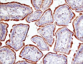
![Immunohistochemistry Galectin-4 Antibody [Unconjugated]](https://images.novusbio.com/images/antibody/af2128_mouse-galectin-4-affinity-purified-polyclonal-ab-immunohistochemistry-29122022154254.jpg)
![N/A IL-10 [Biotin]](https://images.novusbio.com/images/elisa/DATA_IL10_DY417_ELISA_2014.jpg)
![Immunohistochemistry CD45 Antibody [Unconjugated]](https://images.novusbio.com/images/antibody/CD45_AF114_Immunohistochemistry_23525.jpg)
![Immunocytochemistry CD45 Antibody [Unconjugated]](https://images.novusbio.com/images/antibody/af114_mouse-cd45-affinity-purified-polyclonal-ab-immunocytochemistry-6122021145449.jpg)
![Western Blot Galectin-8 Antibody [Unconjugated]](https://images.novusbio.com/images/antibody/Galectin8_AF1305_Western_Blot_16880.jpg)
![Immunohistochemistry Galectin-8 Antibody [Unconjugated]](https://images.novusbio.com/images/antibody/Galectin8_AF1305_Immunohistochemistry_19141.jpg)
![Simple Western Galectin-8 Antibody [Unconjugated]](https://images.novusbio.com/images/antibody/Galectin8_AF1305_Simple_Western_18662.jpg)


![Western Blot ERK2 Antibody [Unconjugated]](https://images.novusbio.com/images/antibody/ERK2_AF1230_Western_Blot_5097.jpg)
![Knockout Validated ERK2 Antibody [Unconjugated]](https://images.novusbio.com/images/antibody/ERK2_AF1230_Knockout_Validated_22864.jpg)
![Immunohistochemistry ERK2 Antibody [Unconjugated]](https://images.novusbio.com/images/antibody/ERK2_AF1230_Immunohistochemistry_20696.jpg)


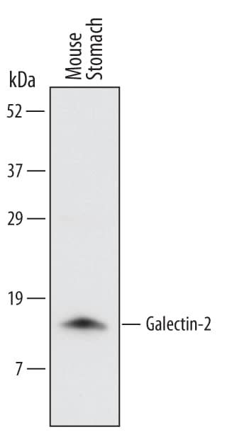
 followed by HRP-conjugated Anti-Goat IgG Secondary Antibody (HAF109). A specific band was detected for Galectin-1 at approximately 14 kDa (as indicated). This experiment was conducted under reducing conditions and using Immunoblot Buffer Group 1.")