Can't find what you are looking for? Use our Antibody Concierge Service & we will help you locate your antibody!
Or feel free to contact us for alternative products.
Datasheet
Reviews & Publications
Protocols & FAQs
Support & Research
Recombinant Human Semaphorin 3E Protein, CF Summary
Details of Functionality
Measured by its ability to inhibit the proliferation of HUVEC human umbilical vein endothelial cells. Moriya, J. et al. (2010) Circ. Res. 106:391. The ED50 for this effect is 0.3-1.5 μg/mL. Measured by its binding ability in a functional ELISA. When Recombinant Human Plexin D1 (Catalog # 4160-PD) is coated at 5 μg/mL, Recombinant Human Semaphorin 3E binds with an apparent KD <2 nM.
Source
Mouse myeloma cell line, NS0-derived human Semaphorin 3E protein Thr25-Ser775 (Arg557Ala and Arg560Ala), with an N-terminal 10-His tag
>90%, by SDS-PAGE under reducing conditions and visualized by silver stain.
Endotoxin Note
<0.10 EU per 1 μg of the protein by the LAL method.
Applications/Dilutions
Dilutions
Binding Activity2
Bioactivity
Theoretical MW
87.9 kDa (monomer). Disclaimer note: The observed molecular weight of the protein may vary from the listed predicted molecular weight due to post translational modifications, post translation cleavages, relative charges, and other experimental factors.
SDS-PAGE
90 kDa, 66 kDa and 25 kDa, reducing conditions
Publications
Read Publications using 3239-S3 in the following applications:
Use a manual defrost freezer and avoid repeated freeze-thaw cycles.
12 months from date of receipt, -20 to -70 °C as supplied.
1 month, 2 to 8 °C under sterile conditions after reconstitution.
3 months, -20 to -70 °C under sterile conditions after reconstitution.
Buffer
Lyophilized from a 0.2 μm filtered solution in PBS and Tween® 20.
Purity
>90%, by SDS-PAGE under reducing conditions and visualized by silver stain.
Reconstitution Instructions
Reconstitute at 100 μg/mL in sterile PBS.
Notes
This product is produced by and ships from R&D Systems, Inc., a Bio-Techne brand.
Alternate Names for Recombinant Human Semaphorin 3E Protein, CF
(semaphorin) 3E
coll-5
KIAA0331M-SEMAH
sema domain, immunoglobulin domain (Ig), short basic domain, secreted
Sema3E
SEMAH
SEMAHM-SemaK
Semaphorin 3E
semaphorin-3E
Background
Semaphorin 3E (Sema3E), previously known as SemaH, is one of six Class 3 (secreted) semaphorins which function in axon guidance and/or vascular tip cell guidance during development (1). Sema3E contains a seven-blade beta -propeller sema domain, a cysteine-knot PSI domain, an Ig-like domain, and a basic region. Dimerization and cleavage within the basic region are required for the repulsing activity of class 3 semaphorins (2). Sema3E can also be cleaved at a furin consensus sequence C-terminal to the sema domain, resulting in a 61 kDa form that does not dimerize and is highly expressed in tumor cell lines with metastatic potential (3, 4). Mature human Sema3E shares 90% aa sequence identity with mouse and rat Sema3E. Alternative splicing generates a short isoform that lacks the signal peptide and the N-terminal 35 residues of the mature protein. Sema3E signaling is transduced by Plexin D1 which may also be associated with Neuropilin 1 and/or VEGF R2 (2, 5, 6). Its interaction with Plexin D1 inhibits axon migration in the neocortex and forebrain (6, 7), although it can attract axons that express both Plexin D1 and Neuropilin 1 (6). Sema3E promotes axonal growth (5), the development of glutamatergic synaptic specificity (8, 9), and the development of GnRH producing neurons (10). Genetic disruption of either Sema3E or Plexin D1 in mouse causes excessive and disorganized vascular growth and branching, indicating the importance of this ligand-receptor pair for vascular guidance (11, 12). In addition, Sema3E is up-regulated by inflammatory macrophages and damaged hepatocytes (13-15). It inhibits smooth muscle cell proliferation and migration in the asthmatic airway (16), promotes hepatic stellate cell activation and wound healing (15), and regulates the migration of developing thymocytes (17).
Oh, W.J. and C. Gu (2013) Semin. Cell Dev. Biol. 24:156.
Adams, R. H. et al. (1997) EMBO J. 16:6077.
Christensen, C. et al. (2005) Cancer Res. 65:6167.
Casazza, A. et al. (2010) J. Clin. Invest. 120:2684.
Bellon, A. et al. (2010) Neuron 66:205.
Chauvet, S. et al. (2007) Neuron 56:807.
Bribian, A. et al. (2014) Nat. Commun. 5:4265.
Ding, J.B. et al. (2011) Nat. Neurosci. 15:215.
Pecho-Vrieseling, E. et al. (2009) Nature 459:842.
Cariboni, A. et al. (2015) J. Clin. Invest. 125:2413.
Gu, C. et al. (2005) Science 307:265.
Gitler, A. D. et al. (2004) Developmental Cell 7:107.
Wanschel, A. et al. (2013) Arterioscler. Thromb. Vasc. Biol. 33:886.
Shimizu, I. et al. (2013) Cell Metab. 18:491.
Yagai, T. et al. (2014) Am. J. Pathol. 184:2250.
Movassagh, H. et al. (2014) J. Allergy Clin. Immunol. 133:560.
Choi, Y.I. et al. (2014) Proc. Natl. Acad. Sci. USA 111:379.
The concentration calculator allows you to quickly calculate the volume, mass or concentration of your vial. Simply enter your mass, volume, or concentration values for your reagent and the calculator will determine the rest.
=
÷
Review this Product
Be the first to review our Recombinant Human Semaphorin 3E Protein, CF and receive a gift card or discount.
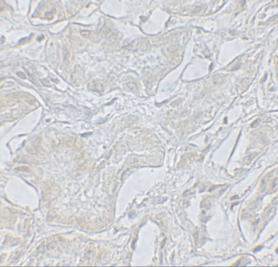
![Immunohistochemistry Neuropilin-1 Antibody [Unconjugated]](https://images.novusbio.com/images2/Neuropilin1_AF566_Immunohistochemistry_21496.jpg)
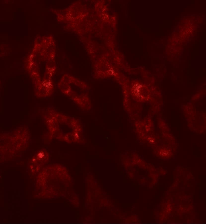
![Immunohistochemistry Neuropilin-1 Antibody [Unconjugated]](https://images.novusbio.com/images2/Neuropilin1_AF566_Immunohistochemistry_21497.jpg)
![Western Blot Neuropilin-1 Antibody [Unconjugated]](https://images.novusbio.com/images2/Neuropilin1_AF566_Western_Blot_21557.jpg)
![Neutralization VEGFR2/KDR/Flk-1 Antibody [Unconjugated]](https://images.novusbio.com/images2/VEGF_R2_AF644_Block_Neutralize_1519.jpg)
![Immunohistochemistry VEGFR2/KDR/Flk-1 Antibody [Unconjugated]](https://images.novusbio.com/images2/VEGF_R2_AF644_Immunohistochemistry_22159.jpg)
![Immunohistochemistry VEGFR2/KDR/Flk-1 Antibody [Unconjugated]](https://images.novusbio.com/images2/VEGF_R2_AF644_Immunohistochemistry_6888.jpg)


![Immunohistochemistry Plexin D1 Antibody [Unconjugated]](https://images.novusbio.com/images2/Plexin_D1_AF4160_Immunohistochemistry_11521.jpg)
![Flow Cytometry Plexin D1 Antibody [Unconjugated]](https://images.novusbio.com/images2/Plexin_D1_AF4160_Flow_Cytometry_8234.jpg)
![Neutralization Plexin D1 Antibody [Unconjugated]](https://images.novusbio.com/images2/Plexin_D1_AF4160_Block_Neutralize_14419.jpg)
![Bioactivity Semaphorin 3A [Unconjugated]](https://images.novusbio.com/images2/Semaphorin_3A_1250S3_1473.jpg)
![Immunohistochemistry Neuropilin-2 Antibody [Unconjugated]](https://images.novusbio.com/images2/Neuropilin2_AF567_Immunohistochemistry_16084.jpg)
![Western Blot Neuropilin-2 Antibody [Unconjugated]](https://images.novusbio.com/images2/Neuropilin2_AF567_Western_Blot_20048.jpg)
![Simple Western Neuropilin-2 Antibody [Unconjugated]](https://images.novusbio.com/images2/Neuropilin2_AF567_Simple_Western_20049.jpg)
![N/A VEGF [HRP]](https://images.novusbio.com/images2/DATA_VEGF_DVE00_ELISA_871.jpg)
![N/A VEGF [HRP]](https://images.novusbio.com/images2/DATA_VEGF_DVE00_ELISA_872.jpg)
![N/A VEGF [HRP]](https://images.novusbio.com/images2/VEGF_DVE00_ELISA_208.jpg)

![Immunohistochemistry Semaphorin 6A Antibody [Unconjugated]](https://images.novusbio.com/images/antibody/af1615_mouse-semaphorin-6a-affinity-purified-polyclonal-ab-immunohistochemistry-92202314926.jpg)
![Immunocytochemistry Semaphorin 3C Antibody (238835) [Unconjugated]](https://images.novusbio.com/images2/Semaphorin_3C_MAB1728_Immunocytochemistry_11554.jpg)
![Immunohistochemistry Semaphorin 3C Antibody (238835) [Unconjugated]](https://images.novusbio.com/images2/Semaphorin_3C_MAB1728_Immunohistochemistry_19277.jpg)
![SDS-PAGE ErbB2/Her2 [Unconjugated]](https://images.novusbio.com/images2/ErbB2_1129-ER_70.jpg)
![Bioactivity ErbB2/Her2 [Unconjugated]](https://images.novusbio.com/images2/1129-er_recombinant-human-erbb2-her2-fc-chimera-protein-cf-bioactivity-7122020142841.jpg)
![N/A ERK1 [p Thr202, p Tyr204] [Biotin]](https://images.novusbio.com/images2/dyc1825-2_phospho-erk1-duoset-ic-economy-pack-15-plate-17122020121349.png)
![N/A ERK1 [p Thr202, p Tyr204] [Biotin]](https://images.novusbio.com/images2/dyc1825-2_phospho-erk1-duoset-ic-economy-pack-15-plate-17122020122737.png)
![N/A ERK1 [p Thr202, p Tyr204] [Biotin]](https://images.novusbio.com/images2/dyc1825-2_phospho-erk1-duoset-ic-economy-pack-15-plate-17122020122919.png)
![Immunocytochemistry ERK2 Antibody (191801) [Unconjugated]](https://images.novusbio.com/images2/ERK2_MAB1230_Immunocytochemistry__Immunofluorescence_20756.jpg)
![Western Blot ERK2 Antibody (191801) [Unconjugated]](https://images.novusbio.com/images2/ERK2_MAB1230_Western_Blot_5959.jpg)
![Simple Western ERK2 Antibody (191801) [Unconjugated]](https://images.novusbio.com/images2/16356.jpg)
![Immunohistochemistry Plexin A3 Antibody [Unconjugated]](https://images.novusbio.com/images2/Plexin_A3_AF4075_Immunohistochemistry_11308.jpg)
![Immunohistochemistry Plexin A3 Antibody [Unconjugated]](https://images.novusbio.com/images2/af4075_mouse-rat-plexin-a3-affinity-purified-polyclonal-ab-immunohistochemistry-1642021131527.jpg)