| Reactivity | HuSpecies Glossary |
| Applications | Binding Activity |
| Format | Carrier-Free |
| Details of Functionality | Measured by its binding ability in a functional ELISA. Immobilized rhEphB6/Fc Chimera at 2 µg/mL (100 µL/well) can bind rmEphrin-B2/Fc Chimera with a linear range of 0.0780-5.00 ng/mL. |
||||||
| Source | Mouse myeloma cell line, NS0-derived human EphB6 protein
|
||||||
| Accession # | |||||||
| N-terminal Sequence | Leu17 |
||||||
| Structure / Form | Disulfide-linked homodimer |
||||||
| Protein/Peptide Type | Recombinant Proteins |
||||||
| Gene | EPHB6 |
||||||
| Purity | >90%, by SDS-PAGE visualized with Silver Staining and quantitative densitometry by Coomassie® Blue Staining |
||||||
| Endotoxin Note | <0.01 EU per 1 μg of the protein by the LAL method. |
| Dilutions |
|
|
| Theoretical MW | 86.7 kDa (monomer). Disclaimer note: The observed molecular weight of the protein may vary from the listed predicted molecular weight due to post translational modifications, post translation cleavages, relative charges, and other experimental factors. |
|
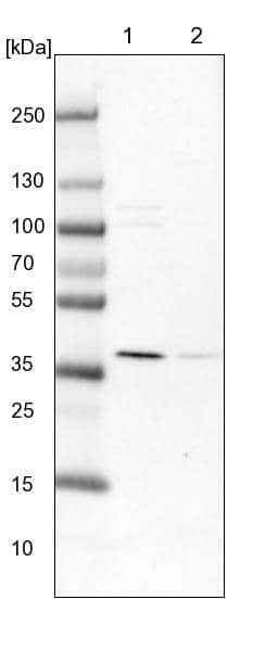
| SDS-PAGE | 104 kDa, under reducing conditions. |
|
| Publications |
|
| Storage | Use a manual defrost freezer and avoid repeated freeze-thaw cycles.
|
| Buffer | Lyophilized from a 0.2 μm filtered solution in PBS. |
| Purity | >90%, by SDS-PAGE visualized with Silver Staining and quantitative densitometry by Coomassie® Blue Staining |
| Reconstitution Instructions | Reconstitute at 100 μg/mL in sterile PBS. |
EphB6, also known as Hep and Mep, is a 110 kDa member of the Eph receptor tyrosine kinase family. The A and B classes of Eph proteins are distinguished by ligand preference and have a common structural organization (1 - 4). The human EphB6 cDNA encodes a 1006 amino acid (aa) precursor that includes a 16 aa signal sequence, a 563 aa extracellular domain (ECD), a 21 aa transmembrane segment, and a 406 aa cytoplasmic domain. The ECD contains serine- and cysteine-rich regions and two fibronectin type-III domains. The cytoplasmic domain contains one non-catalytic protein kinase-like, one proline-rich, one SAM, and one PDZ-binding domain (5, 6). Within the ECD, human EphB6 shares 91% aa sequence identity with mouse and rat EphB6. It shares 38 - 45% aa sequence identity with human EphB1, 2, 3, 4, and 6. Human EphB5 has not been characterized. Two secreted splice variants have been described in mouse but not in human (6). EphB6 is primarily expressed in brain, pancreas, thymus, and peripheral T cells (5, 7, 8). EphB6 forms stable heterodimers with EphB1 and participates in signal transduction by association with other enzymatically active molecules (9 - 11). Ephrin-B2 is the dominant ligand for EphB6, although Ephrin-B1 and Ephrin-B3 can also trigger responses (12 - 14). High concentrations of Ephrin-B2 inhibit cell adhesion and migration as well as tyrosine phosphorylation of EphB6. Conversely, low concentrations of Ephrin-B2 promote adhesion and migration and do not lead to EphB6 phosphorylation (15). The level of EphB6 expression is inversely correlated with tumor aggressiveness in a variety of malignancies (1). EphB6 also functions as a T cell co-stimulatory molecule (8, 11, 13). EphB6 clusters with the T cell receptor and participates in the subsequent attenuation of the T cell response (8, 10, 11, 13).

![Western Blot EphB2 Antibody [Unconjugated]](https://images.novusbio.com/images/af467_human-mouse-ephb2-affinity-purified-polyclonal-ab-41202410485412.jpg)
![Immunohistochemistry EphB2 Antibody [Unconjugated]](https://images.novusbio.com/images/antibody/EphB2_AF467_Immunohistochemistry_20369.jpg)
![Western Blot EphB2 Antibody [Unconjugated]](https://images.novusbio.com/images/antibody/EphB2_AF467_Western_Blot_21511.jpg)


![Immunohistochemistry Albumin Antibody (188835) [Unconjugated] - Serum](https://images.novusbio.com/images/antibody/Albumin_MAB1455_Immunohistochemistry_23317.jpg)
![Immunocytochemistry Albumin Antibody (188835) [Unconjugated] - Serum](https://images.novusbio.com/images/antibody/Albumin_MAB1455_Immunocytochemistry__Immunofluorescence_17777.jpg)
![Intracellular Staining by Flow Cytometry Albumin Antibody (188835) [Unconjugated] - Serum](https://images.novusbio.com/images/antibody/Albumin_MAB1455_Flow_Cytometry_15110.jpg)


| Publication using 3384-B6 | Applications | Species |
|---|---|---|
| Zhang , Gu, Brady , John, Liang , Wei-Chin, Wu , Yan, Henkemeyer , Mark, Yan , Minhong EphB4 forward signalling regulates lymphatic valve development. Nat Commun, 2015-04-13;6(0):6625. 2015-04-13 [PMID: 25865237] (Enzyme Assay, Human) | Enzyme Assay | Human |
The concentration calculator allows you to quickly calculate the volume, mass or concentration of your vial. Simply enter your mass, volume, or concentration values for your reagent and the calculator will determine the rest.
| Gene Symbol | EPHB6 |
| Uniprot |


![Immunohistochemistry Insulin Antibody (182410) [Unconjugated]](https://images.novusbio.com/images/antibody/mab1417_human-bovine-mouse-insulin-mab-clone-182410-immunohistochemistry-308202115145.jpg)
![Immunocytochemistry Insulin Antibody (182410) [Unconjugated]](https://images.novusbio.com/images/antibody/Insulin_MAB1417_Immunocytochemistry_9376.jpg)
![Immunocytochemistry EphA1 Antibody [Unconjugated]](https://images.novusbio.com/images/antibody/EphA1_AF638_Immunocytochemistry__Immunofluorescence_20119.jpg)
![Western Blot EphA1 Antibody [Unconjugated]](https://images.novusbio.com/images/antibody/EphA1_AF638_Western_Blot_17606.jpg)


![N/A Thrombospondin-1 [HRP]](https://images.novusbio.com/images/elisa/Thrombospondin-1_DTSP10_ELISA_199.jpg)
![N/A Thrombospondin-1 [HRP]](https://images.novusbio.com/images/dtsp10_human-thrombospondin-1-quantikine-elisa-kit-23120261145.jpg)
![Western Blot JNK1 Antibody (228601) [Unconjugated]](https://images.novusbio.com/images/antibody/JNK1_MAB17761_Western_Blot_5991.jpg)
![Western Blot JNK1 Antibody (228601) [Unconjugated]](https://images.novusbio.com/images/antibody/JNK1_MAB17761_Western_Blot_6327.jpg)
![Immunocytochemistry JNK1 Antibody (228601) [Unconjugated]](https://images.novusbio.com/images/antibody/JNK1_MAB17761_Immunocytochemistry__Immunofluorescence_20184.jpg)
![N/A IGFBP-1 [Biotin]](https://images.novusbio.com/images/elisa/DATA_IGFBP1_DY871_ELISA_2250.jpg)
![Flow Cytometry Ephrin-B2 Antibody [Unconjugated]](https://images.novusbio.com/images/antibody/EphrinB2_AF496_Flow_Cytometry_18997.jpg)
![Immunocytochemistry Ephrin-B2 Antibody [Unconjugated]](https://images.novusbio.com/images/antibody/Ephrin-B2_AF496_Immunocytochemistry_6513.jpg)
![Western Blot Ephrin-B2 Antibody [Unconjugated]](https://images.novusbio.com/images/antibody/af496_human-mouse-rat-ephrin-b2-affinity-purified-polyclonal-ab-western-blot-2152026132638.jpg)
![N/A IL-6 [HRP]](https://images.novusbio.com/images/elisa/DATA_IL6_M6000_ELISA_936.jpg)
![N/A IL-6 [HRP]](https://images.novusbio.com/images/elisa/IL-6_M6000_ELISA_415.jpg)
![N/A IL-6 [HRP]](https://images.novusbio.com/images/m6000b_mouse-il-6-quantikine-elisa-kit-1752025024034.jpg)
![Bioactivity IGF-I/IGF-1 [Unconjugated]](https://images.novusbio.com/images/protein/IGF-I_291-G1_41.jpg)
![Mass Spectrometry IGF-I/IGF-1 [Unconjugated]](https://images.novusbio.com/images/protein/IGF-I_291-G1_42.jpg)
![SEC-MALS IGF-I/IGF-1 [Unconjugated]](https://images.novusbio.com/images/291-g1_recombinant-human-igf-i-igf-1-protein-cf-sec-mals-224202691859.jpg)
![Western Blot Ephrin-B3 Antibody [Unconjugated]](https://images.novusbio.com/images/antibody/EphrinB3_AF395_Western_Blot_16885.jpg)
![SDS-Page TNF-alpha [Unconjugated]](https://images.novusbio.com/images/protein/TNF-alpha_210-TA_256.jpg)
![Bioactivity TNF-alpha [Unconjugated]](https://images.novusbio.com/images/protein/TNFalpha_210TA_1658.jpg)
![SEC-MALS TNF-alpha [Unconjugated]](https://images.novusbio.com/images/210-ta_recombinant-human-tnf-alpha-protein-sec-mals-35202312244..jpg)