![Western Blot: PMEL17/SILV Antibody (PMEL/1825R) [NBP2-53180] - Western Blot Analysis of COLO-38 cell lysate using PMEL17/SILV Rabbit Recombinant Monoclonal Antibody (PMEL/1825R).](http://images.novusbio.com/fullsize/PMEL17-SILV-Antibody-PMEL-1825R-Western-Blot-NBP2-53180-img0002.jpg "Western Blot: PMEL17/SILV Antibody (PMEL/1825R) [NBP2-53180] - Western Blot Analysis of COLO-38 cell lysate using PMEL17/SILV Rabbit Recombinant Monoclonal Antibody (PMEL/1825R).")
| Reactivity | HuSpecies Glossary |
| Applications | WB, IHC |
| Clone | PMEL/1825R |
| Clonality | Monoclonal |
| Host | Rabbit |
| Conjugate | Unconjugated |
| Concentration | 0.2 mg/ml |
| Description | 200ug/ml of antibody purified from Bioreactor Concentrate by Protein A or G. Prepared in 10 mM PBS with 0.05% BSA & 0.05% azide. Also available WITHOUT BSA & azide at 1.0 mg/ml. (NBP2-54454) Antibody with azide - store at 2 to 8C. Antibody without azide - store at -20 to -80 C. |
| Additional Information | Recombinant Monoclonal Antibody |
| Immunogen | Recombinant full-length human PMEL17/SILV protein (Uniprot: P40967 ) |
| Localization | Cytoplasmic |
| Marker | Melanoma Marker |
| Specificity | The gp100 molecule is a 100kDa glycosylated protein that is cleaved into a small (26kDa) carboxy-terminal fragment and a larger amino- terminal section (60 64 kDa), which is subsequently cleaved to generate 26kDa and 34 38kDa fragments. By immunohistochemistry, it specifically recognizes a protein in melanocytes and melanomas. This monoclonal antibody reacts with junctional and blue nevus cells and variably with fetal and neonatal melanocytes. Intradermal nevi, normal adult melanocytes, and non-melanocytic cells are negative. It does not stain tumor cells of epithelial, lymphoid, glial, or mesenchymal origin. Metastatic amelanotic melanoma can often be confused with a variety of poorly differentiated carcinomas, large cell lymphomas, and sarcomas using H E stains alone. It is also difficult to differentiate melanoma from spindle cell carcinomas and various types of mesenchymal neoplasms. This monoclonal antibody stains fetal and neonatal melanocytes, junctional and blue nevus cells, and malignant melanoma. This monoclonal antibody also stains Angiomyolipoma (PEComa). |
| Isotype | IgG |
| Clonality | Monoclonal |
| Host | Rabbit |
| Gene | PMEL |
| Purity | Protein A or G purified |
| Innovator's Reward | Test in a species/application not listed above to receive a full credit towards a future purchase. |
| Dilutions |
|
| Application Notes | Immunohistochemistry (Formalin-fixed): 1-2ug/ml for 30 minutes at RT. Staining of formalin-fixed tissues requires heating tissue sections in 10mM Tris with 1mM EDTA, pH 9.0, for 45 min at 95C followed by cooling at RT for 20 minutes. Optimal dilution for a specific application should be determined. |
| Storage | Store at 4C. |
| Buffer | 10 mM PBS with 0.05% BSA |
| Preservative | 0.05% Sodium Azide |
| Concentration | 0.2 mg/ml |
| Purity | Protein A or G purified |


Secondary Antibodies |
Isotype Controls |
The concentration calculator allows you to quickly calculate the volume, mass or concentration of your vial. Simply enter your mass, volume, or concentration values for your reagent and the calculator will determine the rest.


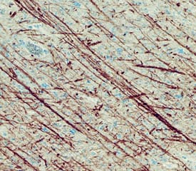
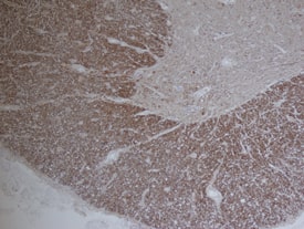
![Immunohistochemistry-Paraffin: PMEL17/SILV Antibody (PMEL/1825R) [NBP2-53180] - Formalin-fixed, paraffin-embedded human Melanoma stained with gp100 Recombinant Rabbit Monoclonal Antibody (PMEL/1825R).](http://images.novusbio.com/fullsize/PMEL17-SILV-Antibody-PMEL-1825R-Immunohistochemistry-Paraffin-NBP2-53180-img0001.jpg "Immunohistochemistry-Paraffin: PMEL17/SILV Antibody (PMEL/1825R) [NBP2-53180] - Formalin-fixed, paraffin-embedded human Melanoma stained with gp100 Recombinant Rabbit Monoclonal Antibody (PMEL/1825R).")


![Immunohistochemistry Insulin Antibody (182410) [Unconjugated]](https://images.novusbio.com/images/antibody/mab1417_human-bovine-mouse-insulin-mab-clone-182410-immunohistochemistry-308202115145.jpg)
![Immunocytochemistry Insulin Antibody (182410) [Unconjugated]](https://images.novusbio.com/images/antibody/Insulin_MAB1417_Immunocytochemistry_9376.jpg)
![Immunocytochemistry CD55/DAF Antibody [Unconjugated]](https://images.novusbio.com/images/antibody/CD55_AF2009_Immunocytochemistry__Immunofluorescence_23052.jpg)
![Immunohistochemistry CD55/DAF Antibody [Unconjugated]](https://images.novusbio.com/images/antibody/CD55_AF2009_Immunohistochemistry_6728.jpg)
![Immunocytochemistry Catalase Antibody [Unconjugated]](https://images.novusbio.com/images/antibody/Catalase_AF3398_Immunocytochemistry__Immunofluorescence_19451.jpg)
![Simple Western Catalase Antibody [Unconjugated]](https://images.novusbio.com/images/antibody/Catalase_AF3398_Simple_Western_16990.jpg)


![SDS-Page TNF-alpha [Unconjugated]](https://images.novusbio.com/images/protein/TNF-alpha_210-TA_256.jpg)
![Bioactivity TNF-alpha [Unconjugated]](https://images.novusbio.com/images/protein/TNFalpha_210TA_1658.jpg)
![SEC-MALS TNF-alpha [Unconjugated]](https://images.novusbio.com/images/210-ta_recombinant-human-tnf-alpha-protein-sec-mals-35202312244..jpg)
![Western Blot: Goat anti-Rabbit IgG (H+L) Secondary Antibody [HRP] [NB7160] - Western blot showing vemurafenib treatment in BRAFV600E CRC cells inhibits fission mediator DRP1 with no significant effect on fusion proteins (Mfn1 & 2) using MFN-1 antibody (NBP1-51841) and corresponding secondary antibody, goat anti-rabbit IgG-HRP (NB7160). Image collected and cropped by CiteAb from the following publication (https://pubmed.ncbi.nlm.nih.gov/33738242).](https://images.novusbio.com/images/Goat-anti-Rabbit-IgG-H+L-Secondary-Antibody-HRP-Western-Blot-NB7160-img0001.jpg "Western Blot: Goat anti-Rabbit IgG (H+L) Secondary Antibody [HRP] [NB7160] - Western blot showing vemurafenib treatment in BRAFV600E CRC cells inhibits fission mediator DRP1 with no significant effect on fusion proteins (Mfn1 & 2) using MFN-1 antibody (NBP1-51841) and corresponding secondary antibody, goat anti-rabbit IgG-HRP (NB7160). Image collected and cropped by CiteAb from the following publication (https://pubmed.ncbi.nlm.nih.gov/33738242).")
 followed by 30 min incubation with Goat anti Rabbit HRP conjugated secondary antibodies (Catalog # HAF008) at 1:20 dilution + DAB chromogen (brown). The tissue was counterstained with Hematoxylin (blue). Control was done by omitting primary antibody.")
![Flow Cytometry: Rabbit IgG Isotype Control [NBP2-24891] - An intracellular stain was performed on Raji cells with Adiponectin antibody NB100-65810 (blue) and a matched isotype control NBP2-24893 (orange). Cells were fixed with 4% PFA and then permeablized with 0.1% saponin. Cells were incubated in an antibody dilution of 1 ug/mL for 30 minutes at room temperature, followed by Dylight488-conjugated anti-rabbit secondary antibody. Image using the Azide Free form of this antibody.](https://images.novusbio.com/images/Rabbit--Mouse-IgG-Isotype-Control-Flow-Cytometry-NBP2-24891-img0006.jpg "Flow Cytometry: Rabbit IgG Isotype Control [NBP2-24891] - An intracellular stain was performed on Raji cells with Adiponectin antibody NB100-65810 (blue) and a matched isotype control NBP2-24893 (orange). Cells were fixed with 4% PFA and then permeablized with 0.1% saponin. Cells were incubated in an antibody dilution of 1 ug/mL for 30 minutes at room temperature, followed by Dylight488-conjugated anti-rabbit secondary antibody. Image using the Azide Free form of this antibody.")