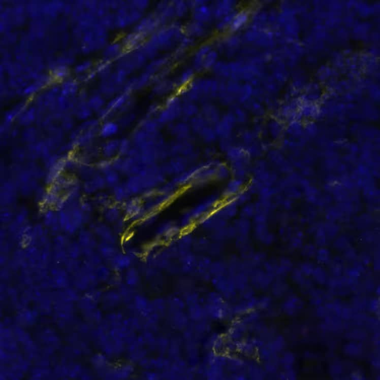
DR647MS) and counterstained with DAPI (blue; Lunaphore Catalog # DR100). Specific staining was localized to the neuropil. Protocol available in COMET™ Panel Builder." title="NCAM-1/CD56 was detected in immersion fixed paraffin-embedded sections of human brain Cortex using Mouse Anti-Human NCAM-1/CD56, Monoclonal Antibody (Catalog # MAB24081) at 25ug/mL at 37° Celsius for 8 minutes. Before incubation with the primary antibody, tissue underwent an all-in-one dewaxing and antigen retrieval preprocessing using PreTreatment Module (PT Module) and Dewax and HIER Buffer H (pH 9; Epredia Catalog # TA-999-DHBH). Tissue was stained using the Alexa Fluor™ 647 Goat anti-Mouse IgG Secondary Antibody at 1:200 at 37 ° Celsius for 2 minutes. (Yellow; Lunaphore Catalog # DR647MS) and counterstained with DAPI (blue; Lunaphore Catalog # DR100). Specific staining was localized to the neuropil. Protocol available in COMET™ Panel Builder." />
| Reactivity | HuSpecies Glossary |
| Applications | WB, Flow, IHC, CyTOF-ready, mIF |
| Clone | 301021 |
| Clonality | Monoclonal |
| Host | Mouse |
| Conjugate | Unconjugated |
| Concentration | LYOPH |
| Immunogen | Mouse myeloma cell line NS0-derived recombinant human NCAM-1/CD56 Leu20-Pro603 and Glu636-Asn741 Accession # NP_001070150 |
| Specificity | Detects human NCAM-1/CD56 in direct ELISAs and Western blots. In direct ELISAs and Western blots, no cross-reactivity with recombinant human (rh) ALCAM, rhBCAM, rhEPCAM, rhMCAM, rhNCAM-1-L1, recombinant mouse (rm) MAdCAM-1, rmNCAM-1-L1, or rmOCAM is observed. |
| Source | N/A |
| Isotype | IgG2b |
| Clonality | Monoclonal |
| Host | Mouse |
| Gene | NCAM1 |
| Purity Statement | Protein A or G purified from hybridoma culture supernatant |
| Innovator's Reward | Test in a species/application not listed above to receive a full credit towards a future purchase. |
| Dilutions |
|
|
| Publications |
|
| Storage | Use a manual defrost freezer and avoid repeated freeze-thaw cycles.
|
| Buffer | Lyophilized from a 0.2 μm filtered solution in PBS with Trehalose. See Certificate of Analysis for details. *Small pack size (-SP) is supplied either lyophilized or as a 0.2 µm filtered solution in PBS. |
| Preservative | No Preservative |
| Concentration | LYOPH |
| Reconstitution Instructions | Reconstitute at 0.5 mg/mL in sterile PBS. For liquid material, refer to CoA for concentration. |
Neural cell adhesion molecule 1 (NCAM-1) is a multifunctional member of the Ig superfamily. It belongs to a family of membrane-bound glycoproteins that are involved in Ca++ independent cell matrix and homophilic or heterophilic cell-cell interactions. NCAM-1 specifically binds to heparan sulfate proteoglycans (1), the extracellular matrix protein agrin (2), and several chondroitin sulfate proteoglycans that include neurocan and phosphocan (3). There are three main forms of human NCAM-1 that arise by alternate splicing. These are designated NCAM-120/NCAM-1 (761 amino acids [aa]), NCAM-140 (848 aa), and NCAM-180 (1120 aa). NCAM-120 is GPI-linked, while NCAM-140 and NCAM-180 are type I transmembrane glycoproteins (4‑6). Additional alternate splicing adds considerable diversity to all three forms, and extracellular proteolytic processing is possible for NCAM-180 (7‑8). NCAM-1 is synthesized as a 761 aa preproprecursor that contains a 19 aa signal sequence, a 722 aa GPI-linked mature region, and a 20 aa C-terminal prosegment (4). The molecule contains five C-2 type Ig-like domains and two fibronectin type-III domains. Human to mouse, NCAM-1 is 93% aa identical. NCAM-1 appears to be highly sialylated. The polysialyation of NCAM-1 reduces its adhesive property and increases its neurite outgrowth promoting features (9). NCAM-1 in the adult brain shows a decline of sialylation relative to earlier developmental periods. In regions that retain a high degree of neuronal plasticity, however, the adult brain continues to express polysialylation-NCAM-1, suggesting sialylation of NCAM-1 is involved in regenerative processes and synaptic plasticity (10‑13).




![NCAM-1/CD56 Antibody (301021) [Unconjugated]](/sites/all/modules/enterprise-tech/et_datasheets/images/novus_guarantee.png "NCAM-1/CD56 Antibody (301021) [Unconjugated]")
Secondary Antibodies |
Isotype Controls |
The concentration calculator allows you to quickly calculate the volume, mass or concentration of your vial. Simply enter your mass, volume, or concentration values for your reagent and the calculator will determine the rest.
| Gene Symbol | NCAM1 |
| Uniprot |


 HAF018). A specific band was detected for NCAM‑1/CD56 at approximately 105-130 kDa (as indicated). This experiment was conducted under reducing conditions and using Western Blot Buffer Group 1." title="Western blot shows lysates of human brain (cerebellum and hypothalamus). PVDF membrane was probed with 2 µg/mL of Mouse Anti-Human NCAM‑1/CD56 Monoclonal Antibody (Catalog # MAB24081) followed by HRP-conjugated Anti-Mouse IgG Secondary Antibody (
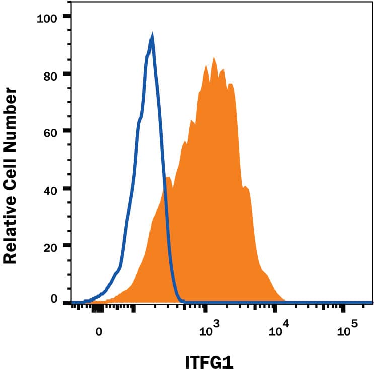
 MAB0041) followed by Anti-Mouse IgG APC-conjugated secondary antibody (
 CLD004) and were stained with either (A) Mouse Anti-Human NCAM-1/CD56 Monoclonal Antibody (Catalog # MAB24081) or (B) Mouse IgG2B Isotype Control (
 VC001) or the HRP-conjugated Anti-Mouse IgG Secondary Antibody (Catalog #
 ![Bioactivity CTLA-4 [Unconjugated]](https://images.novusbio.com/images/protein/CTLA4_7268CT_2293.jpg)
![Bioactivity IL-2 [Unconjugated]](https://images.novusbio.com/images/202-il_recombinant-human-il-2-protein-bioactivity-174202314946.jpg)
![Binding Activity Fc gamma RIIIB/CD16b [Unconjugated]](https://images.novusbio.com/images/protein/Fc_gamma_RIIIB_1597-FC_CF_776.jpg)
![SDS-Page Fc gamma RIIIB/CD16b [Unconjugated]](https://images.novusbio.com/images/protein/Fc_gamma_RIIIB_1597-FC_CF_756.jpg)
![SEC-MALS Fc gamma RIIIA/CD16a [Unconjugated]](https://images.novusbio.com/images/4325-fc_recombinant-human-fc-gamma-riiia-cd16a-protein-cf-sec-mals-2842023172947.jpg)
![Binding Activity Fc gamma RIIIA/CD16a [Unconjugated]](https://images.novusbio.com/images/protein/Fc_gamma_RIIIA_4325-FC_491.jpg)
![SDS-Page Fc gamma RIIIA/CD16a [Unconjugated]](https://images.novusbio.com/images/protein/Fc_gamma_RIIIA_4325-FC_411.jpg)
![Immunohistochemistry CD45 Antibody [Unconjugated]](https://images.novusbio.com/images/antibody/CD45_AF114_Immunohistochemistry_23525.jpg)
![Immunocytochemistry CD45 Antibody [Unconjugated]](https://images.novusbio.com/images/antibody/af114_mouse-cd45-affinity-purified-polyclonal-ab-immunocytochemistry-6122021145449.jpg)
![SDS-Page TNF-alpha [Unconjugated]](https://images.novusbio.com/images/protein/TNF-alpha_210-TA_256.jpg)
![Bioactivity TNF-alpha [Unconjugated]](https://images.novusbio.com/images/protein/TNFalpha_210TA_1658.jpg)
![SEC-MALS TNF-alpha [Unconjugated]](https://images.novusbio.com/images/210-ta_recombinant-human-tnf-alpha-protein-sec-mals-35202312244..jpg)
![SEC-MALS IFN-gamma [Unconjugated]](https://images.novusbio.com/images/485-mi_recombinant-mouse-ifn-gamma-protein-sec-mals-1612202583245.jpg)
![Bioactivity IFN-gamma [Unconjugated]](https://images.novusbio.com/images/protein/IFN-gamma_485-MI_455.jpg)
![SDS-Page IFN-gamma [Unconjugated]](https://images.novusbio.com/images/protein/IFN-gamma_485-MI_407.jpg)


![N/A CD25/IL-2R alpha [HRP]](https://images.novusbio.com/images/elisa/IL-2_R_alpha_DR2A00_ELISA_167.jpg)
![N/A CD25/IL-2R alpha [HRP]](https://images.novusbio.com/images/elisa/DATA_CD25_DR2A00_ELISA_800.jpg)