We're moving to rndsystems.com. Come with us!

After August 17, 2026, Novus Biologicals products and services will no longer be available on this website; you will access all products and services on rndsystems.com. Create your R&D Systems online account today.

IgM Antibody (2123A) [Unconjugated]

Images

 
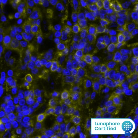
DR555RB) and counterstained with DAPI (blue; Lunaphore Catalog # DR100). Specific staining was localized to the membrane. Protocol available in COMET™ Panel Builder.​​" > IgM was detected in immersion fixed paraffin-embedded sections of human Non-Hodgkins Lymphoma using Rabbit Anti-Human IgM, Monoclonal Antibody (Catalog #MAB9435) at 0.2ug/mL at 37° Celsius for 4 minutes. Before incubation with the primary antibody, tissue underwent an all-in-one dewaxing and antigen retrieval preprocessing using PreTreatment Module (PT Module) and Dewax and HIER Buffer H (pH 9; Epredia Catalog # TA-999-DHBH). Tissue was stained using the Alexa Fluor™ Plus 555 Goat anti-Rabbit IgG Secondary Antibody at 1:100 at 37 ° Celsius for 2 minutes. (Yellow; Lunaphore Catalog # <A class=DR555RB) and counterstained with DAPI (blue; Lunaphore Catalog # DR100). Specific staining was localized to the membrane. Protocol available in COMET™ Panel Builder.​​" title="IgM was detected in immersion fixed paraffin-embedded sections of human Non-Hodgkins Lymphoma using Rabbit Anti-Human IgM, Monoclonal Antibody (Catalog #MAB9435) at 0.2ug/mL at 37° Celsius for 4 minutes. Before incubation with the primary antibody, tissue underwent an all-in-one dewaxing and antigen retrieval preprocessing using PreTreatment Module (PT Module) and Dewax and HIER Buffer H (pH 9; Epredia Catalog # TA-999-DHBH). Tissue was stained using the Alexa Fluor™ Plus 555 Goat anti-Rabbit IgG Secondary Antibody at 1:100 at 37 ° Celsius for 2 minutes. (Yellow; Lunaphore Catalog # DR555RB) and counterstained with DAPI (blue; Lunaphore Catalog # DR100). Specific staining was localized to the membrane. Protocol available in COMET™ Panel Builder.​​" />
IgM was detected in immersion fixed paraffin-embedded sections of human Non-Hodgkins Lymphoma using Rabbit Anti-Human IgM, Monoclonal Antibody (Catalog #MAB9435) at 0.2ug/mL at 37° Celsius for 4 minutes. Before ...read more
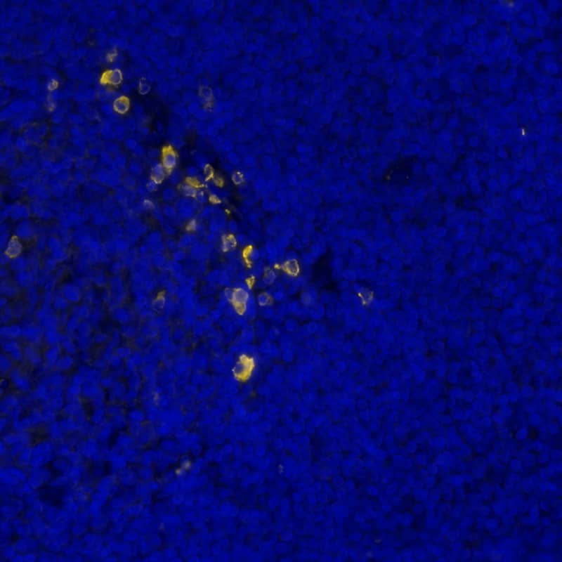
DR555RB) and counterstained with DAPI (blue; Lunaphore Catalog # DR100). Specific staining was localized to the membrane. Protocol available in COMET™ Panel Builder." > DR555RB) and counterstained with DAPI (blue; Lunaphore Catalog # DR100). Specific staining was localized to the membrane. Protocol available in COMET™ Panel Builder." title="IgM was detected in immersion fixed paraffin-embedded sections of human tonsil using Rabbit Anti-Human IgM, Monoclonal Antibody (Catalog #MAB9435) at 0.2ug/mL at 37° Celsius for 4 minutes. Before incubation with the primary antibody, tissue underwent an all-in-one dewaxing and antigen retrieval preprocessing using PreTreatment Module (PT Module)and Dewax and HIER Buffer H (pH 9; Epredia Catalog #TA-999-DHBH). Tissue was stained using the Alexa Fluor™ Plus 555 Goat anti-Rabbit IgG Secondary Antibody at 1:100 at 37 ° Celsius for 2 minutes. (Yellow; Lunaphore Catalog # DR555RB) and counterstained with DAPI (blue; Lunaphore Catalog # DR100). Specific staining was localized to the membrane. Protocol available in COMET™ Panel Builder." />
IgM was detected in immersion fixed paraffin-embedded sections of human tonsil using Rabbit Anti-Human IgM, Monoclonal Antibody (Catalog #MAB9435) at 0.2ug/mL at 37° Celsius for 4 minutes. Before incubation with the ...read more
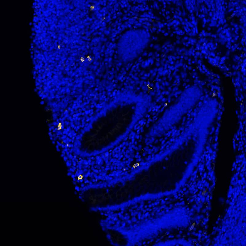
DR555RB) and counterstained with DAPI (blue; Lunaphore Catalog # DR100). Specific staining was localized to the membrane. Protocol available in COMET™ Panel Builder.​" > DR555RB) and counterstained with DAPI (blue; Lunaphore Catalog # DR100). Specific staining was localized to the membrane. Protocol available in COMET™ Panel Builder.​" title="IgM was detected in immersion fixed paraffin-embedded sections of human appendix using Rabbit Anti-Human IgM, Monoclonal Antibody (Catalog #MAB9435) at 0.2ug/mL at 37° Celsius for 4 minutes. Before incubation with the primary antibody, tissue underwent an all-in-one dewaxing and antigen retrieval preprocessing using PreTreatment Module (PT Module) and Dewax and HIER Buffer H (pH 9; Epredia Catalog #TA-999-DHBH). Tissue was stained using the Alexa Fluor™ Plus 555 Goat anti-Rabbit IgG Secondary Antibody at 1:100 at 37 ° Celsius for 2 minutes. (Yellow; Lunaphore Catalog # DR555RB) and counterstained with DAPI (blue; Lunaphore Catalog # DR100). Specific staining was localized to the membrane. Protocol available in COMET™ Panel Builder.​" />
IgM was detected in immersion fixed paraffin-embedded sections of human appendix using Rabbit Anti-Human IgM, Monoclonal Antibody (Catalog #MAB9435) at 0.2ug/mL at 37° Celsius for 4 minutes. Before incubation with the ...read more
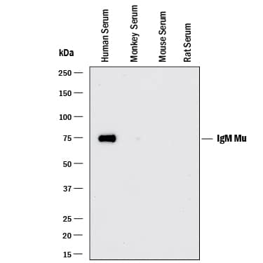
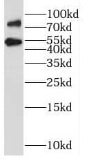
HAF008). A specific band was detected for IgM at approximately 65 kDa (as indicated). This experiment was conducted under reducing conditions and using Immunoblot Buffer Group 1." > HAF008). A specific band was detected for IgM at approximately 65 kDa (as indicated). This experiment was conducted under reducing conditions and using Immunoblot Buffer Group 1." title="Western blot shows lysates of human serum, monkey serum, mouse serum and rat plasma. PVDF membrane was probed with 0.1 µg/mL of Rabbit Anti-Human IgM Monoclonal Antibody (Catalog # MAB9435) followed by HRP-conjugated Anti-Rabbit IgG Secondary Antibody (HAF008). A specific band was detected for IgM at approximately 65 kDa (as indicated). This experiment was conducted under reducing conditions and using Immunoblot Buffer Group 1." />
Western blot shows lysates of human serum, monkey serum, mouse serum and rat plasma. PVDF membrane was probed with 0.1 µg/mL of Rabbit Anti-Human IgM Monoclonal Antibody (Catalog # MAB9435) followed by HRP-conjugated ...read more
Simple Western lane view shows lysates of human serum, loaded at 0.2 mg/mL. A specific band was detected for IgM at approximately 100 kDa (as indicated) using 5 µg/mL of Rabbit Anti-Human IgM Monoclonal ...read more
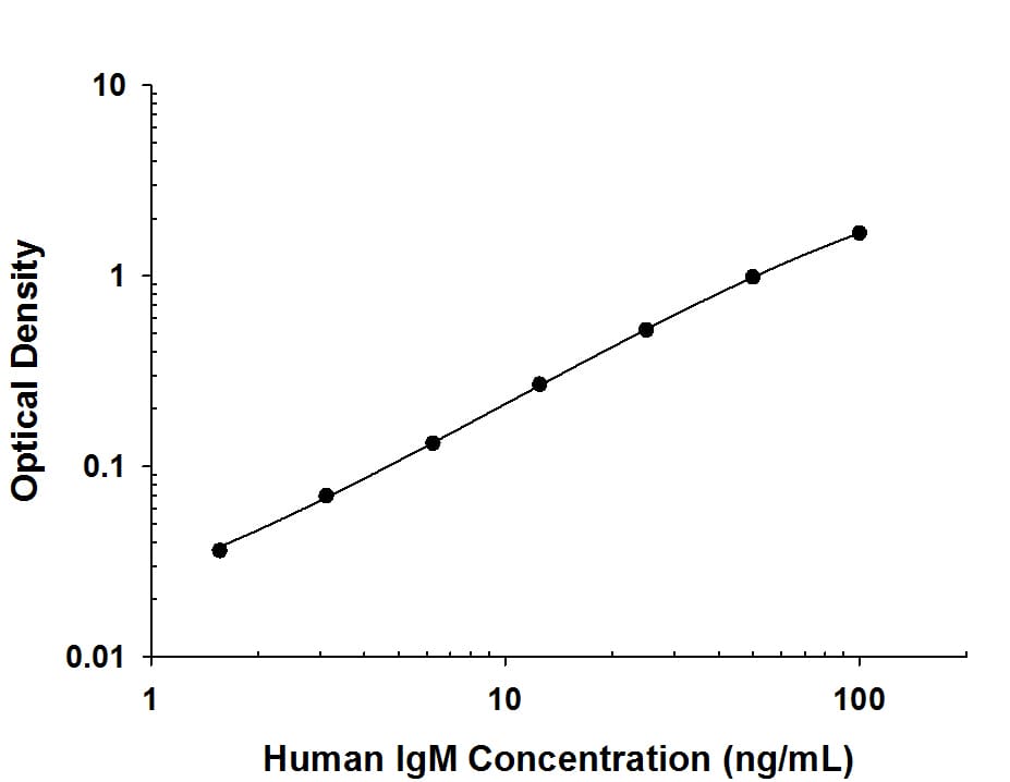
DY990). Rabbit Anti-Human IgM Monoclonal Antibody (MAB94351) was biotinylated and incubated with the protein captured on the plate. Detection of the standard curve was achieved by incubating Streptavidin-HRP (DY998) followed by Substrate Solution (DY999) and stopping the enzymatic reaction with Stop Solution (DY994)." > DY990). Rabbit Anti-Human IgM Monoclonal Antibody (MAB94351) was biotinylated and incubated with the protein captured on the plate. Detection of the standard curve was achieved by incubating Streptavidin-HRP (DY998) followed by Substrate Solution (DY999) and stopping the enzymatic reaction with Stop Solution (DY994)." title="Human IgM protein was serially diluted 2-fold and captured by Rabbit Anti-Human IgM Monoclonal Antibody (Catalog # MAB9435) coated on a Clear Polystyrene Microplate (DY990). Rabbit Anti-Human IgM Monoclonal Antibody (MAB94351) was biotinylated and incubated with the protein captured on the plate. Detection of the standard curve was achieved by incubating Streptavidin-HRP (DY998) followed by Substrate Solution (DY999) and stopping the enzymatic reaction with Stop Solution (DY994)." />
Human IgM protein was serially diluted 2-fold and captured by Rabbit Anti-Human IgM Monoclonal Antibody (Catalog # MAB9435) coated on a Clear Polystyrene Microplate ( ...read more
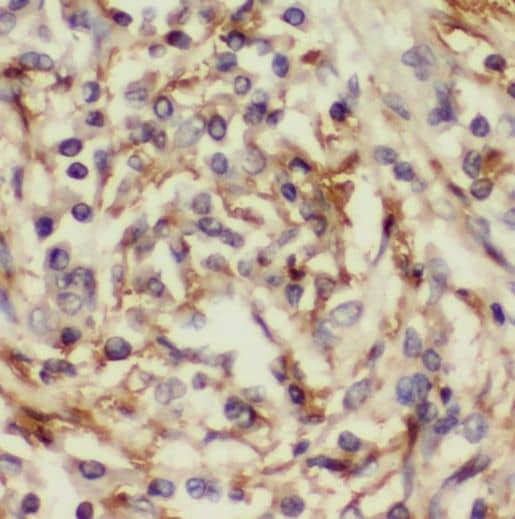
HAF008) or the Anti-Rabbit IgG VisUCyte™ HRP Polymer Antibody (Catalog # VC003). Before incubation with the primary antibody, tissue was subjected to heat-induced epitope retrieval using VisUCyte Antigen Retrieval Reagent-Basic (Catalog # VCTS021). Tissue was stained using DAB (brown) and counterstained with hematoxylin (blue). Specific staining was localized to the membrane of B cells. View our protocol for Chromogenic IHC Staining of Paraffin-embedded Tissue Sections." > HAF008) or the Anti-Rabbit IgG VisUCyte™ HRP Polymer Antibody (Catalog # VC003). Before incubation with the primary antibody, tissue was subjected to heat-induced epitope retrieval using VisUCyte Antigen Retrieval Reagent-Basic (Catalog # VCTS021). Tissue was stained using DAB (brown) and counterstained with hematoxylin (blue). Specific staining was localized to the membrane of B cells. View our protocol for Chromogenic IHC Staining of Paraffin-embedded Tissue Sections." title="IgM was detected in immersion fixed paraffin-embedded sections of human tonsil using Rabbit Anti-Human IgM Monoclonal Antibody (Catalog # MAB9435) at 3 µg/ml for 1 hour at room temperature followed by incubation with the HRP-conjugated Anti-Rabbit IgG Secondary Antibody (Catalog # HAF008) or the Anti-Rabbit IgG VisUCyte™ HRP Polymer Antibody (Catalog # VC003). Before incubation with the primary antibody, tissue was subjected to heat-induced epitope retrieval using VisUCyte Antigen Retrieval Reagent-Basic (Catalog # VCTS021). Tissue was stained using DAB (brown) and counterstained with hematoxylin (blue). Specific staining was localized to the membrane of B cells. View our protocol for Chromogenic IHC Staining of Paraffin-embedded Tissue Sections." />
IgM was detected in immersion fixed paraffin-embedded sections of human tonsil using Rabbit Anti-Human IgM Monoclonal Antibody (Catalog # MAB9435) at 3 µg/ml for 1 hour at room temperature followed by incubation with ...read more

Product Details

Summary
Reactivity HuSpecies Glossary
Applications WB, Simple Western, ELISA, IHC, COMET, mIF
Clone
2123A
Clonality
Monoclonal
Host
Rabbit
Conjugate
Unconjugated

IgM Antibody (2123A) [Unconjugated] Summary

Additional Information
Recombinant Monoclonal Antibody.
Immunogen
Immunoglobulin M (IgM), mu chain from human plasma
Specificity
Detects human IgM in Western blots.
Source
N/A
Isotype
IgG
Clonality
Monoclonal
Host
Rabbit
Purity Statement
Protein A or G purified from cell culture supernatant
Innovator's Reward
Test in a species/application not listed above to receive a full credit towards a future purchase.

Applications/Dilutions

Dilutions
  • COMET 0.2 ug/mL
  • ELISA
  • Immunohistochemistry 1-3 ug/mL
  • Multiplex Immunofluorescence 0.5 ug/mL
  • Simple Western 5 ug/mL
  • Western Blot 0.1 ug/mL

Packaging, Storage & Formulations

Storage
Use a manual defrost freezer and avoid repeated freeze-thaw cycles.
  • 12 months from date of receipt, -20 to -70 °C as supplied.
  • 1 month, 2 to 8 °C under sterile conditions after reconstitution.
  • 6 months, -20 to -70 °C under sterile conditions after reconstitution.
Buffer
Lyophilized from a 0.2 μm filtered solution in PBS with Trehalose. See Certificate of Analysis for details.
*Small pack size (-SP) is supplied either lyophilized or as a 0.2 µm filtered solution in PBS.
Reconstitution Instructions
Reconstitute at 0.5 mg/mL in sterile PBS. For liquid material, refer to CoA for concentration.

Notes

This product is produced by and ships from R&D Systems, Inc., a Bio-Techne brand.

Alternate Names for IgM Antibody (2123A) [Unconjugated]

  • AGM1
  • DKFZp686I15196
  • DKFZp686I15212
  • FLJ00385
  • IgM
  • immunoglobulin heavy constant mu
  • MGC104996
  • MGC52291
  • MU
  • VH

Background

R&D Systems offers a range of secondary antibodies and controls for flow cytometry, immunohistochemistry, and Western blotting. We provide species-specific secondary antibodies that are available with a variety of conjugated labels.

Our NorthernLights fluorescent secondary antibodies are bright and resistant to photobleaching. We are currently offering secondary antibodies recognizing mouse, rat, goat, sheep, and rabbit IgG as well as chicken IgY. These reagents are available with three distinct excitation and emission maxima, making them ideal for multi-color fluorescence microscopy.
Limitations

This product is for research use only and is not approved for use in humans or in clinical diagnosis. Primary Antibodies are guaranteed for 1 year from date of receipt.

Customers Who Viewed This Item Also Viewed...

NBP1-81555
Species: Hu
Applications: IHC,  IHC-P, WB
NBP2-55747
Species: Hu
Applications: ICC/IF
H00001109-M01
Species: Hu
Applications: ELISA, ICC/IF, IHC,  IHC-P, S-ELISA, WB
NBP2-45731
Species: Ca, Hu, Pm
Applications: IHC,  IHC-P, WB
NBP2-75930
Species: Hu, Mu, Rt
Applications: Flow, ICC/IF, IHC,  IHC-P, WB
NB110-58345
Species: Bv, Hu, Mu, Pm
Applications: Flow, ICC/IF, IHC,  IHC-P, WB
NBP2-70360
Species: Hu
Applications: CyTOF-ready, Flow, IMC, ICC/IF, IHC,  IHC-P, WB
7268-CT
Species: Hu
Applications: BA
NBP2-16686
Species: Hu
Applications: ICC/IF, IHC,  IHC-P, KO, WB
NBP3-45895
Species: Hu, Mu
Applications: ELISA, IHC, WB
NBP2-67909
Species: Hu, Mu, Rt
Applications: Flow, IHC,  IHC-P, IP, WB
NBP2-79709
Species: Hu
Applications: ELISA, Flow, ICC/IF, IHC,  IHC-P, PA, WB
NB100-524
Species: Hu, Mu
Applications: Flow-IC, Flow, ICC/IF, IHC, IHC-Fr,  IHC-P, IP, WB
NB200-541
Species: Mu
Applications: IA, ICC/IF, IHC, IHC-Fr, IP, WB
NBP2-19561
Species: Hu, Mu, Rt
Applications: ICC/IF, IHC, IHC-Fr,  IHC-P, WB
NBP1-56929
Species: Hu
Applications: IHC,  IHC-P, WB

Publications for IgM Antibody (MAB9435) (0)

There are no publications for IgM Antibody (MAB9435).
By submitting your publication information earn gift cards and discounts for future purchases.

Reviews for IgM Antibody (MAB9435) (0)

There are no reviews for IgM Antibody (MAB9435). By submitting a review you will receive an Amazon e-Gift Card or Novus Product Discount.
  • Review with no image -- $10/€7/£6/$10 CAD/¥70 Yuan/¥1110 Yen
  • Review with an image -- $25/€18/£15/$25 CAD/¥150 Yuan/¥2500 Yen

Product General Protocols

Find general support by application which include: protocols, troubleshooting, illustrated assays, videos and webinars.

Video Protocols

WB Video Protocol

FAQs for IgM Antibody (MAB9435) (0)

There are no specific FAQs related to this product. Read our general customer & technical service FAQs.

Secondary Antibodies


Isotype Controls

Additional IgM Products

Blogs on IgM.

Early T cell response is associated with mild COVID-19 and rapid SARS-CoV-2 clearance
Jamshed Arslan, Pharm D, PhD SARS-CoV-2 induces both humoral and cellular immunity. A vaccine or natural infection invokes SARS-CoV-2-specific humoral components (antibodies from activated B cells) and cellular resp...  Read full blog post.


  Read full blog post.

How To Identify B Cell Subsets Using Flow Cytometry
By Victoria OsinskiUsing Flow Cytometry to Identify B Cell SubsetsIdentifying cellular subsets by flow cytometry requires careful and thorough planning in order to ensure the correct subset of cells are identified...  Read full blog post.

Understanding the Importance of Human IgG
Human IgG is a component of the immune system that protects the body from infection. It is the most abundantly found antibody isotype within the circulatory system of the human body. All antibody isotypes contain two heavy chains and two light chains ...  Read full blog post.

Customers Who Bought This Also Bought

Contact Information

Product PDFs

Calculators

Concentration Calculator

The concentration calculator allows you to quickly calculate the volume, mass or concentration of your vial. Simply enter your mass, volume, or concentration values for your reagent and the calculator will determine the rest.

=
÷

Review this Product

Be the first to review our IgM Antibody (2123A) [Unconjugated] and receive a gift card or discount.