| Immunogen | human PBL |
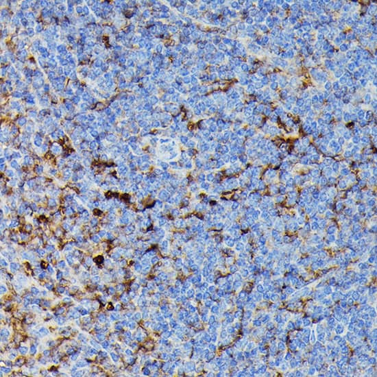
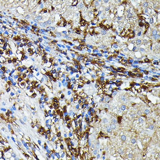
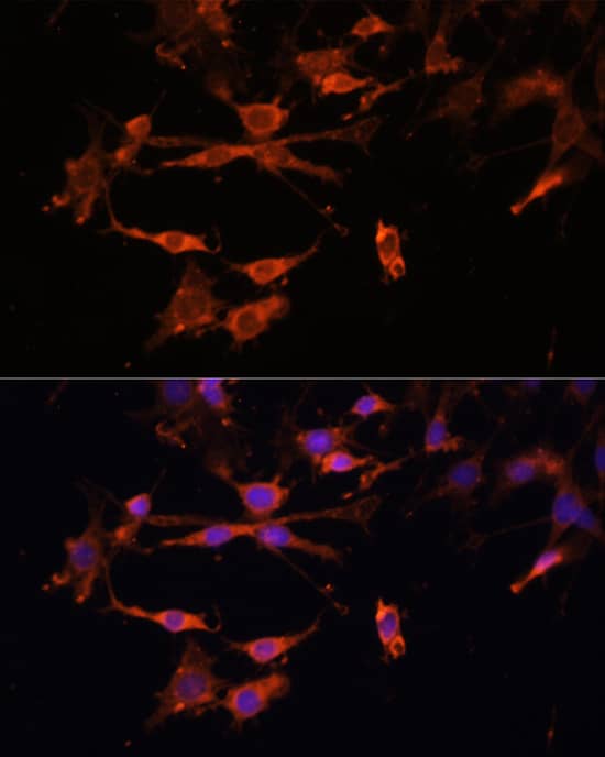
| Specificity | HLA DR (LN3 ) |
| Isotype | IgG2b Kappa |
| Clonality | Monoclonal |
| Host | Mouse |
| Gene | HLA-DRA |
| Purity | Protein A or G purified |
| Innovator's Reward | Test in a species/application not listed above to receive a full credit towards a future purchase. |
| Dilutions |
|
|
| Application Notes | Each lot of this antibody is quality control tested by immunofluorescent staining with flow cytometric analysis. For immunofluorescent staining, the suggested use of this reagent is: 0.5 microgram per one million cells in 100 microliter volume or 100 microliter whole blood. Immunohistochemistry-frozen/paraffin. It is recommended that the reagent be titrated for optimal performance for each application. |
|
| Publications |
|
| Storage | Store at 4C. Do not freeze. |
| Buffer | PBS (pH 7.2) |
| Preservative | 0.09% Sodium Azide |
| Concentration | 0.5 mg/ml |
| Purity | Protein A or G purified |


")


| Publication using NB100-78094 | Applications | Species |
|---|---|---|
| Huynh Jimmy L, Garg Paras, Thin Tin Htwe et al. Epigenome-wide differences in pathology-free regions of multiple sclerosis-affected brains. Nat Neurosci. 2014-01-01 [PMID: 24270187] (IHC-Fr, Human) | IHC-Fr | Human |
The concentration calculator allows you to quickly calculate the volume, mass or concentration of your vial. Simply enter your mass, volume, or concentration values for your reagent and the calculator will determine the rest.


![SDS-PAGE IFN-gamma [Unconjugated]](https://images.novusbio.com/images2/IFN-gamma_485-MI_407.jpg)
![Bioactivity IFN-gamma [Unconjugated]](https://images.novusbio.com/images2/IFN-gamma_485-MI_455.jpg)
![SEC-MALS IFN-gamma [Unconjugated]](https://images.novusbio.com/images/485-mi_recombinant-mouse-ifn-gamma-protein-sec-mals-1612202583245.jpg)


![Western Blot RB1 Antibody (607121) [Unconjugated]](https://images.novusbio.com/images2/RB1_MAB6495_Western_Blot_10216.jpg)
![Immunocytochemistry RB1 Antibody (607121) [Unconjugated]](https://images.novusbio.com/images2/RB1_MAB6495_Immunocytochemistry_10464.jpg)
![Simple Western RB1 Antibody (607121) [Unconjugated]](https://images.novusbio.com/images2/RB1_MAB6495_Simple_Western_16629.jpg)


![SDS-PAGE TNF-alpha [Unconjugated]](https://images.novusbio.com/images2/TNF-alpha_210-TA_256.jpg)
![Bioactivity TNF-alpha [Unconjugated]](https://images.novusbio.com/images2/TNFalpha_210TA_1658.jpg)
![SEC-MALS TNF-alpha [Unconjugated]](https://images.novusbio.com/images/210-ta_recombinant-human-tnf-alpha-protein-sec-mals-35202312244..jpg)
![N/A IL-6 [HRP]](https://images.novusbio.com/images2/DATA_IL6_M6000_ELISA_936.jpg)
![N/A IL-6 [HRP]](https://images.novusbio.com/images2/IL-6_M6000_ELISA_415.jpg)
![N/A IL-6 [HRP]](https://images.novusbio.com/images/m6000b_mouse-il-6-quantikine-elisa-kit-44202415433118.jpg)