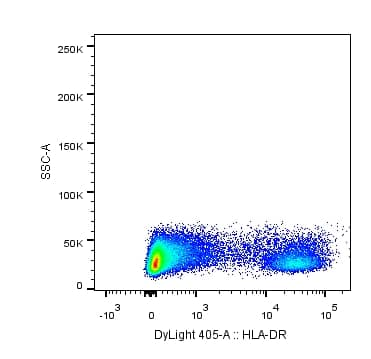
![Flow Cytometry: HLA-DR Antibody (L243) [DyLight 405] [NB100-77855V] - analysis of HLA-DR in human PBMCs using anti-HLA-DR antibody. Image from verified customer review.](http://images.novusbio.com/fullsize/HLA-DR-Antibody-L243-DyLight-405-Flow-Cytometry-NB100-77855V-img0001.jpg "Flow Cytometry: HLA-DR Antibody (L243) [DyLight 405] [NB100-77855V] - analysis of HLA-DR in human PBMCs using anti-HLA-DR antibody. Image from verified customer review.")
| Reactivity | Hu, Ca, Pm, PmSpecies Glossary |
| Applications | WB, ELISA, Flow, Func, ICC/IF, IHC, IP, MS, B/N, CyTOF-ready, Dual ISH-IHC |
| Clone | L243 |
| Clonality | Monoclonal |
| Host | Mouse |
| Conjugate | DyLight 405 |
| Description | This conjugate is made on demand and is not held in inventory. Each order represents a unique lot. All conjugates are generated using validated conjugation protocols and meet strict degree of labeling (F/P) specifications; additional information is available upon request. The antibody concentration is provided on the individual product label. For specialized conjugation needs, like large quantities, specific concentrations, and defined F/P ratios, bulk and custom antibody conjugation services are available. |
| Immunogen | Human lymphoblastoid cell line (RPMI 8866). |
| Isotype | IgG2a Kappa |
| Clonality | Monoclonal |
| Host | Mouse |
| Gene | HLA-DRA |
| Purity | Protein A purified |
| Innovator's Reward | Test in a species/application not listed above to receive a full credit towards a future purchase. |
| Dilutions |
|
|
| Application Notes | Recommended applications are based on validated applications from the unconjugated base product NB100-77855. This conjugated antibody is not kept in inventory and is made to order using validated conjugation protocols. |
|
| Reviewed Applications |
|
| Storage | Store at 4C in the dark. |
| Buffer | 50mM Sodium Borate |
| Preservative | 0.05% Sodium Azide |
| Purity | Protein A purified |







| Images | Ratings | Applications | Species | Date | Details | ||||||
|---|---|---|---|---|---|---|---|---|---|---|---|
Enlarge |
reviewed by:
Verified Customer |
Flow | Human | 06/30/2015 |
Summary
|
The concentration calculator allows you to quickly calculate the volume, mass or concentration of your vial. Simply enter your mass, volume, or concentration values for your reagent and the calculator will determine the rest.
| Gene Symbol | HLA-DRA |


![SEC-MALS IFN-gamma [Unconjugated]](https://images.novusbio.com/images/485-mi_recombinant-mouse-ifn-gamma-protein-sec-mals-1612202583245.jpg)
![Bioactivity IFN-gamma [Unconjugated]](https://images.novusbio.com/images/protein/IFN-gamma_485-MI_455.jpg)
![SDS-Page IFN-gamma [Unconjugated]](https://images.novusbio.com/images/protein/IFN-gamma_485-MI_407.jpg)




![Western Blot RB1 Antibody (607121) [Unconjugated]](https://images.novusbio.com/images/mab6495_human-rb1-mab-clone-607121-812025548577.jpg)
![Simple Western RB1 Antibody (607121) [Unconjugated]](https://images.novusbio.com/images/antibody/RB1_MAB6495_Simple_Western_16629.jpg)
![Western Blot RB1 Antibody (607121) [Unconjugated]](https://images.novusbio.com/images/antibody/RB1_MAB6495_Western_Blot_10216.jpg)


![SDS-Page TNF-alpha [Unconjugated]](https://images.novusbio.com/images/protein/TNF-alpha_210-TA_256.jpg)
![Bioactivity TNF-alpha [Unconjugated]](https://images.novusbio.com/images/protein/TNFalpha_210TA_1658.jpg)
![SEC-MALS TNF-alpha [Unconjugated]](https://images.novusbio.com/images/210-ta_recombinant-human-tnf-alpha-protein-sec-mals-35202312244..jpg)
![N/A IL-6 [HRP]](https://images.novusbio.com/images/elisa/DATA_IL6_M6000_ELISA_936.jpg)
![N/A IL-6 [HRP]](https://images.novusbio.com/images/elisa/IL-6_M6000_ELISA_415.jpg)
![N/A IL-6 [HRP]](https://images.novusbio.com/images/m6000b_mouse-il-6-quantikine-elisa-kit-1752025024034.jpg)
