After August 17, 2026, Novus Biologicals products and services will no longer be available on this website; you will access all products and services on rndsystems.com. Create your R&D Systems online account today.
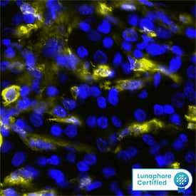
MAB8170) at 20ug/mL at 37 ° Celsius for 4 minutes. Before incubation with the primary antibody, tissue underwent an all-in-one dewaxing and antigen retrieval preprocessing using PreTreatment Module (PT Module) and Dewax and HIER Buffer H (pH 9; Epredia Catalog # TA-999-DHBH).Tissue was stained using the Alexa Fluor™ 647 Goat anti-Mouse IgG Secondary Antibody at 1:200 at 37 ° Celsius for 2 minutes. (Yellow; Lunaphore Catalog # DR647MS) and counterstained with DAPI (blue; Lunaphore Catalog # DR100). Specific staining was localized to the cytoplasm. Protocol available in COMET™ Panel Builder." class="big_lightbox" target="_blank">
HAF018). A specific band was detected for Gelsolin/GSN at approximately 95 kDa (as indicated). This experiment was conducted under reducing conditions and using Immunoblot Buffer Group 1." class="big_lightbox" target="_blank">
Gelsolin is a calcium regulated actin binding protein. Gelsolin is a regulator of actin filament assembly and disassembly and consists of six homologous subdomains (S1-S6). When C-terminal tail (S6) is bound to calcium ion, it straightens and exposes actin binding sites located on S2s helices, leading to Gelsolin actin-serving activity (1). Gelsolin is inhibited by phosphatidylinositol (4,5)-bisphosphate (PIP2), where PIP2 will bind to S1, S2, and S3 sites. Gelsolin is involved in podosome formation and has been linked to inhibition of apoptosis by preventing release of cytochrome C (2).


![Multiplex Immunofluorescence Gelsolin/GSN Antibody (893205) [Unconjugated]](http://images.novusbio.com/images/antibody/mab8170_human-mouse-rat-gelsolin-gsn-mab-clone-893205-multiplex-immunofluorescence-241020248512.jpg)


![Western Blot Gelsolin/GSN Antibody (893205) [Unconjugated]](http://images.novusbio.com/images/antibody/Gelsolin_MAB8170_Western_Blot_14481.jpg)
![Simple Western Gelsolin/GSN Antibody (893205) [Unconjugated]](http://images.novusbio.com/fullsize/antibody/Gelsolin_MAB8170_Simple_Western_17259.jpg)
![Immunohistochemistry Gelsolin/GSN Antibody (893205) [Unconjugated]](http://images.novusbio.com/images/antibody/Gelsolin_MAB8170_Immunohistochemistry_14553.jpg)
![Knockout Validated Gelsolin/GSN Antibody (893205) [Unconjugated]](http://images.novusbio.com/fullsize/mab8170_human-mouse-rat-gelsolin-gsn-mab-clone-893205-knockout-validated-192022105243.jpg)