| Reactivity | HuSpecies Glossary |
| Applications | WB |
| Clonality | Polyclonal |
| Host | Goat |
| Conjugate | Unconjugated |
| Concentration | LYOPH |
| Immunogen | Mouse myeloma cell line NS0-derived recombinant human FLRT3 Lys29-Pro528 Accession # Q9NZU0 |
| Specificity | Detects human FLRT3 in direct ELISAs and Western blots. In direct ELISAs, less than 10% cross-reactivity with recombinant human FLRT1 and rhFLRT2 is observed. |
| Source | N/A |
| Isotype | IgG |
| Clonality | Polyclonal |
| Host | Goat |
| Gene | FLRT3 |
| Purity Statement | Antigen Affinity-purified |
| Innovator's Reward | Test in a species/application not listed above to receive a full credit towards a future purchase. |
| Dilutions |
|
|
| Publications |
|
| Storage | Use a manual defrost freezer and avoid repeated freeze-thaw cycles.
|
| Buffer | Lyophilized from a 0.2 μm filtered solution in PBS with Trehalose. *Small pack size (SP) is supplied either lyophilized or as a 0.2 µm filtered solution in PBS. |
| Preservative | No Preservative |
| Concentration | LYOPH |
| Reconstitution Instructions | Reconstitute at 0.2 mg/mL in sterile PBS. |
FLRT3 is one of three FLRT (fibronectin, leucine rich repeat, transmembrane) glycoproteins expressed in distinct areas of the developing brain and other tissues (1, 2). The 85-95 kDa type I transmembrane (TM) human FLRT3 is synthesized as a 649 amino acid (aa) precursor with a 28 aa signal sequence, a 500 aa extracellular domain (ECD), a 21 aa TM segment and a 100 aa cytoplasmic region. The ECD contains 10 N-terminal leucine-rich repeats flanked by cysteine-rich areas, and a juxtamembrane fibronectin type III domain (1). The human FLRT3 ECD shares 96%, 96%, 97%, 97%, 98%, and 81% aa sequence identity with mouse, rat, canine, bovine, equine, and Xenopus FLRT3 ECD, respectively, and 61% and 48% aa identity to human FLRT2 and FLRT3 ECDs, respectively. The fibronectin domain is responsible for binding to FGF receptors, and is thought to regulate FGF signaling during development (2, 3). The LRR domains are responsible for both the localization in areas of cell contact and homotypic cell-cell association (4). This may be through direct interaction with other FLRT molecules, or alternatively, by regulating internalization of adhesion molecules such as cadherins (4, 5). Developmentally, FLRT3 is located in somitic regions on dermatomyotomal muscle precursors and myotomal cells before their migration to the myotome and syndetome, respectively (2). FLRT3 is also expressed at the midbrain/hindbrain boundary and in the apical ectodermal ridge where it may influence FGF signaling (2). Genetic deletion in mouse embryos results in defective headfold fusion and endoderm migration (6). Postnatally, FLRT3 mRNA is widely expressed (1). It is upregulated and promotes neurite outgrowth following experimental peripheral nerve injury in rats (7, 8).


![Western Blot EphB2 Antibody [Unconjugated]](https://images.novusbio.com/images/af467_human-mouse-ephb2-affinity-purified-polyclonal-ab-41202410485412.jpg)
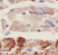
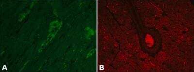
![Immunohistochemistry EphB2 Antibody [Unconjugated]](https://images.novusbio.com/images/antibody/EphB2_AF467_Immunohistochemistry_20369.jpg)
![Western Blot EphB2 Antibody [Unconjugated]](https://images.novusbio.com/images/antibody/EphB2_AF467_Western_Blot_21511.jpg)


![FLRT3 Antibody [Unconjugated]](/sites/all/modules/enterprise-tech/et_datasheets/images/novus_guarantee.png "FLRT3 Antibody [Unconjugated]")
Secondary Antibodies |
Isotype Controls |
The concentration calculator allows you to quickly calculate the volume, mass or concentration of your vial. Simply enter your mass, volume, or concentration values for your reagent and the calculator will determine the rest.


![Simple Western FLRT2 Antibody [Unconjugated]](https://images.novusbio.com/images/antibody/af2877_human-flrt2-affinity-purified-polyclonal-ab-simple-western-2182024142725..jpg)
![Western Blot FLRT2 Antibody [Unconjugated]](https://images.novusbio.com/images/antibody/af2877_human-flrt2-affinity-purified-polyclonal-ab-western-blot-1272024125547..jpg)
![Bioactivity TGF-beta 1 [Unconjugated]](https://images.novusbio.com/images/protein/7754-bhcf_recombinant-human-tgf-beta-1-human-cell-expressed-cf-bioactivity-1811202013946.jpg)
![Immunohistochemistry UNC5H2/UNC5B Antibody [Unconjugated]](https://images.novusbio.com/images/af1006_rat-unc5h2-unc5b-affinity-purified-polyclonal-ab-immunohistochemistry-3152022161010.jpg)
![Western Blot UNC5H2/UNC5B Antibody [Unconjugated]](https://images.novusbio.com/images/antibody/UNC5H2_AF1006_Western_Blot_17311.jpg)


![Cell Culture FGF-8 [Unconjugated]](https://images.novusbio.com/images/423-f8_recombinant-human-mouse-fgf-8b-protein-cell-culture-1052024145159..jpg)
![Bioactivity FGF-8 [Unconjugated]](https://images.novusbio.com/images/protein/FGF8_423F8_1438.jpg)
![Cell Culture FGF-8 [Unconjugated]](https://images.novusbio.com/images/423-f8_recombinant-human-mouse-fgf-8b-protein-cell-culture-1052024145958..jpg)
![Western Blot ERK2 Antibody [Unconjugated]](https://images.novusbio.com/images/antibody/ERK2_AF1230_Western_Blot_5097.jpg)
![Knockout Validated ERK2 Antibody [Unconjugated]](https://images.novusbio.com/images/antibody/ERK2_AF1230_Knockout_Validated_22864.jpg)
![Immunohistochemistry ERK2 Antibody [Unconjugated]](https://images.novusbio.com/images/antibody/ERK2_AF1230_Immunohistochemistry_20696.jpg)
 or Normal Goat IgG Isotype Control Antibody (Catalog # AB-108-C, open histogram), followed by Phycoerythrin-conjugated Anti-Goat IgG Secondary Antibody (Catalog # F0107).")