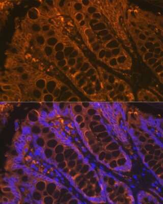
 at 1:500 at 37 ° Celsius for 4 minutes. Before incubation with the primary antibody, tissue underwent an all-in-one dewaxing and antigen retrieval preprocessing using PreTreatment Module (PT Module) and Dewax and HIER Buffer H (pH 9; Epredia Catalog # TA-999-DHBH). Tissue was stained using the Alexa Fluor™ 555 Goat anti-Mouse IgG Secondary Antibody at 1:100 at 37 ° Celsius for 2 minutes. (Yellow; Lunaphore Catalog # DR555MS) and counterstained with DAPI (blue; Lunaphore Catalog # DR100). Specific staining was localized to the cytoplasm. Protocol available in COMET™ Panel Builder.")
| Reactivity | Hu, Mu, Rt, Bv, Ca, Ch, Pm, Rb, Re, ZeSpecies Glossary |
| Applications | WB, Flow, ICC/IF, IHC, COMET, CyTOF-ready, Dual ISH-IHC, mIF, Single-Cell Western |
| Clone | AE-1/AE-3 |
| Clonality | Monoclonal |
| Host | Mouse |
| Conjugate | Unconjugated |
| Concentration | 0.2 mg/ml |
| Description | 200ug/ml of antibody purified from Bioreactor Concentrate by Protein A or G. Prepared in 10 mM PBS with 0.05% BSA & 0.05% azide. Also available WITHOUT BSA & azide at 1.0 mg/ml. (NBP2-33200) Antibody with azide - store at 2 to 8C. Antibody without azide - store at -20 to -80C. |
| Immunogen | Human epidermal keratin |
| Localization | Cytoplasmic |
| Marker | Epithelial Marker |
| Specificity | Twenty human keratins are resolved with two-dimensional gel electrophoresis into acidic (pI 6.0) subfamilies. This antibody cocktail recognizes acidic (Type I or LMW) and basic (Type II or HMW) cytokeratins, which 67kDa (CK1); 64kDa (CK3); 59kDa (CK4); 58kDa (CK5); 56kDa (CK6); 52kDa (CK8); 56.5kDa (CK10); 50kDa (CK14); 50kDa (CK15); 48kDa (CK16); 40kDa (CK19). Many studies have shown the usefulness of keratins as markers in cancer research and tumor diagnosis. AE-1/AE-3 is a broad spectrum anti pan-cytokeratin antibody cocktail, which differentiates epithelial tumors from non-epithelial tumors e.g. squamous vs. adenocarcinoma of the lung, liver carcinoma, breast cancer, and esophageal cancer. It has been used to characterize the source of various neoplasms and to study the distribution of cytokeratin containing cells in epithelia during normal development and during the development of epithelial neoplasms. This antibody stains cytokeratins present in normal and abnormal human tissues and has shown high sensitivity in the recognition of epithelial cells and carcinomas. |
| Isotype | IgG1 Kappa/IgG1 Kappa |
| Clonality | Monoclonal |
| Host | Mouse |
| Gene | KRT1 |
| Purity | Protein A or G purified |
| Innovator's Reward | Test in a species/application not listed above to receive a full credit towards a future purchase. |
| Dilutions |
|
|
| Application Notes | Use in ICC/IF reported in scientific literature (PMID:34376789) Immunohistochemistry (Formalin-fixed): 0.25-0.5ug/ml for 30 min at RT. Staining of formalin-fixed tissues requires heating tissue sections in 10mM Tris with 1mM EDTA, pH 9.0, for 45 min at 95C followed by cooling at RT for 20 minutes. Optimal dilution for a specific application should be determined. Western Blot: 1-2ug/ml for 2 hours at RT. The staining pattern of the pan cytokeratin antibody cocktail may be different than that of either antibody separately. This antibody cocktail recognizes acidic (Type I or LMW) and basic (Type II or HMW) cytokeratins, which 67 kDa (CK1) ; 64 kDa (CK3) ; 59 kDa (CK4) ; 58 kDa (CK5) ; 56 kDa (CK6) ; 52 kDa (CK8) ; 56.5 kDa (CK10) ; 50k Da (CK14) ; 50 kDa (CK15) ; 48 kDa (CK16) ; 40 kDa (CK19) . The pan cytokeratin cocktail does not react with keratin 18, which is also expressed in carcinomas. As such, negative staining with NBP2-29429 in of itself may not be sufficient evidence to rule out the possibility of a carcinoma (Ordonez, 2013) . For example, hepatocellular, adrenal cortical, clear cell renal and chromophobe renal cell carcinomas have been reported to be negative for the pan cytokeratin antibody. In this regard, the pan cytokeratin antibody can be used as part of a screening panel to more extensively define the tumor cell lineages. The pan cytokeratin antibody may cross-react with GFAP, leading to aberrant positive staining of glial tumors such as ependymoma, glioblastoma, or schwannoma (Ordonez, 2013) . Use in Immunohistochemistry reported in scientific literature (PMID: 29169625) . This Cytokeratin, pan Antibody (AE-1/AE-3) is validated for CyTOF from a verified customer review. |
|
| Reviewed Applications |
|
|
| Publications |
|
| Storage | Store at 4C. |
| Buffer | 10 mM PBS with 0.05% BSA |
| Preservative | 0.05% Sodium Azide |
| Concentration | 0.2 mg/ml |
| Purity | Protein A or G purified |


![Simple Western Albumin Antibody (188835) [Unconjugated] - Serum](https://images.novusbio.com/images2/Albumin_MAB1455_Simple_Western_21888.jpg)
![Immunocytochemistry Albumin Antibody (188835) [Unconjugated] - Serum](https://images.novusbio.com/images2/Albumin_MAB1455_Immunocytochemistry__Immunofluorescence_17777.jpg)
![Immunohistochemistry Albumin Antibody (188835) [Unconjugated] - Serum](https://images.novusbio.com/images2/Albumin_MAB1455_Immunohistochemistry_23317.jpg)


| Images | Ratings | Applications | Species | Date | Details | ||||||||||
|---|---|---|---|---|---|---|---|---|---|---|---|---|---|---|---|

Enlarge |
reviewed by:
Toni johanson |
CyTOF | Human | 03/31/2021 |
Summary
Comments
|
Secondary Antibodies |
Isotype Controls |
Research Areas for Cytokeratin, pan Antibody (NBP2-29429)Find related products by research area.
|
The concentration calculator allows you to quickly calculate the volume, mass or concentration of your vial. Simply enter your mass, volume, or concentration values for your reagent and the calculator will determine the rest.


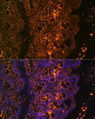
 at 1:200 dilution at 37 ° Celsius for 4 minutes. Before incubation with the primary antibody, tissue underwent an all-in-one dewaxing and antigen retrieval preprocessing using PreTreatment Module (PT Module) and Dewax and HIER Buffer H (pH 9; Epredia Catalog # TA-999-DHBH).Tissue was stained using the Alexa Fluor™ 647 Goat anti-Mouse IgG Secondary Antibody at 1:200 at 37 ° Celsius for 2 minutes. (Yellow; Lunaphore Catalog # DR647MS) and counterstained with DAPI (blue; Lunaphore Catalog # DR100). Specific staining was localized to the membrane. Protocol available in COMET™ Panel Builder.")
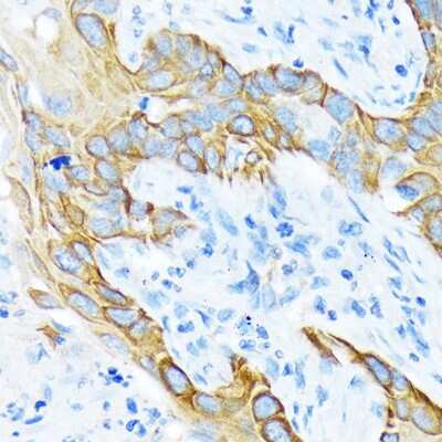
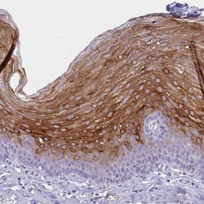
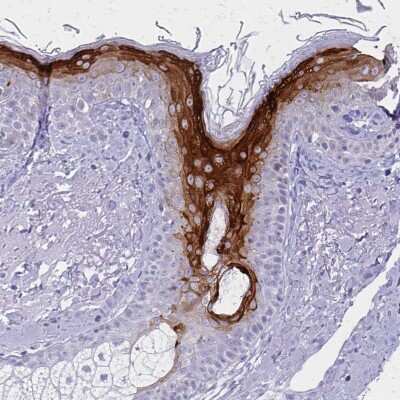
. Adjacent tissue section was processes for IHC using mouse monoclonal antibody (Novus catalog # NBP2-29429) at 0.3 ug/mL for 1 hour at room temperature followed by incubation with the anti-mouse IgG VisUCyte HRP Polymer Antibody (Novus Catalog # VC001) and DAB chromogen (yellow-brown). Tissue was counterstained with hematoxylin (blue).")
 SAv-A488 only at a dilution of 1:500, B) pan CK at a dilution of 1:200, C) pan CK at a dilution of 1:400, D) pan CK at a dilution of 1:800. ICC/IF image submitted by a verified customer review.")
.")
 NBP2-33200R (blue) and a matched isotype control (orange). Cells were fixed with 4% PFA and then permeabilized with 0.1% saponin. Cells were incubated in an antibody dilution of 10 ug/mL for 30 minutes at room temperature. Both antibodies were conjugated to Dylight 550.")
")

![Flow Cytometry: Cytokeratin, pan Antibody (AE-1/AE-3) [NBP2-29429] - An intracellular stain was performed on HeLa cells with pan Cytokeratin Antibody (AE1 + AE3) NBP2-33200AF647 (blue) and a matched isotype control (orange). Cells were fixed with 4% PFA and then permeabilized with 0.1% saponin. Cells were incubated in an antibody dilution of 5 ug/mL for 30 minutes at room temperature. Both antibodies were conjugated to Alexa Fluor 647. Image from the AF647 version of this antibody.](http://images.novusbio.com/fullsize/nbp2-29429_mouse-monoclonal-cytokeratin-pan-antibody-ae-1-ae-3-imgenex-nbo-135a-20ug-17720249362612.jpg "Flow Cytometry: Cytokeratin, pan Antibody (AE-1/AE-3) [NBP2-29429] - An intracellular stain was performed on HeLa cells with pan Cytokeratin Antibody (AE1 + AE3) NBP2-33200AF647 (blue) and a matched isotype control (orange). Cells were fixed with 4% PFA and then permeabilized with 0.1% saponin. Cells were incubated in an antibody dilution of 5 ug/mL for 30 minutes at room temperature. Both antibodies were conjugated to Alexa Fluor 647. Image from the AF647 version of this antibody.")
![Flow Cytometry: Cytokeratin, pan Antibody (AE-1/AE-3) [NBP2-29429] - An intracellular stain was performed on HeLa cells with pan Cytokeratin [AE1 + AE3] Antibody NBP2-33200AF488 (blue) and a matched isotype control (orange). Cells were fixed with 4% PFA and then permeabilized with 0.1% saponin. Cells were incubated in an antibody dilution of 5 ug/mL for 30 minutes at room temperature. Both antibodies were conjugated to Alexa Fluor 488. Image from the AF488 version of this antibody.](http://images.novusbio.com/fullsize/nbp2-29429_mouse-monoclonal-cytokeratin-pan-antibody-ae-1-ae-3-imgenex-nbo-135a-20ug-17720241003217.jpg "Flow Cytometry: Cytokeratin, pan Antibody (AE-1/AE-3) [NBP2-29429] - An intracellular stain was performed on HeLa cells with pan Cytokeratin [AE1 + AE3] Antibody NBP2-33200AF488 (blue) and a matched isotype control (orange). Cells were fixed with 4% PFA and then permeabilized with 0.1% saponin. Cells were incubated in an antibody dilution of 5 ug/mL for 30 minutes at room temperature. Both antibodies were conjugated to Alexa Fluor 488. Image from the AF488 version of this antibody.")
![Flow Cytometry: Cytokeratin, pan Antibody (AE-1/AE-3) [NBP2-29429] - An intracellular stain was performed on HeLa cells with pan Cytokeratin Antibody (AE1 + AE3) NBP2-33200PE (blue) and a matched isotype control (orange). Cells were fixed with 4% PFA and then permeabilized with 0.1% saponin. Cells were incubated in an antibody dilution of 2.5 ug/mL for 30 minutes at room temperature. Both antibodies were conjugated to phycoerythrin. Image from the PE version of this antibody.](http://images.novusbio.com/fullsize/nbp2-29429_mouse-monoclonal-cytokeratin-pan-antibody-ae-1-ae-3-imgenex-nbo-135a-20ug-1772024944230.jpg "Flow Cytometry: Cytokeratin, pan Antibody (AE-1/AE-3) [NBP2-29429] - An intracellular stain was performed on HeLa cells with pan Cytokeratin Antibody (AE1 + AE3) NBP2-33200PE (blue) and a matched isotype control (orange). Cells were fixed with 4% PFA and then permeabilized with 0.1% saponin. Cells were incubated in an antibody dilution of 2.5 ug/mL for 30 minutes at room temperature. Both antibodies were conjugated to phycoerythrin. Image from the PE version of this antibody.")


![N/A Endostatin [HRP]](https://images.novusbio.com/images2/DATA_Endostatin_DNST0_ELISA_778.jpg)
![N/A Endostatin [HRP]](https://images.novusbio.com/images2/Endostatin_DNST0_ELISA_152.jpg)


 using a 1:1000 dilution of HRP-conjugated Anti-Mouse IgG Secondary Antibody (Catalog # HAF007). This experiment was conducted under reducing conditions and using Immunoblot Buffer Group 1.")
![Immunohistochemistry-Paraffin: Goat anti-Mouse IgG (H+L) Secondary Antibody [HRP] [NB7539] - FFPE serial sections of human stomach carcinoma. Primary Antibody: Mouse anti-p53 (Clone DO-1) used at a dilution of 1:100. Secondary Antibody: Goat anti-Mouse IgG (H+L) Secondary Antibody [HRP] Conjugated used at a dilution of 1:200 (5ug/ml). Detection: DAB](https://images.novusbio.com/images/Goat-anti-Mouse-IgG-H+L-Secondary-Antibody-HRP-Immunohistochemistry-Paraffin-NB7539-img0014.jpg "Immunohistochemistry-Paraffin: Goat anti-Mouse IgG (H+L) Secondary Antibody [HRP] [NB7539] - FFPE serial sections of human stomach carcinoma. Primary Antibody: Mouse anti-p53 (Clone DO-1) used at a dilution of 1:100. Secondary Antibody: Goat anti-Mouse IgG (H+L) Secondary Antibody [HRP] Conjugated used at a dilution of 1:200 (5ug/ml). Detection: DAB")
