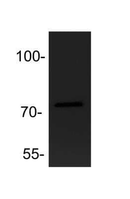
 HAF017). Specific bands were detected for ADAM9 at approximately 110 and 80 kDa (as indicated). This experiment was conducted under reducing conditions and using Immunoblot Buffer Group 1." title="Western blot shows lysates of C2C12 mouse myoblast cell line and WI-38 human lung fibroblast cell line. PVDF membrane was probed with 1 µg/mL of Goat Anti-Human/Mouse ADAM9 Ectodomain Antigen Affinity-purified Polyclonal Antibody (Catalog # AF949) followed by HRP-conjugated Anti-Goat IgG Secondary Antibody (HAF017). Specific bands were detected for ADAM9 at approximately 110 and 80 kDa (as indicated). This experiment was conducted under reducing conditions and using Immunoblot Buffer Group 1." />
| Applications | WB, Flow, IHC, IP, CyTOF-ready, KO |
| Clonality | Polyclonal |
| Host | Goat |
| Conjugate | Unconjugated |
| Concentration | LYOPH |
| Immunogen | Mouse myeloma cell line NS0-derived recombinant mouse ADAM9 Ala206-Asp697 Accession # Q61072 |
| Specificity | Detects mouse ADAM9 Ectodomain in direct ELISAs and Western blots. In Western blots no cross-reactivity with the Ectodomain of recombinant mouse ADAM10 and rhADAM8, 15, and 17 (TACE) is observed. |
| Source | N/A |
| Isotype | IgG |
| Clonality | Polyclonal |
| Host | Goat |
| Gene | ADAM9 |
| Purity Statement | Antigen Affinity-purified |
| Innovator's Reward | Test in a species/application not listed above to receive a full credit towards a future purchase. |
| Dilutions |
|
|
| Publications |
|
| Storage | Use a manual defrost freezer and avoid repeated freeze-thaw cycles.
|
| Buffer | Lyophilized from a 0.2 μm filtered solution in PBS with Trehalose. See Certificate of Analysis for details. *Small pack size (-SP) is supplied either lyophilized or as a 0.2 µm filtered solution in PBS. |
| Preservative | No Preservative |
| Concentration | LYOPH |
| Reconstitution Instructions | Reconstitute at 0.2 mg/mL in sterile PBS. For liquid material, refer to CoA for concentration. |
ADAM9, also known as MDC9 or meltrin gamma , is a member of the ADAM family that contains a disintegrin and metalloprotease-like domain (1). Like other membrane‑anchored ADAMs, ADAM9 consists of a pro domain with a cysteine switch and furin cleavage sequence, a catalytic domain with the zinc-binding site and Met-turn expected for reprolysins, a disintegrin-like domain, a cysteine-rich domain, an EGF-like domain, a transmembrane domain, and the cytoplasmic domain. ADAM9 is able to cleave peptides corresponding to cleavage sites of tumor necrosis factor-alpha (TNF-alpha ), the p75-TNF receptor, the beta -amyloid protein precursor, and the c-kit ligand-1, implying that it may participate in shedding of these membrane proteins (2). In fact, ADAM9 has been shown to shed membrane‑anchored heparin‑binding EGF-like growth factor (3). In addition, it also cleaves oxidized insulin B-chain and fibronectin (2, 4). Besides its catalytic activity, ADAM9 functions as an adhesion molelcule through binding of its disintegrin domain to integrins such as alpha v beta 5 and alpha 6 beta 1 (5, 6). The cytoplasmic domain of ADAM9 interacts with Src homology 3
(SH3)‑containing proteins and protein kinase C, and may mediate different signaling pathways (3, 7). ADAM9 is widely expressed in tissues (8).


![Immunocytochemistry EGFR Antibody [Unconjugated]](https://images.novusbio.com/images/antibody/EGF_R_AF231_Immunocytochemistry__Immunofluorescence_21143.jpg)
![Flow Cytometry EGFR Antibody [Unconjugated]](https://images.novusbio.com/images/antibody/EGF_R_AF231_Flow_Cytometry_20401.jpg)
![Western Blot EGFR Antibody [Unconjugated]](https://images.novusbio.com/images/antibody/EGF_R_AF231_Western_Blot_19925.jpg)


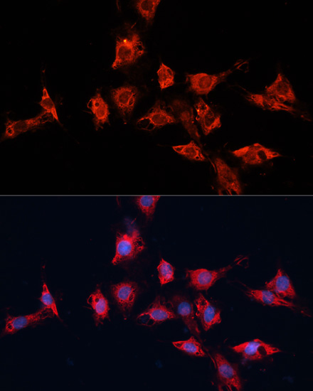
![Immunocytochemistry E-Cadherin Antibody [Unconjugated]](https://images.novusbio.com/images/antibody/ECadherin_AF748_Immunocytochemistry_16573.jpg)
![Western Blot E-Cadherin Antibody [Unconjugated]](https://images.novusbio.com/images/antibody/ECadherin_AF748_Western_Blot_19846.jpg)
![Immunocytochemistry E-Cadherin Antibody [Unconjugated]](https://images.novusbio.com/images/antibody/ECadherin_AF748_Immunocytochemistry_16572.jpg)
![ADAM9 Antibody [Unconjugated]](/sites/all/modules/enterprise-tech/et_datasheets/images/novus_guarantee.png "ADAM9 Antibody [Unconjugated]")
Secondary Antibodies |
Isotype Controls |
The concentration calculator allows you to quickly calculate the volume, mass or concentration of your vial. Simply enter your mass, volume, or concentration values for your reagent and the calculator will determine the rest.


 HAF017). Specific bands were detected for ADAM9 at approximately 78 and 110 kDa (as indicated) in the parental HeLa cell line, but is not detectable in knockout HeLa cell line. GAPDH (
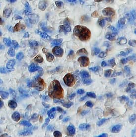
 VC004). Before incubation with the primary antibody, tissue was subjected to heat-induced epitope retrieval using VisUCyte Antigen Retrieval Reagent-Basic (Catalog #
  AB-108-C, open histogram) followed by Allophycocyanin-conjugated Anti-Goat IgG Secondary Antibody (Catalog #
 ![SEC-MALS TACE/ADAM17 [Unconjugated]](https://images.novusbio.com/images/930-adb_recombinant-human-tace-adam17-protein-cf-sec-mals-134202611180.jpg)
![Immunohistochemistry ADAM15 Antibody (23G9) [Unconjugated]](https://images.novusbio.com/images/antibody/ADAM15_MAB935_Immunohistochemistry_7054.jpg)
![Immunocytochemistry ADAM15 Antibody (23G9) [Unconjugated]](https://images.novusbio.com/images/antibody/mab935_human-adam15-mab-clone-23g9-immunocytochemistry-1292023231852.jpg)
![Flow Cytometry ADAM15 Antibody (23G9) [Unconjugated]](https://images.novusbio.com/images/mab935_human-adam15-mab-clone-23g9-41202412511316.jpg)


![Bioactivity EGF [Unconjugated]](https://images.novusbio.com/images/protein/EGF_236EG_1570.jpg)
![Cell Culture EGF [Unconjugated]](https://images.novusbio.com/images/236-eg_recombinant-human-egf-protein-cf-25202394526.jpg)
![Cell Culture EGF [Unconjugated]](https://images.novusbio.com/images/236-eg_recombinant-human-egf-protein-cf-bioactivity-25202394053.jpg)
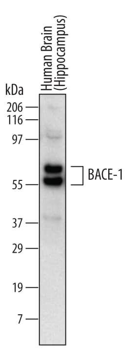
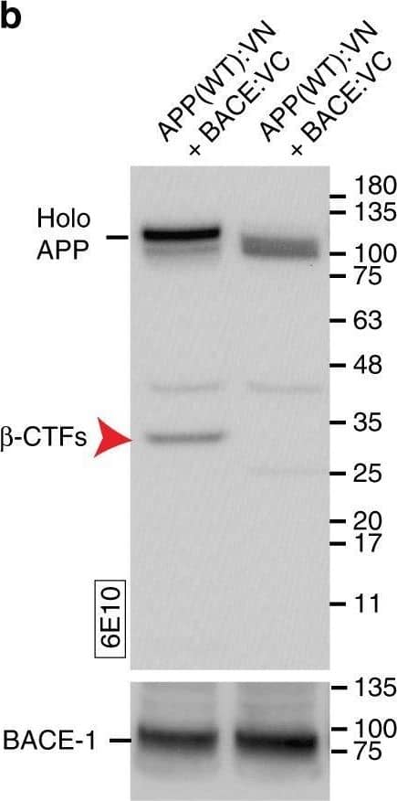
![Immunocytochemistry ADAM8 Antibody [Unconjugated]](https://images.novusbio.com/images/antibody/ADAM8_AF1031_Immunocytochemistry__Immunofluorescence_17417.jpg)
![SDS-Page TNF-alpha [Unconjugated]](https://images.novusbio.com/images/protein/TNF-alpha_210-TA_256.jpg)
![Bioactivity TNF-alpha [Unconjugated]](https://images.novusbio.com/images/protein/TNFalpha_210TA_1658.jpg)
![SEC-MALS TNF-alpha [Unconjugated]](https://images.novusbio.com/images/210-ta_recombinant-human-tnf-alpha-protein-sec-mals-35202312244..jpg)


![Immunohistochemistry HB-EGF Antibody [Unconjugated]](https://images.novusbio.com/images/antibody/HB-EGF_AF-259-NA_Immunohistochemistry_9357.jpg)
![Western Blot HB-EGF Antibody [Unconjugated]](https://images.novusbio.com/images/af-259-na_human-hb-egf-affinity-purified-polyclonal-ab-1752025024018.jpg)
![Western Blot HB-EGF Antibody [Unconjugated]](https://images.novusbio.com/images/af-259-na_human-hb-egf-affinity-purified-polyclonal-ab-41202410484345.jpg)


