HAF019). A specific band was detected for EMMPRIN/CD147 at approximately 38-50 kDa (as indicated). This experiment was conducted under reducing conditions and using
Immunoblot Buffer Group 1." class="big_lightbox">
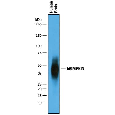
HAF019). A specific band was detected for EMMPRIN/CD147 at approximately 38-50 kDa (as indicated). This experiment was conducted under reducing conditions and using
Immunoblot Buffer Group 1." alt="Western blot shows lysates of human brain tissue. PVDF membrane was probed with 1 µg/mL of Goat Anti-Human EMMPRIN/CD147 Antigen Affinity-purified Polyclonal Antibody (Catalog # AF972) followed by HRP-conjugated Anti-Goat IgG Secondary Antibody (Catalog #
HAF019). A specific band was detected for EMMPRIN/CD147 at approximately 38-50 kDa (as indicated). This experiment was conducted under reducing conditions and using
Immunoblot Buffer Group 1." class="big_thumb" />
HAF109). This experiment was conducted under reducing conditions and using the 12-230 kDa separation system." class="big_lightbox">
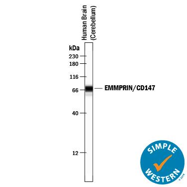
HAF109). This experiment was conducted under reducing conditions and using the 12-230 kDa separation system." class="big_thumb" />
HAF017). A specific band was detected for EMMPRIN/CD147 at approximately 51 kDa (as indicated) in the parental HEK293T cell line, but is not detectable in knockout HEK293T cell line. GAPDH (Catalog #
AF5718) is shown as a loading control. This experiment was conducted under reducing conditions and using
Immunoblot Buffer Group 1." class="big_lightbox">
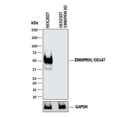
HAF017). A specific band was detected for EMMPRIN/CD147 at approximately 51 kDa (as indicated) in the parental HEK293T cell line, but is not detectable in knockout HEK293T cell line. GAPDH (Catalog #
AF5718) is shown as a loading control. This experiment was conducted under reducing conditions and using
Immunoblot Buffer Group 1." class="big_thumb" />
MAB972) coated on a Clear Polystyrene Microplate (
DY990). Goat Anti-Human EMMPRIN/CD147 Antigen Affinity-purified Polyclonal Antibody (Catalog # AF972) was biotinylated and incubated with the protein captured on the plate. Detection of the standard curve was achieved by incubating Streptavidin-HRP (
DY998) followed by Substrate Solution (
DY999) and stopping the enzymatic reaction with Stop Solution (
DY994)." class="big_lightbox">
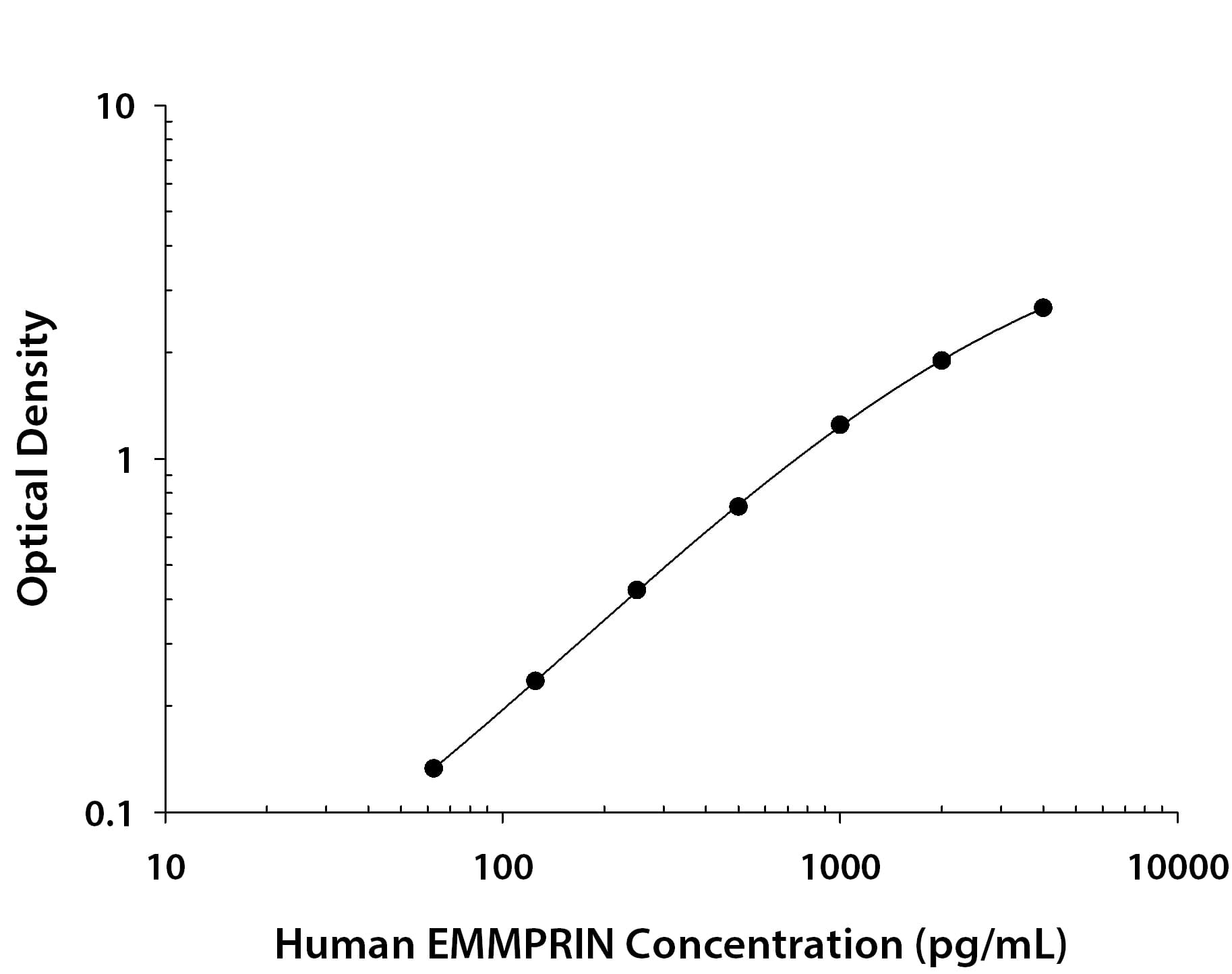
MAB972) coated on a Clear Polystyrene Microplate (
DY990). Goat Anti-Human EMMPRIN/CD147 Antigen Affinity-purified Polyclonal Antibody (Catalog # AF972) was biotinylated and incubated with the protein captured on the plate. Detection of the standard curve was achieved by incubating Streptavidin-HRP (
DY998) followed by Substrate Solution (
DY999) and stopping the enzymatic reaction with Stop Solution (
DY994)." class="big_thumb" />
972-EMN) was serially diluted and captured by Mouse Anti-Human EMMPRIN/CD147 Monoclonal Antibody (Catalog #
MAB972) coated on a Clear Polystyrene Microplate (Catalog #
DY990). Goat Anti-Human EMMPRIN/CD147 Antigen Affinity-purified Polyclonal Antibody (Catalog # AF972) was biotinylated and incubated with the protein captured on the plate. Detection of the standard curve was achieved by incubating Streptavidin-HRP (Catalog #
DY998)" class="big_lightbox">

972-EMN) was serially diluted and captured by Mouse Anti-Human EMMPRIN/CD147 Monoclonal Antibody (Catalog #
MAB972) coated on a Clear Polystyrene Microplate (Catalog #
DY990). Goat Anti-Human EMMPRIN/CD147 Antigen Affinity-purified Polyclonal Antibody (Catalog # AF972) was biotinylated and incubated with the protein captured on the plate. Detection of the standard curve was achieved by incubating Streptavidin-HRP (Catalog #
DY998)" class="big_thumb" />
 HAF019). A specific band was detected for EMMPRIN/CD147 at approximately 38-50 kDa (as indicated). This experiment was conducted under reducing conditions and using Immunoblot Buffer Group 1." alt="Western blot shows lysates of human brain tissue. PVDF membrane was probed with 1 µg/mL of Goat Anti-Human EMMPRIN/CD147 Antigen Affinity-purified Polyclonal Antibody (Catalog # AF972) followed by HRP-conjugated Anti-Goat IgG Secondary Antibody (Catalog # HAF019). A specific band was detected for EMMPRIN/CD147 at approximately 38-50 kDa (as indicated). This experiment was conducted under reducing conditions and using Immunoblot Buffer Group 1." class="big_thumb" />
 HAF018). A specific band was detected for TRA-1-85/CD147 at approximately 40-60 kDa (as indicated). This experiment was conducted under reducing conditions and using Immunoblot Buffer Group 1." alt="Western blot shows lysates of human brain (cerebellum) tissue, human heart tissue, and human liver tissue. PVDF membrane was probed with 1 µg/mL of Mouse Anti-Human TRA-1-85/CD147 Monoclonal Antibody (Catalog # MAB3195) followed by HRP-conjugated Anti-Mouse IgG Secondary Antibody (Catalog # HAF018). A specific band was detected for TRA-1-85/CD147 at approximately 40-60 kDa (as indicated). This experiment was conducted under reducing conditions and using Immunoblot Buffer Group 1." class="big_thumb" />
 HAF018). A specific band was detected for EMMPRIN/CD147 at approximately 40-60 kDa (as indicated). This experiment was conducted under reducing conditions and using Immunoblot Buffer Group 1." alt="Western blot shows lysates of human brain (cerebellum) tissue, human heart tissue, and human liver tissue. PVDF membrane was probed with 0.2 µg/mL of Mouse Anti-Human EMMPRIN/CD147 Monoclonal Antibody (Catalog # MAB972) followed by HRP-conjugated Anti-Mouse IgG Secondary Antibody (Catalog # HAF018). A specific band was detected for EMMPRIN/CD147 at approximately 40-60 kDa (as indicated). This experiment was conducted under reducing conditions and using Immunoblot Buffer Group 1." class="big_thumb" />
 HAF017). A specific band was detected for EMMPRIN/CD147 at approximately 45-60 kDa (as indicated). This experiment was conducted under reducing conditions and using Immunoblot Buffer Group 1." alt="Western blot shows lysates of mouse brain tissue, mouse heart tissue, and mouse small intestine tissue. PVDF membrane was probed with 0.2 µg/mL of Goat Anti-Mouse EMMPRIN/CD147 Antigen Affinity-purified Polyclonal Antibody (Catalog # AF772) followed by HRP-conjugated Anti-Goat IgG Secondary Antibody (Catalog # HAF017). A specific band was detected for EMMPRIN/CD147 at approximately 45-60 kDa (as indicated). This experiment was conducted under reducing conditions and using Immunoblot Buffer Group 1." class="big_thumb" />


 CTS008) and counterstained with hematoxylin (blue). Specific labeling was localized to the plasma membrane of epithelial cells. View our protocol for
  CTS008) and counterstained with hematoxylin (blue). Specific staining was localized to plasma membranes. View our protocol for
  HAF109). This experiment was conducted under reducing conditions and using the 12-230 kDa separation system." class="big_thumb" />
 HAF017). A specific band was detected for EMMPRIN/CD147 at approximately 51 kDa (as indicated) in the parental HEK293T cell line, but is not detectable in knockout HEK293T cell line. GAPDH (Catalog #
  MAB972) coated on a Clear Polystyrene Microplate (

 972-EMN) was serially diluted and captured by Mouse Anti-Human EMMPRIN/CD147 Monoclonal Antibody (Catalog #
  MAB002) followed by Allophycocyanin-conjugated Anti-Mouse IgG Secondary Antibody (Catalog #
  NL007) and counterstained with DAPI (blue). Specific staining was localized to cytoplasm. View our protocol for
  HAF018). A specific band was detected for TRA-1-85/CD147 at approximately 51 kDa (as indicated) in the parental HEK293T cell line, but is not detectable in knockout HEK293T cell line. GAPDH (Catalog #
 ![Immunohistochemistry-Paraffin: EMMPRIN/CD147 Antibody (MEM-M6/1) [NB500-430] - Staining of human prostate. Immunohistochemistry of formalin-fixed, paraffin-embedded tissue after heat-induced antigen retrieval. Antibody concentration 10 ug/ml.](http://images.novusbio.com/fullsize/EMMPRIN-CD147-Antibody-MEM-M6-1-Immunohistochemistry-Paraffin-NB500-430-img0004.jpg "Immunohistochemistry-Paraffin: EMMPRIN/CD147 Antibody (MEM-M6/1) [NB500-430] - Staining of human prostate. Immunohistochemistry of formalin-fixed, paraffin-embedded tissue after heat-induced antigen retrieval. Antibody concentration 10 ug/ml.")
![Flow Cytometry: EMMPRIN/CD147 Antibody (MEM-M6/1) [NB500-430] - Surface staining of human peripheral blood with anti-human CD147 (MEM-M6/1) purified, GAM-APC.](http://images.novusbio.com/fullsize/EMMPRIN-CD147-Antibody-MEM-M6-1-Flow-Cytometry-NB500-430-img0010.jpg)
![Immunohistochemistry-Paraffin: EMMPRIN/CD147 Antibody (MEM-M6/1) [NB500-430] - Staining of human testis. Immunohistochemistry of formalin-fixed, paraffin-embedded tissue after heat-induced antigen retrieval. Antibody concentration 10 ug/ml.](http://images.novusbio.com/fullsize/EMMPRIN-CD147-Antibody-MEM-M6-1-Immunohistochemistry-Paraffin-NB500-430-img0003.jpg)
![Flow Cytometry: EMMPRIN/CD147 Antibody (MEM-M6/1) [NB500-430] - Surface staining of human peripheral blood with anti-human CD147 (MEM-M6/1) biotin.](http://images.novusbio.com/fullsize/EMMPRIN-CD147-Antibody-MEM-M6-1-Flow-Cytometry-NB500-430-img0005.jpg)
![Flow Cytometry: EMMPRIN/CD147 Antibody (MEM-M6/1) [NB500-430] - Surface staining of human peripheral blood with anti-human CD147 (MEM-M6/1) FITC.](http://images.novusbio.com/fullsize/EMMPRIN-CD147-Antibody-MEM-M6-1-Flow-Cytometry-NB500-430-img0006.jpg)
![Flow Cytometry: EMMPRIN/CD147 Antibody (MEM-M6/1) [NB500-430] - Staining of human peripheral blood with anti-human CD147 (MEM-M6/1) Alexa Fluor® 488.](http://images.novusbio.com/fullsize/EMMPRIN-CD147-Antibody-MEM-M6-1-Flow-Cytometry-NB500-430-img0007.jpg)
![Flow Cytometry: EMMPRIN/CD147 Antibody (MEM-M6/1) [NB500-430] - Staining of human peripheral blood with anti-human CD147 (MEM-M6/1) PE.](http://images.novusbio.com/fullsize/EMMPRIN-CD147-Antibody-MEM-M6-1-Flow-Cytometry-NB500-430-img0008.jpg)
![Flow Cytometry: EMMPRIN/CD147 Antibody (MEM-M6/1) [NB500-430] - Staining of human peripheral blood with anti-human CD147 (MEM-M6/1) APC.](http://images.novusbio.com/fullsize/EMMPRIN-CD147-Antibody-MEM-M6-1-Flow-Cytometry-NB500-430-img0009.jpg)
 HAF018). A specific band was detected for EMMPRIN/CD147 at approximately 51 kDa (as indicated) in the parental HEK293T cell line, but is not detectable in knockout HEK293T cell line. GAPDH (Catalog #
  DY990). Goat Anti-Human EMMPRIN/CD147 Antigen Affinity-purified Polyclonal Antibody (


 972-EMN) was serially diluted and captured by Mouse Anti-Human EMMPRIN/CD147 Monoclonal Antibody (Catalog # MAB972) coated on a Clear Polystyrene Microplate (Catalog #
 ![Flow Cytometry: EMMPRIN/CD147 Antibody (OX-47) [NB100-64848] - Rat Splenocytes were stained with EMMPRIN/CD147 (OX-47) antibody NB100-64848 (blue) and a matched isotype control NBP2-27287 (orange). Cells were incubated in an antibody dilution of 1 ug/mL for 20 minutes at room temperature. The antibodies were directly conjugated to Alexa Fluor 488.](http://images.novusbio.com/fullsize/EMMPRIN-CD147-Antibody-OX-47-Flow-Cytometry-NB100-64848-img0001.jpg "Flow Cytometry: EMMPRIN/CD147 Antibody (OX-47) [NB100-64848] - Rat Splenocytes were stained with EMMPRIN/CD147 (OX-47) antibody NB100-64848 (blue) and a matched isotype control NBP2-27287 (orange). Cells were incubated in an antibody dilution of 1 ug/mL for 20 minutes at room temperature. The antibodies were directly conjugated to Alexa Fluor 488.")
![Immunohistochemistry-Paraffin: EMMPRIN/CD147 Antibody (OX-47) [NB100-64848] - Analysis of FFPE tissue section of rat kidney using 1:100 dilution of CD147 antibody (clone OX-47). The staining was detected using HRP-labelled secondary antibody and DAB reagent which followed counterstaining of sections with hematoxylin. This antibody generated primarily a membrane staining with some cytoplasmic signal in the cuboidal epithelial cells of various tubules.](http://images.novusbio.com/fullsize/EMMPRIN-CD147-Antibody-OX-47-Immunohistochemistry-Paraffin-NB100-64848-img0002.jpg)
 HAF109). This experiment was conducted under reducing conditions and using the 12-230 kDa separation system." class="big_thumb" />
![Immunocytochemistry/Immunofluorescence: EMMPRIN/CD147 Antibody (JF1-045) [NBP2-66820] - Staining CD147 in SKOV-3 cells (red). The nuclear counter stain is DAPI (blue). Cells were fixed in paraformaldehyde, permeabilised with 0.25% Triton X-100/PBS.](http://images.novusbio.com/fullsize/EMMPRIN-CD147-Antibody-JF1-045-Immunocytochemistry-Immunofluorescence-NBP2-66820-img0003.jpg "Immunocytochemistry/Immunofluorescence: EMMPRIN/CD147 Antibody (JF1-045) [NBP2-66820] - Staining CD147 in SKOV-3 cells (red). The nuclear counter stain is DAPI (blue). Cells were fixed in paraformaldehyde, permeabilised with 0.25% Triton X-100/PBS.")
![Immunohistochemistry-Paraffin: EMMPRIN/CD147 Antibody (JF1-045) [NBP2-66820] - Analysis of paraffin-embedded rat brain tissue using anti-CD147 antibody. Counter stained with hematoxylin.](http://images.novusbio.com/fullsize/EMMPRIN-CD147-Antibody-JF1-045-Immunohistochemistry-Paraffin-NBP2-66820-img0008.jpg)
![Immunocytochemistry/Immunofluorescence: EMMPRIN/CD147 Antibody (JF1-045) [NBP2-66820] - Staining CD147 in A431 cells (red). The nuclear counter stain is DAPI (blue). Cells were fixed in paraformaldehyde, permeabilised with 0.25% Triton X-100/PBS.](http://images.novusbio.com/fullsize/EMMPRIN-CD147-Antibody-JF1-045-Immunocytochemistry-Immunofluorescence-NBP2-66820-img0001.jpg)
![Immunocytochemistry/Immunofluorescence: EMMPRIN/CD147 Antibody (JF1-045) [NBP2-66820] - Staining CD147 in HeLa cells (red). The nuclear counter stain is DAPI (blue). Cells were fixed in paraformaldehyde, permeabilised with 0.25% Triton X-100/PBS.](http://images.novusbio.com/fullsize/EMMPRIN-CD147-Antibody-JF1-045-Immunocytochemistry-Immunofluorescence-NBP2-66820-img0002.jpg)
![Immunohistochemistry-Paraffin: EMMPRIN/CD147 Antibody (JF1-045) [NBP2-66820] - Analysis of paraffin-embedded human colon cancer tissue using anti-CD147 antibody. Counter stained with hematoxylin.](http://images.novusbio.com/fullsize/EMMPRIN-CD147-Antibody-JF1-045-Immunohistochemistry-Paraffin-NBP2-66820-img0004.jpg)
![Immunohistochemistry-Paraffin: EMMPRIN/CD147 Antibody (JF1-045) [NBP2-66820] - Analysis of paraffin-embedded mouse brain tissue using anti-CD147 antibody. Counter stained with hematoxylin.](http://images.novusbio.com/fullsize/EMMPRIN-CD147-Antibody-JF1-045-Immunohistochemistry-Paraffin-NBP2-66820-img0005.jpg)
![Immunohistochemistry-Paraffin: EMMPRIN/CD147 Antibody (JF1-045) [NBP2-66820] - Analysis of paraffin-embedded mouse heart tissue using anti-CD147 antibody. Counter stained with hematoxylin.](http://images.novusbio.com/fullsize/EMMPRIN-CD147-Antibody-JF1-045-Immunohistochemistry-Paraffin-NBP2-66820-img0006.jpg)
![Immunohistochemistry-Paraffin: EMMPRIN/CD147 Antibody (JF1-045) [NBP2-66820] - Analysis of paraffin-embedded mouse testis tissue using anti-CD147 antibody. Counter stained with hematoxylin.](http://images.novusbio.com/fullsize/EMMPRIN-CD147-Antibody-JF1-045-Immunohistochemistry-Paraffin-NBP2-66820-img0007.jpg)
![Western blot analysis on different lysates with NBP2-66820 at 1/5,000 dilution. Lane 1: HepG2 cell lysate (15 µg/Lane) Lane 2: NCI-H226 cell lysate (15 µg/Lane) Lane 3: 786-0 cell lysate (15 µg/Lane) Lane 4: RAW264.7 cell lysate (15 µg/Lane) Lane 5: Mouse liver tissue lysate (20 µg/Lane) Lane 6: Rat liver tissue lysate (20 µg/Lane) Predicted band size: 42 kDa Observed band size: 45-60 kDa Exposure time: 3 minutes; 4-20% SDS-PAGE gel. Proteins were transferred to a PVDF membrane and blocked with 5% NFDM/TBST for 1 hour at room temperature. NBP2-66820 at 1/5,000 dilution was used in 5% NFDM/TBST at 4℃ overnight. Goat Anti-Rabbit IgG [HRP] Secondary Antibody at 1/50,000 dilution was used for 1 hour at room temperature.](http://images.novusbio.com/fullsize/nbp2-66820_-western-blot-639204586676093216.jpg)
![N/A EMMPRIN/CD147 [HRP]](http://images.novusbio.com/fullsize/elisa/DATA_EMMPRIN_DEMP00_ELISA_657.jpg "N/A EMMPRIN/CD147 [HRP]")
![N/A EMMPRIN/CD147 [HRP]](http://images.novusbio.com/fullsize/elisa/DATA_EMMPRIN_DEMP00_ELISA_658.jpg)
![N/A EMMPRIN/CD147 [HRP]](http://images.novusbio.com/fullsize/elisa/EMMPRIN_DEMP00_ELISA_82.jpg)
![Western Blot: EMMPRIN/CD147 Antibody (9H5) [NBP2-02534] - Analysis of extracts (10ug) from 10 Human tissue by using anti-BSG monoclonal antibody at 1:500 (1: Testis; 2: Omentum; 3: Uterus; 4: Breast; 5: Brain; 6: Liver; 7: Ovary; 8: Thyroid gland; 9: colon;10: spleen).](http://images.novusbio.com/fullsize/EMMPRIN-CD147-Antibody-9H5-Western-Blot-NBP2-02534-img0010.jpg "Western Blot: EMMPRIN/CD147 Antibody (9H5) [NBP2-02534] - Analysis of extracts (10ug) from 10 Human tissue by using anti-BSG monoclonal antibody at 1:500 (1: Testis; 2: Omentum; 3: Uterus; 4: Breast; 5: Brain; 6: Liver; 7: Ovary; 8: Thyroid gland; 9: colon;10: spleen).")
![Immunocytochemistry/Immunofluorescence: CD147 Antibody (9H5) [NBP2-02534] - Staining of COS7 cells transiently transfected by pCMV6-ENTRY CD147.](http://images.novusbio.com/fullsize/CD147-Antibody-9H5-Immunocytochemistry-Immunofluorescence-NBP2-02534-img0006.jpg)
![Immunohistochemistry-Paraffin: CD147 Antibody (9H5) [NBP2-02534] - Staining of paraffin-embedded Human prostate tissue using anti-CD147 mouse monoclonal antibody.](http://images.novusbio.com/fullsize/CD147-Antibody-9H5-Immunohistochemistry-Paraffin-NBP2-02534-img0005.jpg)
![Flow Cytometry: CD147 Antibody (9H5) [NBP2-02534] - HEK293T cells transfected with either overexpression plasmid (Red) or empty vector control plasmid (Blue) were immunostained by anti-CD147 antibody, and then analyzed by flow cytometry.](http://images.novusbio.com/fullsize/CD147-Antibody-9H5-Flow-Cytometry-NBP2-02534-img0001.jpg)
![Western Blot: CD147 Antibody (9H5) [NBP2-02534] - HEK293T cells were transfected with the pCMV6-ENTRY control (Left lane) or pCMV6-ENTRY CD147 (Right lane) cDNA for 48 hrs and lysed. Equivalent amounts of cell lysates (5 ug per lane) were separated by SDS-PAGE and immunoblotted with anti-CD147.](http://images.novusbio.com/fullsize/CD147-Antibody-9H5-Western-Blot-NBP2-02534-img0002.jpg)
![Western Blot: CD147 Antibody (9H5) [NBP2-02534] Analysis of extracts (35ug) from 9 different cell lines by using anti-CD147 monoclonal antibody.](http://images.novusbio.com/fullsize/CD147-Antibody-9H5-Western-Blot-NBP2-02534-img0007.jpg)
![Immunocytochemistry/Immunofluorescence: CD147 Antibody (9H5) [NBP2-02534] - Immunofluorescent staining of HT29 cells using anti-CD147 mouse monoclonal antibody.](http://images.novusbio.com/fullsize/CD147-Antibody-9H5-Immunocytochemistry-Immunofluorescence-NBP2-02534-img0003.jpg)
![Immunohistochemistry-Paraffin: CD147 Antibody (9H5) [NBP2-02534] - Staining of paraffin-embedded Human Kidney tissue using anti-CD147 mouse monoclonal antibody.](http://images.novusbio.com/fullsize/CD147-Antibody-9H5-Immunohistochemistry-Paraffin-NBP2-02534-img0004.jpg)
![Western Blot: EMMPRIN/CD147 Antibody (125) [NBP2-89320] - Lane A: A431 Whole Cell Lysate Lysates/proteins at 30 ug per lane.Secondary Goat Anti-Rabbit IgG H&L (Dylight800) at 1/10000 dilution.Developed using the Odyssey technique. Performed under reducing conditions.Predicted band size:42 kDa Observed band size:54 kDa](http://images.novusbio.com/fullsize/EMMPRIN-CD147-Antibody-125-Western-Blot-NBP2-89320-img0011.jpg "Western Blot: EMMPRIN/CD147 Antibody (125) [NBP2-89320] - Lane A: A431 Whole Cell Lysate Lysates/proteins at 30 ug per lane.Secondary Goat Anti-Rabbit IgG H&L (Dylight800) at 1/10000 dilution.Developed using the Odyssey technique. Performed under reducing conditions.Predicted band size:42 kDa Observed band size:54 kDa")
![Immunocytochemistry/Immunofluorescence: EMMPRIN/CD147 Antibody (125) [NBP2-89320] - Staining of CD147 in monkey stomach with rabbit monoclonal antibody (1:1000, frozen section). The image showing membrane staining of gastric gland cell. The right panel: merge with DAPI](http://images.novusbio.com/fullsize/EMMPRIN-CD147-Antibody-125-Immunocytochemistry-Immunofluorescence-NBP2-89320-img0002.jpg)
![Immunohistochemistry-Paraffin: EMMPRIN/CD147 Antibody (125) [NBP2-89320] - Staining of human CD147 in human esophagus with rabbit monoclonal antibody (1:5000). The image showing membrane staining of squamous epithelium cell. The left panel: tissue incubated with primary antibody; The right panel: tissue incubated with the mixture of primary antibody and antigen (recombinant protein).](http://images.novusbio.com/fullsize/EMMPRIN-CD147-Antibody-125-Immunohistochemistry-Paraffin-NBP2-89320-img0008.jpg)
![Flow Cytometry: EMMPRIN/CD147 Antibody (125) [NBP2-89320] - Analysis of anti-CD147 reactivity on HeLa cells.](http://images.novusbio.com/fullsize/EMMPRIN-CD147-Antibody-125-Flow-Cytometry-NBP2-89320-img0009.jpg)
![Immunocytochemistry/Immunofluorescence: EMMPRIN/CD147 Antibody (125) [NBP2-89320] - Staining of human CD147 in HeLa cells with rabbit monoclonal antibody (1:200). The image showing membrane staining of HeLa cells.](http://images.novusbio.com/fullsize/EMMPRIN-CD147-Antibody-125-Immunocytochemistry-Immunofluorescence-NBP2-89320-img0001.jpg)
![Immunohistochemistry-Paraffin: EMMPRIN/CD147 Antibody (125) [NBP2-89320] - Staining of human CD147 in human stomach with rabbit monoclonal antibody (1:5000). The image showing membrane staining of epithelium cell.](http://images.novusbio.com/fullsize/EMMPRIN-CD147-Antibody-125-Immunohistochemistry-Paraffin-NBP2-89320-img0003.jpg)
![Immunohistochemistry-Paraffin: EMMPRIN/CD147 Antibody (125) [NBP2-89320] - Staining of human CD147 in human rectal cancer with rabbit monoclonal antibody (1:5000). The image showing membrane staining of epithelium cell in intestinal gland.](http://images.novusbio.com/fullsize/EMMPRIN-CD147-Antibody-125-Immunohistochemistry-Paraffin-NBP2-89320-img0004.jpg)
![Immunohistochemistry-Paraffin: EMMPRIN/CD147 Antibody (125) [NBP2-89320] - Staining of human CD147 in human placenta with rabbit monoclonal antibody (1:5000). The image showing positive staining of trophoblast.](http://images.novusbio.com/fullsize/EMMPRIN-CD147-Antibody-125-Immunohistochemistry-Paraffin-NBP2-89320-img0005.jpg)
![Immunohistochemistry-Paraffin: EMMPRIN/CD147 Antibody (125) [NBP2-89320] - Staining of human CD147 in human liver with rabbit monoclonal antibody (1:5000). The image showing membrane staining of hepatocyte.](http://images.novusbio.com/fullsize/EMMPRIN-CD147-Antibody-125-Immunohistochemistry-Paraffin-NBP2-89320-img0006.jpg)
![Immunohistochemistry-Paraffin: EMMPRIN/CD147 Antibody (125) [NBP2-89320] - Staining of human CD147 in human gastric cancer with rabbit monoclonal antibody (1:5000). The image showing membrane staining of epithelium cell.](http://images.novusbio.com/fullsize/EMMPRIN-CD147-Antibody-125-Immunohistochemistry-Paraffin-NBP2-89320-img0007.jpg)
![<b>Genetic Strategies Validation. </b>Western Blot: EMMPRIN/CD147 Antibody (125) [NBP2-89320] - NBP2-89320 at 1:500 dilution. Lane A: Hela Whole Cell Lysate. Lane B: CD147 knockout Hela Whole Cell Lysate. Lysates/proteins at 30 ug per lane. Secondary Goat Anti-Rabbit IgG (H+L) [HRP] at 1/10000 dilution. Developed using the ECL technique. Performed under reducing conditions. Predicted band size:42 kDa. Observed band size:35-55 kDa](http://images.novusbio.com/fullsize/nbp2-89320_rabbit-monoclonal-emmprin-cd147-antibody-125-136202422345057.png)
![N/A EMMPRIN/CD147 [Biotin]](http://images.novusbio.com/fullsize/elisa/DATA_EMMPRIN_DY972_ELISA_2286.jpg "N/A EMMPRIN/CD147 [Biotin]")
![Flow Cytometry: EMMPRIN/CD147 Antibody (OX114) [NB100-65518] - Analysis using the FITC conjugate of NB100-65518. Staining of mouse peripheral blood lymphocytes with Rat anti Mouse CD147: FITC.](http://images.novusbio.com/fullsize/EMMPRIN-CD147-Antibody-OX114-Flow-Cytometry-NB100-65518-img0008.jpg "Flow Cytometry: EMMPRIN/CD147 Antibody (OX114) [NB100-65518] - Analysis using the FITC conjugate of NB100-65518. Staining of mouse peripheral blood lymphocytes with Rat anti Mouse CD147: FITC.")
![Flow Cytometry: EMMPRIN/CD147 Antibody (OX114) [NB100-65518] - Staining of mouse peripheral blood lymphocytes with RAT ANTI MOUSE CD147.](http://images.novusbio.com/fullsize/EMMPRIN-CD147-Antibody-OX114-Flow-Cytometry-NB100-65518-img0001.jpg)
![Flow Cytometry: EMMPRIN/CD147 Antibody (OX114) [NB100-65518] - Staining of mouse peripheral blood lymphocytes with Rat anti MouseE CD147: FITC.](http://images.novusbio.com/fullsize/EMMPRIN-CD147-Antibody-OX114-Flow-Cytometry-NB100-65518-img0003.jpg)
![Flow Cytometry: EMMPRIN/CD147 Antibody (OX114) [NB100-65518] - Staining of mouse spleen cells with Rat anti Mouse CD147: Alexa Fluor 488.](http://images.novusbio.com/fullsize/EMMPRIN-CD147-Antibody-OX114-Flow-Cytometry-NB100-65518-img0005.jpg)
![Flow Cytometry: EMMPRIN/CD147 Antibody (OX114) [NB100-65518] - Analysis using the Biotin conjugate of NB100-65518. Staining of mouse spleen cells with RAT ANTI MOUSE CD147: BIOTIN.](http://images.novusbio.com/fullsize/EMMPRIN-CD147-Antibody-OX114-Flow-Cytometry-NB100-65518-img0007.jpg)


