 HAF019). A specific band was detected for Thioredoxin-1 at approximately 12 kDa (as indicated). This experiment was conducted under reducing conditions and using Immunoblot Buffer Group 1." title="Western blot shows lysates of HeLa human cervical epithelial carcinoma cell line, HepG2 human hepatocellular carcinoma cell line, Jurkat human acute T cell leukemia cell line, K562 human chronic myelogenous leukemia cell line, and Raji human Burkitt's lymphoma cell line. PVDF membrane was probed with 0.1 µg/mL of Goat Anti-Human Thioredoxin-1 Antigen Affinity-purified Polyclonal Antibody (Catalog # AF1970) followed by HRP-conjugated Anti-Goat IgG Secondary Antibody (Catalog # HAF019). A specific band was detected for Thioredoxin-1 at approximately 12 kDa (as indicated). This experiment was conducted under reducing conditions and using Immunoblot Buffer Group 1." />
| Reactivity | HuSpecies Glossary |
| Applications | WB, Simple Western, IHC |
| Clonality | Polyclonal |
| Host | Goat |
| Conjugate | Unconjugated |
| Concentration | LYOPH |
| Immunogen | E. coli-derived recombinant human Thioredoxin-1 Val2-Val105 Accession # P10599 |
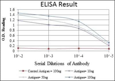
| Specificity | Detects human Thioredoxin-1 in direct ELISAs and Western blots. In direct ELISAs, less than 25% cross-reactivity with recombinant mouse Thioredoxin-1 is observed. |
| Source | N/A |
| Isotype | IgG |
| Clonality | Polyclonal |
| Host | Goat |
| Gene | TXN |
| Purity Statement | Antigen Affinity-purified |
| Innovator's Reward | Test in a species/application not listed above to receive a full credit towards a future purchase. |
| Dilutions |
|
|
| Publications |
|
| Storage | Use a manual defrost freezer and avoid repeated freeze-thaw cycles.
|
| Buffer | Lyophilized from a 0.2 μm filtered solution in PBS with Trehalose. See Certificate of Analysis for details. *Small pack size (-SP) is supplied either lyophilized or as a 0.2 µm filtered solution in PBS. |
| Preservative | No Preservative |
| Concentration | LYOPH |
| Reconstitution Instructions | Reconstitute at 0.2 mg/mL in sterile PBS. For liquid material, refer to CoA for concentration. |


Secondary Antibodies |
Isotype Controls |
The concentration calculator allows you to quickly calculate the volume, mass or concentration of your vial. Simply enter your mass, volume, or concentration values for your reagent and the calculator will determine the rest.


 (NBP3-11685) and Jurkat human acute T cell leukemia cell line, loaded at 0.5 mg/ml. A specific band was detected for Thioredoxin‑1 at approximately 10 kDa (as indicated) using 10 µg/mL of Goat Anti-Human Thioredoxin‑1 Antigen Affinity-purified Polyclonal Antibody (Catalog # AF1970). This experiment was conducted under reducing conditions and using the 2-40kDa separation system.")
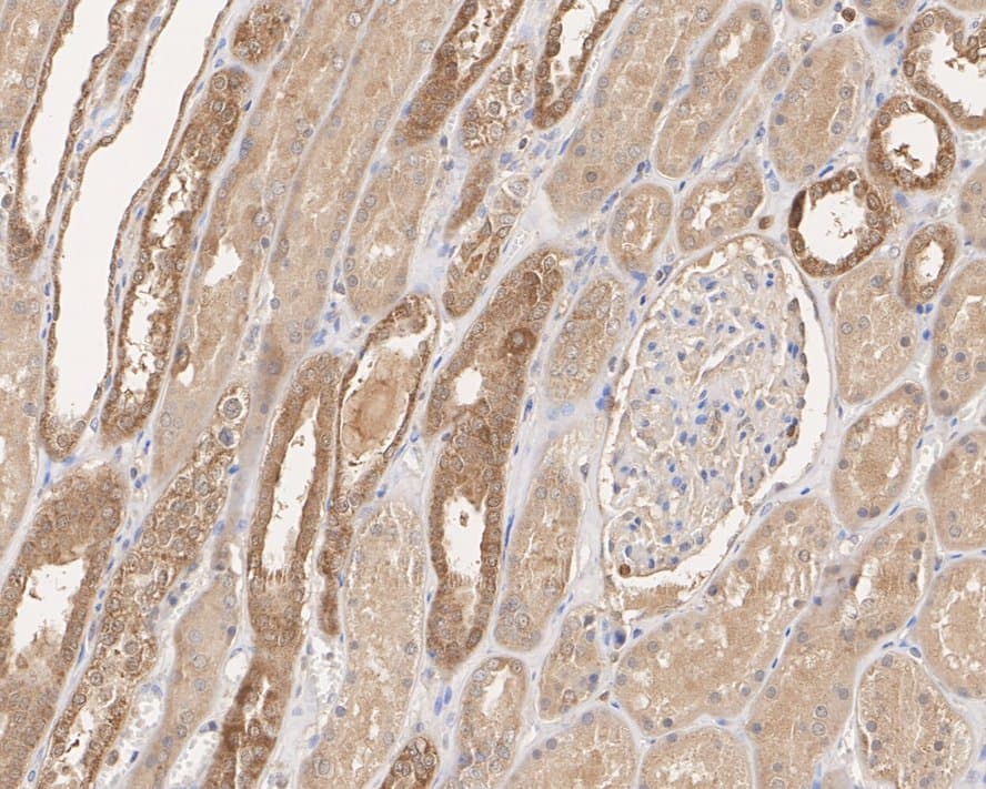
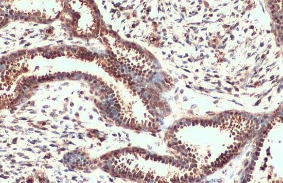
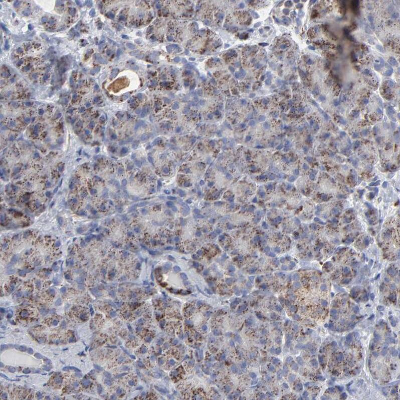
 CTS008) and counterstained with hematoxylin (blue). View our protocol for
  CTS008) and counterstained with hematoxylin (blue). View our protocol for
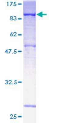
![SDS-Page Neuregulin-1 beta 1/NRG1 beta 1 [Unconjugated]](https://images.novusbio.com/images/protein/Neuregulin-1_beta_1_396-HB_222.jpg)
![SEC-MALS Neuregulin-1 beta 1/NRG1 beta 1 [Unconjugated]](https://images.novusbio.com/images/396-hb_recombinant-human-nrg1-beta-1-hrg1-beta-1-egf-domain-protein-sec-mals-51220258550.jpg)
![Bioactivity Neuregulin-1 beta 1/NRG1 beta 1 [Unconjugated]](https://images.novusbio.com/images/protein/Neuregulin-1_beta_1_396-HB_626.jpg)


![SDS-Page TNF-alpha [Unconjugated]](https://images.novusbio.com/images/protein/TNF-alpha_210-TA_256.jpg)
![Bioactivity TNF-alpha [Unconjugated]](https://images.novusbio.com/images/protein/TNFalpha_210TA_1658.jpg)
![SEC-MALS TNF-alpha [Unconjugated]](https://images.novusbio.com/images/210-ta_recombinant-human-tnf-alpha-protein-sec-mals-35202312244..jpg)
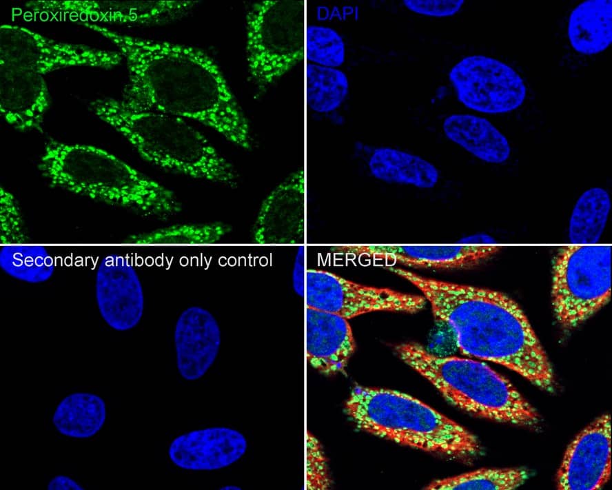
![Immunocytochemistry Catalase Antibody [Unconjugated]](https://images.novusbio.com/images/antibody/Catalase_AF3398_Immunocytochemistry__Immunofluorescence_19451.jpg)
![Simple Western Catalase Antibody [Unconjugated]](https://images.novusbio.com/images/antibody/Catalase_AF3398_Simple_Western_16990.jpg)

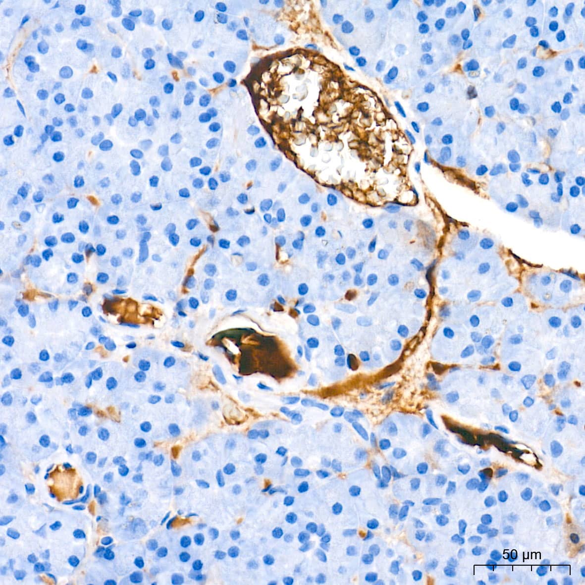
![Immunohistochemistry Insulin Antibody (182410) [Unconjugated]](https://images.novusbio.com/images/antibody/mab1417_human-bovine-mouse-insulin-mab-clone-182410-immunohistochemistry-308202115145.jpg)
![Immunocytochemistry Insulin Antibody (182410) [Unconjugated]](https://images.novusbio.com/images/antibody/Insulin_MAB1417_Immunocytochemistry_9376.jpg)
![Immunocytochemistry/ Immunofluorescence c-jun [p Ser63] Antibody (SY0297)](https://images.novusbio.com/images/c-jun-p-Ser63-Antibody-SY0297-Immunocytochemistry-Immunofluorescence-NBP2-67471-img0006.jpg)
![Western Blot c-jun [p Ser63] Antibody (SY0297)](https://images.novusbio.com/images/c-jun-p-Ser63-Antibody-SY0297-Western-Blot-NBP2-67471-img0008.jpg)
![Western Blot c-jun [p Ser63] Antibody (SY0297)](https://images.novusbio.com/images/c-jun-p-Ser63-Antibody-SY0297-Western-Blot-NBP2-67471-img0009.jpg)

![Bioactivity IL-2 [Unconjugated]](https://images.novusbio.com/images/202-il_recombinant-human-il-2-protein-bioactivity-174202314946.jpg)
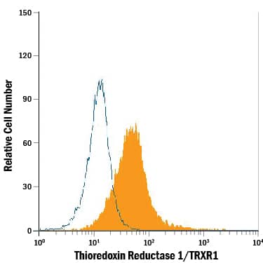
 or Normal Goat IgG Isotype Control Antibody (Catalog # AB-108-C, open histogram), followed by Phycoerythrin-conjugated Anti-Goat IgG Secondary Antibody (Catalog # F0107).")