![Synapsin-I-[p-Ser603]-Antibody-Western-Blot-NB300-181-img0002.jpg](http://images.novusbio.com/fullsize/Synapsin-I-[p-Ser603]-Antibody-Western-Blot-NB300-181-img0002.jpg "Synapsin-I-[p-Ser603]-Antibody-Western-Blot-NB300-181-img0002.jpg")
| Reactivity | Hu, Mu, Rt, Gp, Bv, Xp, ZeSpecies Glossary |
| Applications | WB |
| Clonality | Polyclonal |
| Host | Rabbit |
| Conjugate | Unconjugated |
| Format | Azide Free |
| Immunogen | Synthetic phospho-peptide corresponding to amino acid residues surrounding Ser603 conjugated to KLH. Accession # P17599 |
| Modification | p Ser603 |
| Localization | Cell junction; synapse |
| Marker | pre-Synaptic Marker |
| Specificity | Specific for endogenous levels of the ~78 kDa synapsin I doublet phosphorylated at Ser603 . Immunolabeling is completely eliminated by treatment with lambda-phosphatase. |
| Predicted Species | Bovine (100%), Zebrafish (100%), Xenopus (100%). Backed by our 100% Guarantee. |
| Isotype | IgG |
| Clonality | Polyclonal |
| Host | Rabbit |
| Gene | SYN1 |
| Purity | Antigen Affinity-purified |
| Innovator's Reward | Test in a species/application not listed above to receive a full credit towards a future purchase. |
| Dilutions |
|
|
| Theoretical MW | 78 kDa. Disclaimer note: The observed molecular weight of the protein may vary from the listed predicted molecular weight due to post translational modifications, post translation cleavages, relative charges, and other experimental factors. |
|
| Publications |
|
| Storage | Store at -20C. Avoid freeze-thaw cycles. |
| Buffer | 10mM HEPES (pH 7.5), 0.15M NaCl, 0.1 mg/ml BSA and 50% Glycerol |
| Preservative | No Preservative |
| Purity | Antigen Affinity-purified |


![Immunohistochemistry-Paraffin Tau [p Thr181] Antibody - BSA Free](https://images.novusbio.com/images/Tau-[p-Thr181]-Antibody-Immunohistochemistry-Paraffin-NB100-82245-img0005.jpg)
![Simple Western Tau [p Thr181] Antibody - BSA Free](https://images.novusbio.com/images/Tau-[p-Thr181]-Antibody-Simple-Western-NB100-82245-img0004.jpg)
![Western Blot Tau [p Thr181] Antibody - BSA Free](https://images.novusbio.com/images/Tau-[p-Thr181]-Antibody-Western-Blot-NB100-82245-img0001.jpg)
Secondary Antibodies |
Isotype Controls |
Research Areas for Synapsin I Antibody (NB300-181)Find related products by research area.
|
|
Synapsin I, a pre-synaptic marker Synapsin-I, also called Synapsin 1/Syn1, is an ~80 kDa protein (predicted mol. wt. 74.1 kDa) which belongs to the Synapsin family (Synapsin I, Synapsin II, Synapsin III). Synapsins are the evolutionarily conserved phospho-proteins which are associ... Read full blog post. |
The concentration calculator allows you to quickly calculate the volume, mass or concentration of your vial. Simply enter your mass, volume, or concentration values for your reagent and the calculator will determine the rest.


![Western Blot: Synapsin I [p Ser603] Antibody [NB300-181] - Rat cortical lysate showing specific immunolabeling of the ~78 kDa synapsin I phosphorylated at Ser603 in the first lane (-). Phosphospecificity is shown in thesecond lane (+) where the immunolabeling is completely eliminated by blot treatment with lambda phosphatase (l-Ptase, 1200 units for 30 minutes).](http://images.novusbio.com/fullsize/Synapsin-I-[p-Ser603]-Antibody-Western-Blot-NB300-181-img0001.jpg "Western Blot: Synapsin I [p Ser603] Antibody [NB300-181] - Rat cortical lysate showing specific immunolabeling of the ~78 kDa synapsin I phosphorylated at Ser603 in the first lane (-). Phosphospecificity is shown in thesecond lane (+) where the immunolabeling is completely eliminated by blot treatment with lambda phosphatase (l-Ptase, 1200 units for 30 minutes).")
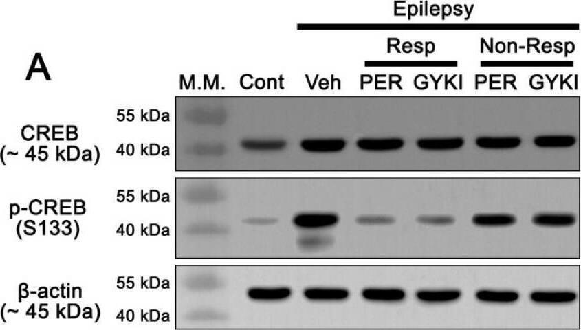
 are consistent with splice variants as confirmed by manufacturer. P-CAMKII and CAMKII were detected around 50 kDa in accordance with previous findings.")
![Western Blot: Synapsin I [p Ser603] Antibody [NB300-181] - Examples of Western blots of p-/synapsin 1 & p-/Ca2+-calmodulin-dependent kinase II. The figure depicts examples of the Western blots of the four synaptic plasticity markers investigated. The two bands seen in the p-synapsin & synapsin Western blots (70 & 74 kDa respectively) are consistent with splice variants as confirmed by manufacturer. P-CAMKII & CAMKII were detected around 50 kDa in accordance with previous findings. Image collected & cropped by CiteAb from the following publication (//pubmed.ncbi.nlm.nih.gov/29890692), licensed under a CC-BY license. Not internally tested by Novus Biologicals.](http://images.novusbio.com/fullsize/nb300-181_rabbit-polyclonal-synapsin-i-p-ser603-antibody-imgenex-img-683-31020241539736.jpg "Western Blot: Synapsin I [p Ser603] Antibody [NB300-181] - Examples of Western blots of p-/synapsin 1 & p-/Ca2+-calmodulin-dependent kinase II. The figure depicts examples of the Western blots of the four synaptic plasticity markers investigated. The two bands seen in the p-synapsin & synapsin Western blots (70 & 74 kDa respectively) are consistent with splice variants as confirmed by manufacturer. P-CAMKII & CAMKII were detected around 50 kDa in accordance with previous findings. Image collected & cropped by CiteAb from the following publication (//pubmed.ncbi.nlm.nih.gov/29890692), licensed under a CC-BY license. Not internally tested by Novus Biologicals.")
![N/A BDNF [HRP]](https://images.novusbio.com/images/elisa/BDNF_DBD00_ELISA_36.jpg)
![N/A BDNF [HRP]](https://images.novusbio.com/images/elisa/DATA_BDNF_DBD00_ELISA_587.jpg)
![N/A BDNF [HRP]](https://images.novusbio.com/images/elisa/DATA_BDNF_DBD00_ELISA_588.jpg)


![Western Blot ERK2 Antibody [Unconjugated]](https://images.novusbio.com/images/antibody/ERK2_AF1230_Western_Blot_5097.jpg)
![Knockout Validated ERK2 Antibody [Unconjugated]](https://images.novusbio.com/images/antibody/ERK2_AF1230_Knockout_Validated_22864.jpg)
![Immunohistochemistry ERK2 Antibody [Unconjugated]](https://images.novusbio.com/images/antibody/ERK2_AF1230_Immunohistochemistry_20696.jpg)
![Western Blot Carbonic Anhydrase III/CA3 Antibody [Unconjugated]](https://images.novusbio.com/images/antibody/Carbonic_Anhydrase_III_AF2185_Western_Blot_20454.jpg)
![Immunocytochemistry EGR1 Antibody [Unconjugated]](https://images.novusbio.com/images/antibody/af2818_human-egr1-affinity-purified-polyclonal-ab-immunocytochemistry-3102024831..jpg)


![Immunocytochemistry Synaptotagmin 1 Antibody (ASV48) [Unconjugated]](https://images.novusbio.com/images/antibody/Synaptotagmin-1_MAB4364_Immunocytochemistry_11323.jpg)
![Knockout Validated Synaptotagmin 1 Antibody (ASV48) [Unconjugated]](https://images.novusbio.com/images/antibody/mab4364_rat-synaptotagmin-1-mab-clone-asv48-knockout-validated-2862024122433..png)
![Knockout Validated Synaptotagmin 1 Antibody (ASV48) [Unconjugated]](https://images.novusbio.com/images/antibody/mab4364_rat-synaptotagmin-1-mab-clone-asv48-knockout-validated-286202412275.jpg)
![Western Blot: Goat anti-Rabbit IgG (H+L) Secondary Antibody [HRP] [NB7160] - Western blot showing vemurafenib treatment in BRAFV600E CRC cells inhibits fission mediator DRP1 with no significant effect on fusion proteins (Mfn1 & 2) using MFN-1 antibody (NBP1-51841) and corresponding secondary antibody, goat anti-rabbit IgG-HRP (NB7160). Image collected and cropped by CiteAb from the following publication (https://pubmed.ncbi.nlm.nih.gov/33738242).](https://images.novusbio.com/images/Goat-anti-Rabbit-IgG-H+L-Secondary-Antibody-HRP-Western-Blot-NB7160-img0001.jpg "Western Blot: Goat anti-Rabbit IgG (H+L) Secondary Antibody [HRP] [NB7160] - Western blot showing vemurafenib treatment in BRAFV600E CRC cells inhibits fission mediator DRP1 with no significant effect on fusion proteins (Mfn1 & 2) using MFN-1 antibody (NBP1-51841) and corresponding secondary antibody, goat anti-rabbit IgG-HRP (NB7160). Image collected and cropped by CiteAb from the following publication (https://pubmed.ncbi.nlm.nih.gov/33738242).")
 followed by 30 min incubation with Goat anti Rabbit HRP conjugated secondary antibodies (Catalog # HAF008) at 1:20 dilution + DAB chromogen (brown). The tissue was counterstained with Hematoxylin (blue). Control was done by omitting primary antibody.")
![Flow Cytometry: Rabbit IgG Isotype Control [NBP2-24891] - An intracellular stain was performed on Raji cells with Adiponectin antibody NB100-65810 (blue) and a matched isotype control NBP2-24893 (orange). Cells were fixed with 4% PFA and then permeablized with 0.1% saponin. Cells were incubated in an antibody dilution of 1 ug/mL for 30 minutes at room temperature, followed by Dylight488-conjugated anti-rabbit secondary antibody. Image using the Azide Free form of this antibody.](https://images.novusbio.com/images/Rabbit--Mouse-IgG-Isotype-Control-Flow-Cytometry-NBP2-24891-img0006.jpg "Flow Cytometry: Rabbit IgG Isotype Control [NBP2-24891] - An intracellular stain was performed on Raji cells with Adiponectin antibody NB100-65810 (blue) and a matched isotype control NBP2-24893 (orange). Cells were fixed with 4% PFA and then permeablized with 0.1% saponin. Cells were incubated in an antibody dilution of 1 ug/mL for 30 minutes at room temperature, followed by Dylight488-conjugated anti-rabbit secondary antibody. Image using the Azide Free form of this antibody.")