| Reactivity | HuSpecies Glossary |
| Applications | Bioactivity |
| Format | Carrier-Free |
| Details of Functionality | Measured by the ability of the immobilized protein to support the adhesion of human red blood cells. Kelm, S. et al. (1994) Current Biology 4:965. The ED50 for this effect is 0.6‑3 µg/mL. |
| Source | Mouse myeloma cell line, NS0-derived human Siglec-1/CD169 protein Ser20-Gln1641, with a C-terminal 6-His tag |
| Accession # | |
| N-terminal Sequence | Ser20 |
| Protein/Peptide Type | Recombinant Proteins |
| Gene | SIGLEC1 |
| Purity | >80%, by SDS-PAGE visualized with Silver Staining and quantitative densitometry by Coomassie® Blue Staining |
| Endotoxin Note | <0.10 EU per 1 μg of the protein by the LAL method. |
| Dilutions |
|
|
| Theoretical MW | 173.9 kDa. Disclaimer note: The observed molecular weight of the protein may vary from the listed predicted molecular weight due to post translational modifications, post translation cleavages, relative charges, and other experimental factors. |
|
| SDS-PAGE | 175 kDa-190 kDa, reducing conditions |
|
| Publications |
|
| Storage | Use a manual defrost freezer and avoid repeated freeze-thaw cycles.
|
| Buffer | Lyophilized from a 0.2 μm filtered solution in PBS. |
| Purity | >80%, by SDS-PAGE visualized with Silver Staining and quantitative densitometry by Coomassie® Blue Staining |
| Reconstitution Instructions | Reconstitute at 100 μg/mL in sterile PBS. |
Siglecs are sialic acid specific I-type lectins that belong to the immunoglobulin superfamily. Structurally, they are transmembrane proteins with an N-terminal Ig-like V‑set domain followed by varying numbers of Ig-like C2-set domains (1, 2). Human Siglec-1, also known as sialoadhesin and CD169, is a 175-185 kDa glycoprotein. It contains a 1622 amino acid (aa) extracellular domain (ECD) with one Ig-like V‑set domain and 16 Ig-like C2-set domains, a 21 aa transmembrane segment, and a 44 aa cytoplasmic domain (3). Within the ECD, human Siglec-1 shares approximately 70% aa sequence identity with mouse and rat Siglec-1. Alternate splicing generates a potentially soluble form of the ECD, and a second isoform with a substituted cytoplasmic domain. Siglec-1 expression is restricted to lymph node and splenic macrophages, plus some tissue macrophages (3). The adhesive function of Siglec-1 is supported by the N-terminal Ig-like domain which shows a selectivity for alpha 2,3-linked sialic acid residues (3-5). Siglec-1 binds a number of sialylated molecules including the mannose receptor, MGL1, MUC1, PSGL-1, and different glycoforms of CD43 (6-9). Its binding capacity can be masked by endogenous sialylated molecules (10, 11). The sialylated and sulfated N-linked carbohydrates that modify Siglec-1 itself are required for ligand binding (6, 7). Siglec-1 is expressed on dendritic cells following rhinovirus exposure, and these DC promote T cell anergy (12). It is also induced on circulating monocytes during systemic sclerosis and HIV-1 infection (13 - 15). Siglec-1 can trap HIV-1 particles for trans infection of permissive cells (14).


The concentration calculator allows you to quickly calculate the volume, mass or concentration of your vial. Simply enter your mass, volume, or concentration values for your reagent and the calculator will determine the rest.


![Bioactivity CTLA-4 [Unconjugated]](https://images.novusbio.com/images/protein/CTLA4_7268CT_2293.jpg)


![Immunocytochemistry CD55/DAF Antibody [Unconjugated]](https://images.novusbio.com/images/antibody/CD55_AF2009_Immunocytochemistry__Immunofluorescence_23052.jpg)
![Immunohistochemistry CD55/DAF Antibody [Unconjugated]](https://images.novusbio.com/images/antibody/CD55_AF2009_Immunohistochemistry_6728.jpg)


![SDS-Page TNF-alpha [Unconjugated]](https://images.novusbio.com/images/protein/TNF-alpha_210-TA_256.jpg)
![Bioactivity TNF-alpha [Unconjugated]](https://images.novusbio.com/images/protein/TNFalpha_210TA_1658.jpg)
![SEC-MALS TNF-alpha [Unconjugated]](https://images.novusbio.com/images/210-ta_recombinant-human-tnf-alpha-protein-sec-mals-35202312244..jpg)
![Bioactivity IL-2 [Unconjugated]](https://images.novusbio.com/images/202-il_recombinant-human-il-2-protein-bioactivity-174202314946.jpg)
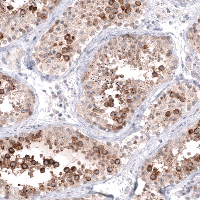
![Immunohistochemistry TFF2 Antibody (366508) [Unconjugated]](https://images.novusbio.com/images/antibody/TFF2_MAB4077_Immunohistochemistry_21485.jpg)

![Flow Cytometry: Siglec-1/CD169 Antibody (HSn 7D2) [NB600-534] - Human CD14+ PBMC differentiated to M1 macrophages with rhGM-CSF were stained with Mouse Anti-Siglec-1/CD169 Monoclonal Antibody (NB600-534, filled histogram), or Mouse IgG1 isotype control (MAB002, open histogram) followed by APC-conjugated Anti-Mouse IgG Secondary Antibody (F0101B).](https://images.novusbio.com/images/Siglec-1-CD169-Antibody-HSn-7D2-Flow-Cytometry-NB600-534-img0002.jpg "Flow Cytometry: Siglec-1/CD169 Antibody (HSn 7D2) [NB600-534] - Human CD14+ PBMC differentiated to M1 macrophages with rhGM-CSF were stained with Mouse Anti-Siglec-1/CD169 Monoclonal Antibody (NB600-534, filled histogram), or Mouse IgG1 isotype control (MAB002, open histogram) followed by APC-conjugated Anti-Mouse IgG Secondary Antibody (F0101B).")