| Reactivity | HuSpecies Glossary |
| Applications | Bioactivity |
| Format | Carrier-Free |
| Details of Functionality | Measured by the ability of the immobilized protein to support the adhesion of Neuro‑2A mouse neuroblastoma cells. Recombinant Human FLRT3 immobilized at 5 μg/mL, 100 μL/well, will mediate >50% Neuro‑2A cell adhesion. Optimal dilutions should be determined by each laboratory for each application. |
| Source | Mouse myeloma cell line, NS0-derived human FLRT3 protein Lys29-Pro528, with a C-terminal 6-His tag |
| Accession # | |
| N-terminal Sequence | Lys29 |
| Protein/Peptide Type | Recombinant Proteins |
| Gene | FLRT3 |
| Purity | >95%, by SDS-PAGE under reducing conditions and visualized by silver stain |
| Endotoxin Note | <0.01 EU per 1 μg of the protein by the LAL method. |
| Dilutions |
|
|
| Theoretical MW | 57.3 kDa . Disclaimer note: The observed molecular weight of the protein may vary from the listed predicted molecular weight due to post translational modifications, post translation cleavages, relative charges, and other experimental factors. |
|
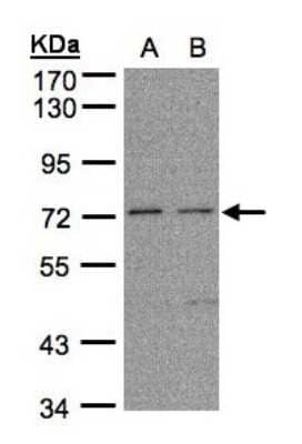
| SDS-PAGE | 70-90 kDa, under reducing conditions |
|
| Publications |
|
| Storage | Use a manual defrost freezer and avoid repeated freeze-thaw cycles.
|
| Buffer | Lyophilized from a 0.2 μm filtered solution in PBS. |
| Purity | >95%, by SDS-PAGE under reducing conditions and visualized by silver stain |
| Reconstitution Instructions | Reconstitute at 200 μg/mL in sterile PBS. |
FLRT3 is one of three FLRT (fibronectin, leucine rich repeat, transmembrane) glycoproteins expressed in distinct areas of the developing brain and other tissues (1, 2). The 85-95 kDa type I transmembrane (TM) human FLRT3 is synthesized as a 649 amino acid (aa) precursor with a 28 aa signal sequence, a 500 aa extracellular domain (ECD), a 21 aa TM segment and a 100 aa cytoplasmic region. The ECD contains 10 N-terminal leucine-rich repeats flanked by cysteine-rich areas, and a juxtamembrane fibronectin type III domain (1). The human FLRT3 ECD shares 96%, 96%, 97%, 97%, 98% and 81% aa sequence identity with mouse, rat, canine, bovine, equine and Xenopus FLRT3 ECD, respectively, and 61% and 48% aa identity to human FLRT2 and FLRT3 ECDs, respectively. The fibronectin domain is responsible for binding to FGF receptors, and is thought to regulate FGF signaling during development (2, 3). The LRR domains are responsible for both the localization in areas of cell contact and homotypic cell-cell association (4). This may be through direct interaction with other FLRT molecules, or alternatively, by regulating internalization of adhesion molecules such as cadherins (4, 5). Developmentally, FLRT3 is located in somitic regions on dermatomyotomal muscle precursors and myotomal cells before their migration to the myotome and syndetome, respectively (2). FLRT3 is also expressed at the midbrain/hindbrain boundary and in the apical ectodermal ridge where it may influence FGF signaling (2). Genetic deletion in mouse embryos results in defective headfold fusion and endoderm migration (6). Postnatally, FLRT3 mRNA is widely expressed (1). It is up-regulated and promotes neurite outgrowth following experimental peripheral nerve injury in rats (7, 8).


![Western Blot EphB2 Antibody [Unconjugated]](https://images.novusbio.com/images/af467_human-mouse-ephb2-affinity-purified-polyclonal-ab-41202410485412.jpg)
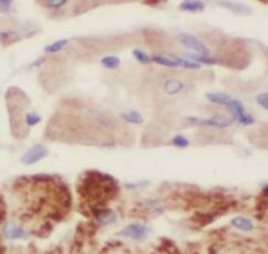
![Immunohistochemistry EphB2 Antibody [Unconjugated]](https://images.novusbio.com/images/antibody/EphB2_AF467_Immunohistochemistry_20369.jpg)
![Western Blot EphB2 Antibody [Unconjugated]](https://images.novusbio.com/images/antibody/EphB2_AF467_Western_Blot_21511.jpg)


The concentration calculator allows you to quickly calculate the volume, mass or concentration of your vial. Simply enter your mass, volume, or concentration values for your reagent and the calculator will determine the rest.


![Simple Western FLRT2 Antibody [Unconjugated]](https://images.novusbio.com/images/antibody/af2877_human-flrt2-affinity-purified-polyclonal-ab-simple-western-2182024142725..jpg)
![Western Blot FLRT2 Antibody [Unconjugated]](https://images.novusbio.com/images/antibody/af2877_human-flrt2-affinity-purified-polyclonal-ab-western-blot-1272024125547..jpg)
![Bioactivity TGF-beta 1 [Unconjugated]](https://images.novusbio.com/images/protein/7754-bhcf_recombinant-human-tgf-beta-1-human-cell-expressed-cf-bioactivity-1811202013946.jpg)
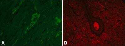
![Immunohistochemistry UNC5H2/UNC5B Antibody [Unconjugated]](https://images.novusbio.com/images/af1006_rat-unc5h2-unc5b-affinity-purified-polyclonal-ab-immunohistochemistry-3152022161010.jpg)
![Western Blot UNC5H2/UNC5B Antibody [Unconjugated]](https://images.novusbio.com/images/antibody/UNC5H2_AF1006_Western_Blot_17311.jpg)


![Cell Culture FGF-8 [Unconjugated]](https://images.novusbio.com/images/423-f8_recombinant-human-mouse-fgf-8b-protein-cell-culture-1052024145159..jpg)
![Bioactivity FGF-8 [Unconjugated]](https://images.novusbio.com/images/protein/FGF8_423F8_1438.jpg)
![Cell Culture FGF-8 [Unconjugated]](https://images.novusbio.com/images/423-f8_recombinant-human-mouse-fgf-8b-protein-cell-culture-1052024145958..jpg)
![Western Blot ERK2 Antibody [Unconjugated]](https://images.novusbio.com/images/antibody/ERK2_AF1230_Western_Blot_5097.jpg)
![Knockout Validated ERK2 Antibody [Unconjugated]](https://images.novusbio.com/images/antibody/ERK2_AF1230_Knockout_Validated_22864.jpg)
![Immunohistochemistry ERK2 Antibody [Unconjugated]](https://images.novusbio.com/images/antibody/ERK2_AF1230_Immunohistochemistry_20696.jpg)