 HAF016). A specific band was detected for Neurogranin at approximately 14-16 kDa (as indicated). This experiment was conducted under reducing conditions and using Immunoblot Buffer Group 1." title="Western blot shows lysates of mouse brain (cortex) tissue, human brain (motor cortex) tissue, and rat brain (cortex) tissue. PVDF membrane was probed with 0.5 µg/mL of Sheep Anti-Human/Mouse/Rat Neurogranin Antigen Affinity-purified Polyclonal Antibody (Catalog # AF7947) followed by HRP-conjugated Anti-Sheep IgG Secondary Antibody (HAF016). A specific band was detected for Neurogranin at approximately 14-16 kDa (as indicated). This experiment was conducted under reducing conditions and using Immunoblot Buffer Group 1." />
| Reactivity | Hu, Mu, RtSpecies Glossary |
| Applications | WB, Simple Western, ELISA, IHC |
| Clonality | Polyclonal |
| Host | Sheep |
| Conjugate | Unconjugated |
| Concentration | LYOPH |
| Immunogen | E. coli-derived recombinant human Neurogranin Met1-Asp78 Accession # Q92686 |
| Specificity | Detects human, mouse, and rat Neurogranin Western blots. |
| Source | N/A |
| Isotype | IgG |
| Clonality | Polyclonal |
| Host | Sheep |
| Gene | NRGN |
| Purity Statement | Antigen Affinity-purified |
| Innovator's Reward | Test in a species/application not listed above to receive a full credit towards a future purchase. |
| Dilutions |
|
|
| Publications |
|
| Storage | Use a manual defrost freezer and avoid repeated freeze-thaw cycles.
|
| Buffer | Lyophilized from a 0.2 μm filtered solution in PBS with Trehalose. *Small pack size (SP) is supplied either lyophilized or as a 0.2 µm filtered solution in PBS. |
| Preservative | No Preservative |
| Concentration | LYOPH |
| Reconstitution Instructions | Sterile PBS to a final concentration of 0.2 mg/mL. |












The concentration calculator allows you to quickly calculate the volume, mass or concentration of your vial. Simply enter your mass, volume, or concentration values for your reagent and the calculator will determine the rest.


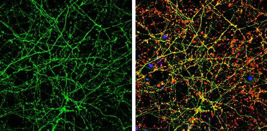
 NL010) and counterstained with DAPI (blue). Specific staining was localized to pyramidal neurons in the hippocampus. View our protocol for
  HAF016). This experiment was conducted under reducing conditions and using the 2-40 kDa separation system." title="Simple Western lane view shows lysates of human brain (motor cortex), mouse brain, and rat brain, loaded at 0.2 mg/mL. A specific band was detected for Neurogranin at approximately 12 kDa (as indicated) using 20 µg/mL of Sheep Anti-Human/Mouse/Rat Neurogranin Antigen Affinity-purified Polyclonal Antibody (Catalog # AF7947) followed by 1:50 dilution of HRP-conjugated Anti-Sheep IgG Secondary Antibody (
 MAB79473) coated on a Clear Polystyrene Microplate (Catalog #




![SDS-Page TNF-alpha [Unconjugated]](https://images.novusbio.com/images/protein/TNF-alpha_210-TA_256.jpg)
![Bioactivity TNF-alpha [Unconjugated]](https://images.novusbio.com/images/protein/TNFalpha_210TA_1658.jpg)
![SEC-MALS TNF-alpha [Unconjugated]](https://images.novusbio.com/images/210-ta_recombinant-human-tnf-alpha-protein-sec-mals-35202312244..jpg)


![Western Blot ERK2 Antibody [Unconjugated]](https://images.novusbio.com/images/antibody/ERK2_AF1230_Western_Blot_5097.jpg)
![Knockout Validated ERK2 Antibody [Unconjugated]](https://images.novusbio.com/images/antibody/ERK2_AF1230_Knockout_Validated_22864.jpg)
![Immunohistochemistry ERK2 Antibody [Unconjugated]](https://images.novusbio.com/images/antibody/ERK2_AF1230_Immunohistochemistry_20696.jpg)


